|
Columnar Epithelial Cell
Epithelium or epithelial tissue is one of the four basic types of animal tissue, along with connective tissue, muscle tissue and nervous tissue. It is a thin, continuous, protective layer of compactly packed cells with a little intercellular matrix. Epithelial tissues line the outer surfaces of organs and blood vessels throughout the body, as well as the inner surfaces of cavities in many internal organs. An example is the epidermis, the outermost layer of the skin. There are three principal shapes of epithelial cell: squamous (scaly), columnar, and cuboidal. These can be arranged in a singular layer of cells as simple epithelium, either squamous, columnar, or cuboidal, or in layers of two or more cells deep as stratified (layered), or ''compound'', either squamous, columnar or cuboidal. In some tissues, a layer of columnar cells may appear to be stratified due to the placement of the nuclei. This sort of tissue is called pseudostratified. All glands are made up of epithelia ... [...More Info...] [...Related Items...] OR: [Wikipedia] [Google] [Baidu] |
Animal
Animals are multicellular, eukaryotic organisms in the Kingdom (biology), biological kingdom Animalia. With few exceptions, animals Heterotroph, consume organic material, Cellular respiration#Aerobic respiration, breathe oxygen, are Motility, able to move, can Sexual reproduction, reproduce sexually, and go through an ontogenetic stage in which their body consists of a hollow sphere of Cell (biology), cells, the blastula, during Embryogenesis, embryonic development. Over 1.5 million Extant taxon, living animal species have been Species description, described—of which around 1 million are Insecta, insects—but it has been estimated there are over 7 million animal species in total. Animals range in length from to . They have Ecology, complex interactions with each other and their environments, forming intricate food webs. The scientific study of animals is known as zoology. Most living animal species are in Bilateria, a clade whose members have a Symmetry in biology#Bilate ... [...More Info...] [...Related Items...] OR: [Wikipedia] [Google] [Baidu] |
Transcellular Transport
Transcellular transport involves the transportation of solutes by a cell ''through'' a cell. Transcellular transport can occur in three different ways active transport, passive transport, and transcytosis. Active Transport Main article: Active transport Active transport is the process of moving molecules from an area of low concentrations to an area of high concentration. There are two types of active transport, primary active transport and secondary active transport. Primary active transport uses adenosine triphosphate (ATP) to move specific molecules and solutes against its concentration gradient. Examples of molecules that follow this process are potassium K+, sodium Na+, and calcium Ca2+. A place in the human body where this occurs is in the intestines with the uptake of glucose. Secondary active transport is when one solute moves down the electrochemical gradient to produce enough energy to force the transport of another solute from low concentration to high concentration. ... [...More Info...] [...Related Items...] OR: [Wikipedia] [Google] [Baidu] |
Mesothelium
The mesothelium is a membrane composed of simple squamous epithelial cells of mesodermal origin, which forms the lining of several body cavities: the pleura (pleural cavity around the lungs), peritoneum (abdominopelvic cavity including the mesentery, omenta, falciform ligament and the perimetrium) and pericardium (around the heart). Mesothelial tissue also surrounds the male testis (as the tunica vaginalis) and occasionally the spermatic cord (in a patent processus vaginalis). Mesothelium that covers the internal organs is called visceral mesothelium, while one that covers the surrounding body walls is called the parietal mesothelium. The mesothelium that secretes serous fluid as a main function is also known as a serosa. Origin Mesothelium derives from the embryonic mesoderm cell layer, that lines the coelom (body cavity) in the embryo. It develops into the layer of cells that covers and protects most of the internal organs of the body. Structure The mesothelium forms a ... [...More Info...] [...Related Items...] OR: [Wikipedia] [Google] [Baidu] |
Pericardium
The pericardium, also called pericardial sac, is a double-walled sac containing the heart and the roots of the great vessels. It has two layers, an outer layer made of strong connective tissue (fibrous pericardium), and an inner layer made of serous membrane (serous pericardium). It encloses the pericardial cavity, which contains pericardial fluid, and defines the middle mediastinum. It separates the heart from interference of other structures, protects it against infection and blunt trauma, and lubricates the heart's movements. The English name originates from the Ancient Greek prefix "''peri-''" (περί; "around") and the suffix "''-cardion''" (κάρδιον; "heart"). Anatomy The pericardium is a tough fibroelastic sac which covers the heart from all sides except at the cardiac root (where the great vessels join the heart) and the bottom (where only the serous pericardium exists to cover the upper surface of the central tendon of diaphragm). The fibrous pericardiu ... [...More Info...] [...Related Items...] OR: [Wikipedia] [Google] [Baidu] |
Endothelium
The endothelium is a single layer of squamous endothelial cells that line the interior surface of blood vessels and lymphatic vessels. The endothelium forms an interface between circulating blood or lymph in the lumen and the rest of the vessel wall. Endothelial cells form the barrier between vessels and tissue and control the flow of substances and fluid into and out of a tissue. Endothelial cells in direct contact with blood are called vascular endothelial cells whereas those in direct contact with lymph are known as lymphatic endothelial cells. Vascular endothelial cells line the entire circulatory system, from the heart to the smallest capillaries. These cells have unique functions that include fluid filtration, such as in the glomerulus of the kidney, blood vessel tone, hemostasis, neutrophil recruitment, and hormone trafficking. Endothelium of the interior surfaces of the heart chambers is called endocardium. An impaired function can lead to serious health issues throug ... [...More Info...] [...Related Items...] OR: [Wikipedia] [Google] [Baidu] |
Type 1 Pneumocyte
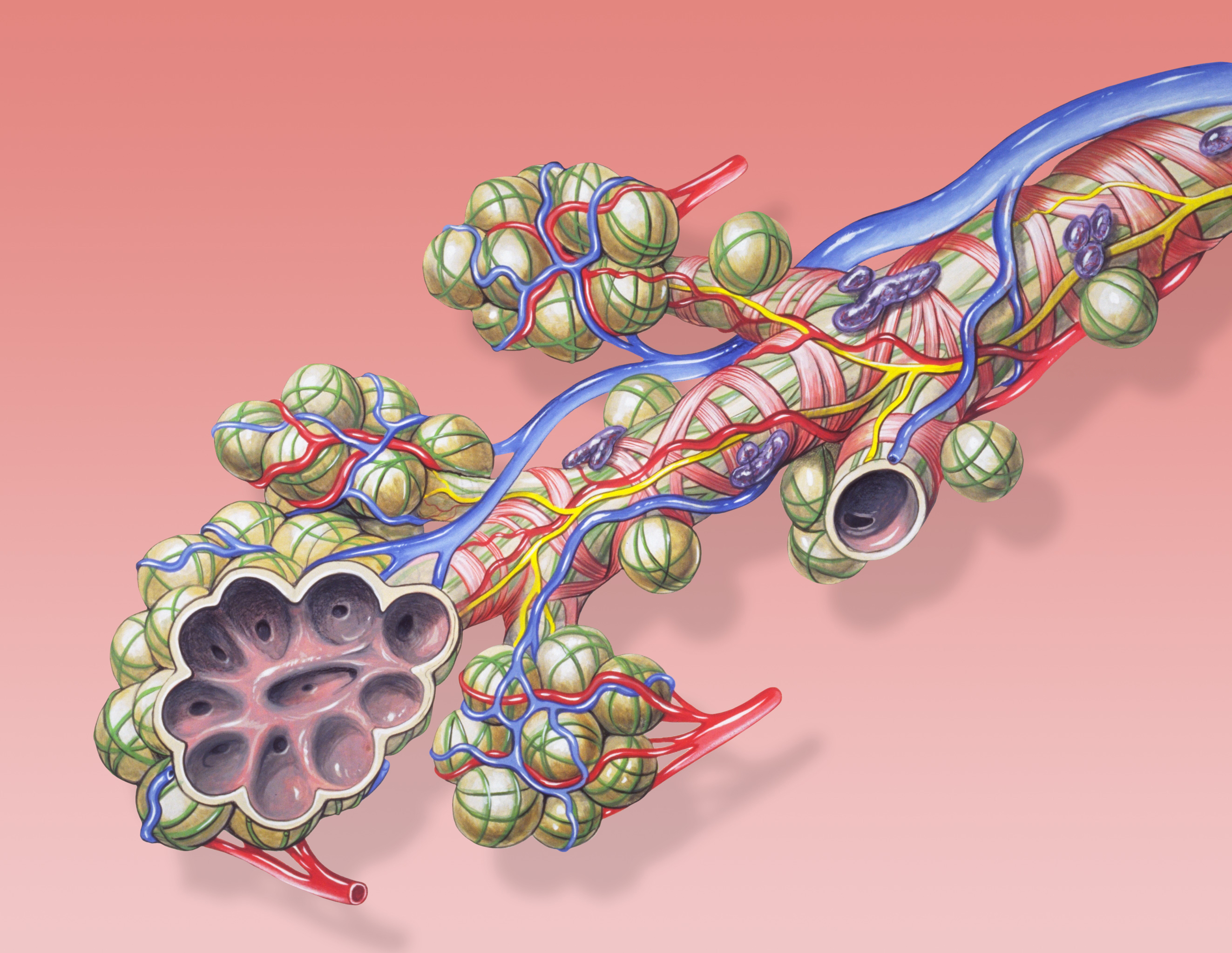
A pulmonary alveolus (plural: alveoli, from Latin ''alveolus'', "little cavity"), also known as an air sac or air space, is one of millions of hollow, distensible cup-shaped cavities in the lungs where oxygen is exchanged for carbon dioxide. Alveoli make up the functional tissue of the mammalian lungs known as the lung parenchyma, which takes up 90 percent of the total lung volume. Alveoli are first located in the respiratory bronchioles that mark the beginning of the respiratory zone. They are located sparsely in these bronchioles, line the walls of the alveolar ducts, and are more numerous in the blind-ended alveolar sacs. The acini are the basic units of respiration, with gas exchange taking place in all the alveoli present. The alveolar membrane is the gas exchange surface, surrounded by a network of capillaries. Across the membrane oxygen is diffused into the capillaries and carbon dioxide released from the capillaries into the alveoli to be breathed out. Alveoli are part ... [...More Info...] [...Related Items...] OR: [Wikipedia] [Google] [Baidu] |
Lung
The lungs are the primary organs of the respiratory system in humans and most other animals, including some snails and a small number of fish. In mammals and most other vertebrates, two lungs are located near the backbone on either side of the heart. Their function in the respiratory system is to extract oxygen from the air and transfer it into the bloodstream, and to release carbon dioxide from the bloodstream into the atmosphere, in a process of gas exchange. Respiration is driven by different muscular systems in different species. Mammals, reptiles and birds use their different muscles to support and foster breathing. In earlier tetrapods, air was driven into the lungs by the pharyngeal muscles via buccal pumping, a mechanism still seen in amphibians. In humans, the main muscle of respiration that drives breathing is the diaphragm. The lungs also provide airflow that makes vocal sounds including human speech possible. Humans have two lungs, one on the left and on ... [...More Info...] [...Related Items...] OR: [Wikipedia] [Google] [Baidu] |
Pulmonary Alveolus
A pulmonary alveolus (plural: alveoli, from Latin ''alveolus'', "little cavity"), also known as an air sac or air space, is one of millions of hollow, distensible cup-shaped cavities in the lungs where oxygen is exchanged for carbon dioxide. Alveoli make up the functional tissue of the mammalian lungs known as the lung parenchyma, which takes up 90 percent of the total lung volume. Alveoli are first located in the respiratory bronchioles that mark the beginning of the respiratory zone. They are located sparsely in these bronchioles, line the walls of the alveolar ducts, and are more numerous in the blind-ended alveolar sacs. The acini are the basic units of respiration, with gas exchange taking place in all the alveoli present. The alveolar membrane is the gas exchange surface, surrounded by a network of capillaries. Across the membrane oxygen is diffused into the capillaries and carbon dioxide released from the capillaries into the alveoli to be breathed out. Alveoli are pa ... [...More Info...] [...Related Items...] OR: [Wikipedia] [Google] [Baidu] |
Human Skin
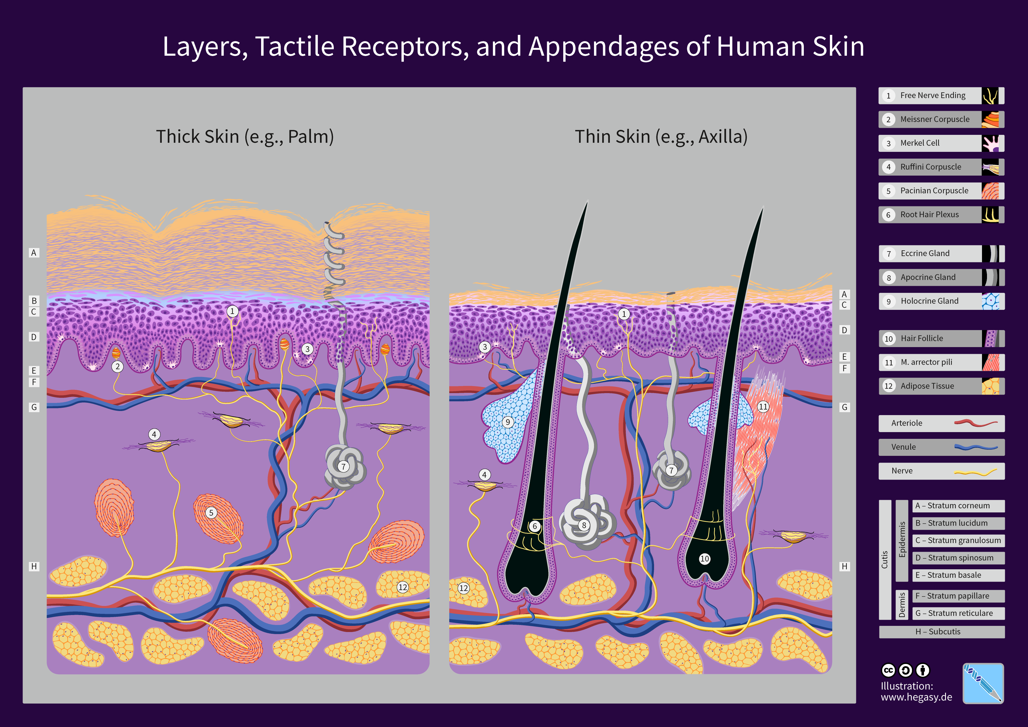
The human skin is the outer covering of the body and is the largest organ of the integumentary system. The skin has up to seven layers of ectodermal tissue guarding muscles, bones, ligaments and internal organs. Human skin is similar to most of the other mammals' skin, and it is very similar to pig skin. Though nearly all human skin is covered with hair follicles, it can appear hairless. There are two general types of skin, hairy and glabrous skin (hairless). The adjective ''cutaneous'' literally means "of the skin" (from Latin ''cutis'', skin). Because it interfaces with the environment, skin plays an important immunity role in protecting the body against pathogens and excessive water loss. Its other functions are insulation, temperature regulation, sensation, synthesis of vitamin D, and the protection of vitamin B folates. Severely damaged skin will try to heal by forming scar tissue. This is often discoloured and depigmented. In humans, skin pigmentation (affected b ... [...More Info...] [...Related Items...] OR: [Wikipedia] [Google] [Baidu] |
Morphology (biology)
Morphology is a branch of biology dealing with the study of the form and structure of organisms and their specific structural features. This includes aspects of the outward appearance (shape, structure, colour, pattern, size), i.e. external morphology (or eidonomy), as well as the form and structure of the internal parts like bones and organs, i.e. internal morphology (or anatomy). This is in contrast to physiology, which deals primarily with function. Morphology is a branch of life science dealing with the study of gross structure of an organism or taxon and its component parts. History The etymology of the word "morphology" is from the Ancient Greek (), meaning "form", and (), meaning "word, study, research". While the concept of form in biology, opposed to function, dates back to Aristotle (see Aristotle's biology), the field of morphology was developed by Johann Wolfgang von Goethe (1790) and independently by the German anatomist and physiologist Karl Friedrich Burdach ... [...More Info...] [...Related Items...] OR: [Wikipedia] [Google] [Baidu] |
423 Table 04 02 Summary Of Epithelial Tissue CellsN
4 (four) is a number, numeral and digit. It is the natural number following 3 and preceding 5. It is the smallest semiprime and composite number, and is considered unlucky in many East Asian cultures. In mathematics Four is the smallest composite number, its proper divisors being and . Four is the sum and product of two with itself: 2 + 2 = 4 = 2 x 2, the only number b such that a + a = b = a x a, which also makes four the smallest squared prime number p^. In Knuth's up-arrow notation, , and so forth, for any number of up arrows. By consequence, four is the only square one more than a prime number, specifically three. The sum of the first four prime numbers two + three + five + seven is the only sum of four consecutive prime numbers that yields an odd prime number, seventeen, which is the fourth super-prime. Four lies between the first proper pair of twin primes, three and five, which are the first two Fermat primes, like seventeen, which is the third. On the other hand, ... [...More Info...] [...Related Items...] OR: [Wikipedia] [Google] [Baidu] |
Cell Junction
Cell junctions (or intercellular bridges) are a class of cellular structures consisting of multiprotein complexes that provide contact or adhesion between neighboring cells or between a cell and the extracellular matrix in animals. They also maintain the paracellular barrier of epithelia and control paracellular transport. Cell junctions are especially abundant in epithelial tissues. Combined with cell adhesion molecules and extracellular matrix, cell junctions help hold animal cells together. Cell junctions are also especially important in enabling communication between neighboring cells via specialized protein complexes called communicating (gap) junctions. Cell junctions are also important in reducing stress placed upon cells. In plants, similar communication channels are known as plasmodesmata, and in fungi they are called septal pores. Types In vertebrates, there are three major types of cell junction: *Adherens junctions, desmosomes and hemidesmosomes (anchoring junct ... [...More Info...] [...Related Items...] OR: [Wikipedia] [Google] [Baidu] |


