|
Clotting Disorders
Coagulopathy (also called a bleeding disorder) is a condition in which the blood's ability to coagulate (form clots) is impaired. This condition can cause a tendency toward prolonged or excessive bleeding (bleeding diathesis), which may occur spontaneously or following an injury or medical and dental procedures. Coagulopathies are sometimes erroneously referred to as "clotting disorders", but a clotting disorder is the opposite, defined as a predisposition to excessive clot formation (thrombus), also known as a hypercoagulable state or thrombophilia. Signs and symptoms Coagulopathy may cause uncontrolled internal or external bleeding. Left untreated, uncontrolled bleeding may cause damage to joints, muscles, or internal organs and may be life-threatening. People should seek immediate medical care for serious symptoms, including heavy external bleeding, blood in the urine or stool, double vision, severe head or neck pain, repeated vomiting, difficulty walking, convulsions, or sei ... [...More Info...] [...Related Items...] OR: [Wikipedia] [Google] [Baidu] |
Platelets
Platelets, also called thrombocytes (from Greek θρόμβος, "clot" and κύτος, "cell"), are a component of blood whose function (along with the coagulation factors) is to react to bleeding from blood vessel injury by clumping, thereby initiating a blood clot. Platelets have no cell nucleus; they are fragments of cytoplasm that are derived from the megakaryocytes of the bone marrow or lung, which then enter the circulation. Platelets are found only in mammals, whereas in other vertebrates (e.g. birds, amphibians), thrombocytes circulate as intact mononuclear cells. One major function of platelets is to contribute to hemostasis: the process of stopping bleeding at the site of interrupted endothelium. They gather at the site and, unless the interruption is physically too large, they plug the hole. First, platelets attach to substances outside the interrupted endothelium: ''adhesion''. Second, they change shape, turn on receptors and secrete chemical messengers: ''activatio ... [...More Info...] [...Related Items...] OR: [Wikipedia] [Google] [Baidu] |
Partial Thromboplastin Time
The partial thromboplastin time (PTT), also known as the activated partial thromboplastin time (aPTT or APTT), is a blood test that characterizes coagulation of the blood. A historical name for this measure is the kaolin-cephalin clotting time (KCCT), reflecting kaolinite, kaolin and phosphatidylethanolamine, cephalin as materials historically used in the test. Apart from detecting abnormalities in blood clotting, partial thromboplastin time is also used to monitor the treatment effect of heparin, a widely prescribed Anticoagulant, drug that reduces blood's tendency to clot. The PTT measures the overall speed at which blood clots form by means of two consecutive series of biochemical reactions known as the Coagulation#Intrinsic pathway, ''intrinsic'' pathway and Coagulation#Common pathway, common pathway of coagulation. The PTT indirectly measures action of the following coagulation factors: fibrinogen, I (fibrinogen), thrombin, II (prothrombin), Factor V, V (proaccelerin), Facto ... [...More Info...] [...Related Items...] OR: [Wikipedia] [Google] [Baidu] |
Prothrombin Time
The prothrombin time (PT) – along with its derived measures of prothrombin ratio (PR) and international normalized ratio (INR) – is an assay for evaluating the ''extrinsic'' pathway and common pathway of coagulation. This blood test is also called ''protime INR'' and ''PT/INR''. They are used to determine the clotting tendency of blood, in such things as the measure of warfarin dosage, liver damage, and vitamin K status. PT measures the following coagulation factors: I (fibrinogen), II (prothrombin), V (proaccelerin), VII (proconvertin), and X (Stuart–Prower factor). PT is often used in conjunction with the activated partial thromboplastin time (aPTT) which measures the ''intrinsic'' pathway and common pathway of coagulation. Laboratory measurement The reference range for prothrombin time depends on the analytical method used, but is usually around 12–13 seconds (results should always be interpreted using the reference range from the laboratory that performed ... [...More Info...] [...Related Items...] OR: [Wikipedia] [Google] [Baidu] |
Venous Thromboembolism
Venous thrombosis is blockage of a vein caused by a thrombus (blood clot). A common form of venous thrombosis is deep vein thrombosis (DVT), when a blood clot forms in the deep veins. If a thrombus breaks off (embolizes) and flows to the lungs to lodge there, it becomes a pulmonary embolism (PE), a blood clot in the lungs. The conditions of DVT only, DVT with PE, and PE only, are all captured by the term venous thromboembolism (VTE). The initial treatment for VTE is typically either low-molecular-weight heparin (LMWH) or unfractionated heparin, or increasingly with direct acting oral anticoagulants (DOAC). Those initially treated with heparins can be switched to other anticoagulants (warfarin, DOACs), although pregnant women and some people with cancer receive ongoing heparin treatment. Superficial venous thrombosis or phlebitis affects the superficial veins of the upper or lower extremity and only require anticoagulation in specific situations, and may be treated with anti-inf ... [...More Info...] [...Related Items...] OR: [Wikipedia] [Google] [Baidu] |
Hemorrhage
Bleeding, hemorrhage, haemorrhage or blood loss, is blood escaping from the circulatory system from damaged blood vessels. Bleeding can occur internally, or externally either through a natural opening such as the mouth, nose, ear, urethra, vagina or anus, or through a puncture in the skin. Hypovolemia is a massive decrease in blood volume, and death by excessive loss of blood is referred to as exsanguination. Typically, a healthy person can endure a loss of 10–15% of the total blood volume without serious medical difficulties (by comparison, blood donation typically takes 8–10% of the donor's blood volume). The stopping or controlling of bleeding is called hemostasis and is an important part of both first aid and surgery. Types * Upper head ** Intracranial hemorrhage – bleeding in the skull. ** Cerebral hemorrhage – a type of intracranial hemorrhage, bleeding within the brain tissue itself. ** Intracerebral hemorrhage – bleeding in the brain caused by the ruptu ... [...More Info...] [...Related Items...] OR: [Wikipedia] [Google] [Baidu] |
Multiple Organ Dysfunction Syndrome
Multiple organ dysfunction syndrome (MODS) is altered organ (anatomy), organ function in an acutely ill patient requiring medicine, medical intervention to achieve homeostasis. Although Irwin and Rippe cautioned in 2005 that the use of "multiple organ failure" or "multisystem organ failure" should be avoided, both Harrison's (2015) and Cecil's (2012) medical textbooks still use the terms "multi-organ failure" and "multiple organ failure" in several chapters and do not use "multiple organ dysfunction syndrome" at all. There are different stages of organ dysfunction for certain different organs, both in acute and in chronic onset, whether or not there are one or more organs affected. Each stage of dysfunction (whether it be the heart, lung, liver, or kidney) has defined parameters, in terms of laboratory values based on blood and other tests, as to what it is (each of these organs' levels of failure is divided into stage I, II, III, IV, and V). The word "failure" is commonly used t ... [...More Info...] [...Related Items...] OR: [Wikipedia] [Google] [Baidu] |
Acute Respiratory Distress Syndrome
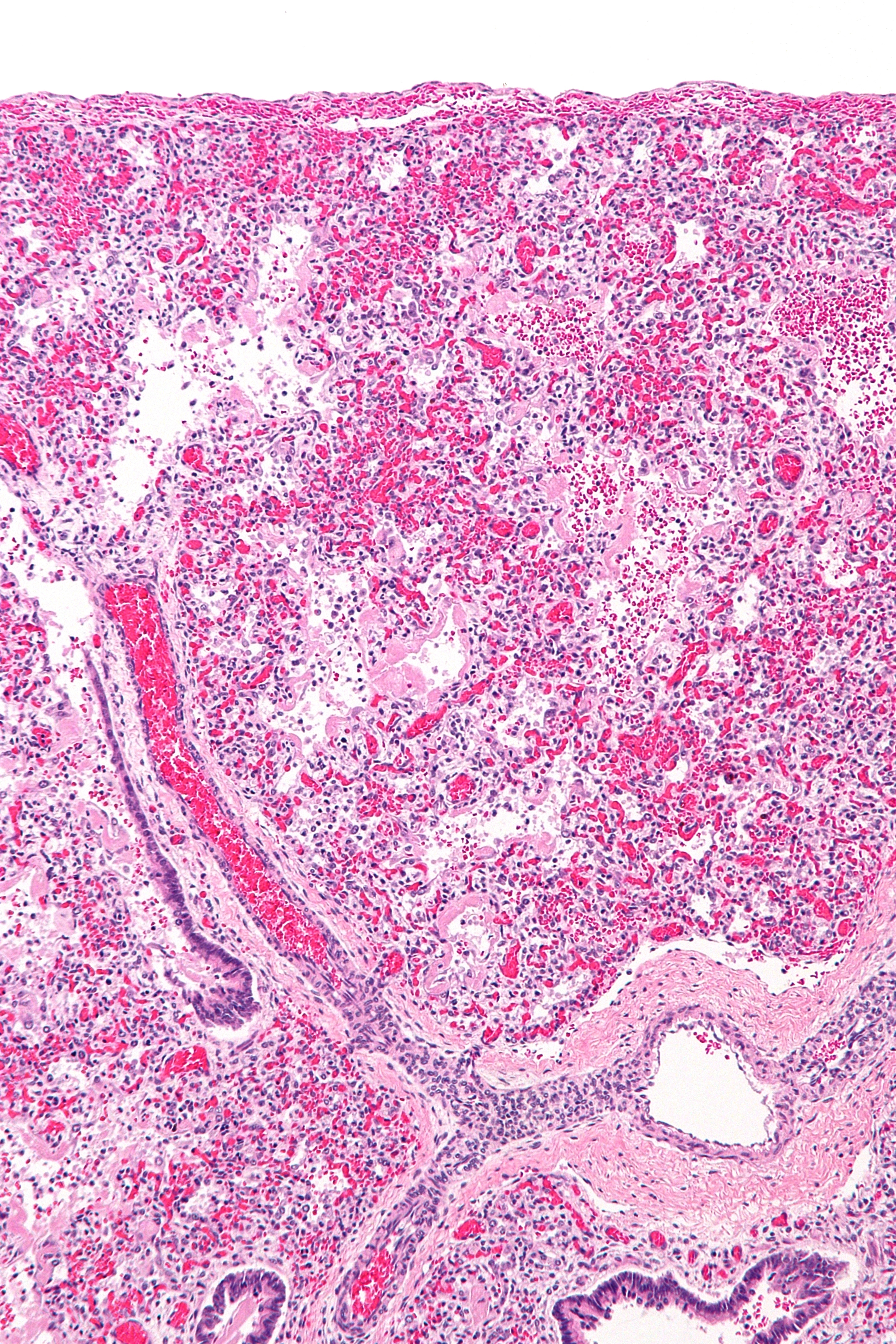
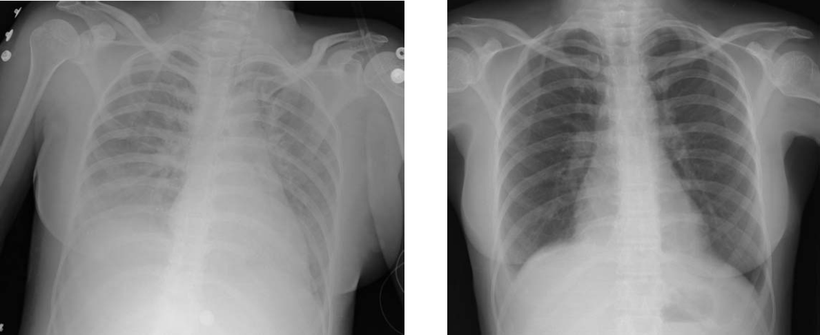
Acute respiratory distress syndrome (ARDS) is a type of respiratory failure characterized by rapid onset of widespread inflammation in the lungs. Symptoms include shortness of breath (dyspnea), rapid breathing (tachypnea), and bluish skin coloration (cyanosis). For those who survive, a decreased quality of life is common. Causes may include sepsis, pancreatitis, trauma, pneumonia, and aspiration. The underlying mechanism involves diffuse injury to cells which form the barrier of the microscopic air sacs of the lungs, surfactant dysfunction, activation of the immune system, and dysfunction of the body's regulation of blood clotting. In effect, ARDS impairs the lungs' ability to exchange oxygen and carbon dioxide. Adult diagnosis is based on a PaO2/FiO2 ratio (ratio of partial pressure arterial oxygen and fraction of inspired oxygen) of less than 300 mm Hg despite a positive end-expiratory pressure (PEEP) of more than 5 cm H2O. Cardiogenic pulmonary edema, a ... [...More Info...] [...Related Items...] OR: [Wikipedia] [Google] [Baidu] |
Acute Lung Injury
Acute respiratory distress syndrome (ARDS) is a type of respiratory failure characterized by rapid onset of widespread inflammation in the lungs. Symptoms include shortness of breath (dyspnea), rapid breathing (tachypnea), and bluish skin coloration (cyanosis). For those who survive, a decreased quality of life is common. Causes may include sepsis, pancreatitis, trauma, pneumonia, and aspiration. The underlying mechanism involves diffuse injury to cells which form the barrier of the microscopic air sacs of the lungs, surfactant dysfunction, activation of the immune system, and dysfunction of the body's regulation of blood clotting. In effect, ARDS impairs the lungs' ability to exchange oxygen and carbon dioxide. Adult diagnosis is based on a PaO2/FiO2 ratio (ratio of partial pressure arterial oxygen and fraction of inspired oxygen) of less than 300 mm Hg despite a positive end-expiratory pressure (PEEP) of more than 5 cm H2O. Cardiogenic pulmonary edema, as ... [...More Info...] [...Related Items...] OR: [Wikipedia] [Google] [Baidu] |
Tranexamic Acid
Tranexamic acid (TXA) is a medication used to treat or prevent excessive blood loss from major trauma, postpartum bleeding, surgery, tooth removal, nosebleeds, and heavy menstruation. It is also used for hereditary angioedema. It is taken either orally or by injection into a vein. Mechanism of action Tranexamic acid is a synthetic analog of the amino acid lysine. It serves as an antifibrinolytic by reversibly binding four to five lysine receptor sites on plasminogen. This decreases the conversion of plasminogen to plasmin, preventing fibrin degradation and preserving the framework of fibrin's matrix structure. Tranexamic acid has roughly eight times the antifibrinolytic activity of an older analogue, ε-aminocaproic acid. Tranexamic acid also directly inhibits the activity of plasmin with weak potency ( IC50 = 87 mM), and it can block the active-site of urokinase plasminogen activator (uPA) with high specificity ( Ki = 2 mM), one of the highest among all the serine proteases. ... [...More Info...] [...Related Items...] OR: [Wikipedia] [Google] [Baidu] |
Fibrinogen
Fibrinogen (factor I) is a glycoprotein complex, produced in the liver, that circulates in the blood of all vertebrates. During tissue and vascular injury, it is converted enzymatically by thrombin to fibrin and then to a fibrin-based blood clot. Fibrin clots function primarily to occlude blood vessels to stop bleeding. Fibrin also binds and reduces the activity of thrombin. This activity, sometimes referred to as antithrombin I, limits clotting. Fibrin also mediates blood platelet and endothelial cell spreading, tissue fibroblast proliferation, capillary tube formation, and angiogenesis and thereby promotes revascularization and wound healing. Reduced and/or dysfunctional fibrinogens occur in various congenital and acquired human fibrinogen-related disorders. These disorders represent a group of rare conditions in which individuals may present with severe episodes of pathological bleeding and thrombosis; these conditions are treated by supplementing blood fibrinogen levels an ... [...More Info...] [...Related Items...] OR: [Wikipedia] [Google] [Baidu] |
Factor XIII
Factor XIII or fibrin stabilizing factor is a zymogen found in blood of humans and some other animals. It is activated by thrombin to factor XIIIa. Factor XIIIa is an enzyme of the blood coagulation system that crosslinks fibrin. Deficiency of XIII worsens clot stability and increases bleeding tendency. Human XIII is a heterotetramer. It consists of 2 enzymatic A peptides and 2 non-enzymatic B peptides. XIIIa is a dimer of activated A peptides. Function Within blood, thrombins cleave fibrinogens to fibrins during coagulation and a fibrin-based blood clot forms. Factor XIII is a transglutaminase that circulates in human blood as a heterotetramer of two A and two B subunits. Factor XIII binds to the clot via their B units. In the presence of fibrins, thrombin efficiently cleaves the R37– G38 peptide bond of each A unit within a XIII tetramer. A units release their N-terminal activation peptides. Both of the non-covalently bound B units are now able to dissociate from the te ... [...More Info...] [...Related Items...] OR: [Wikipedia] [Google] [Baidu] |


