|
Ciliospinal Center
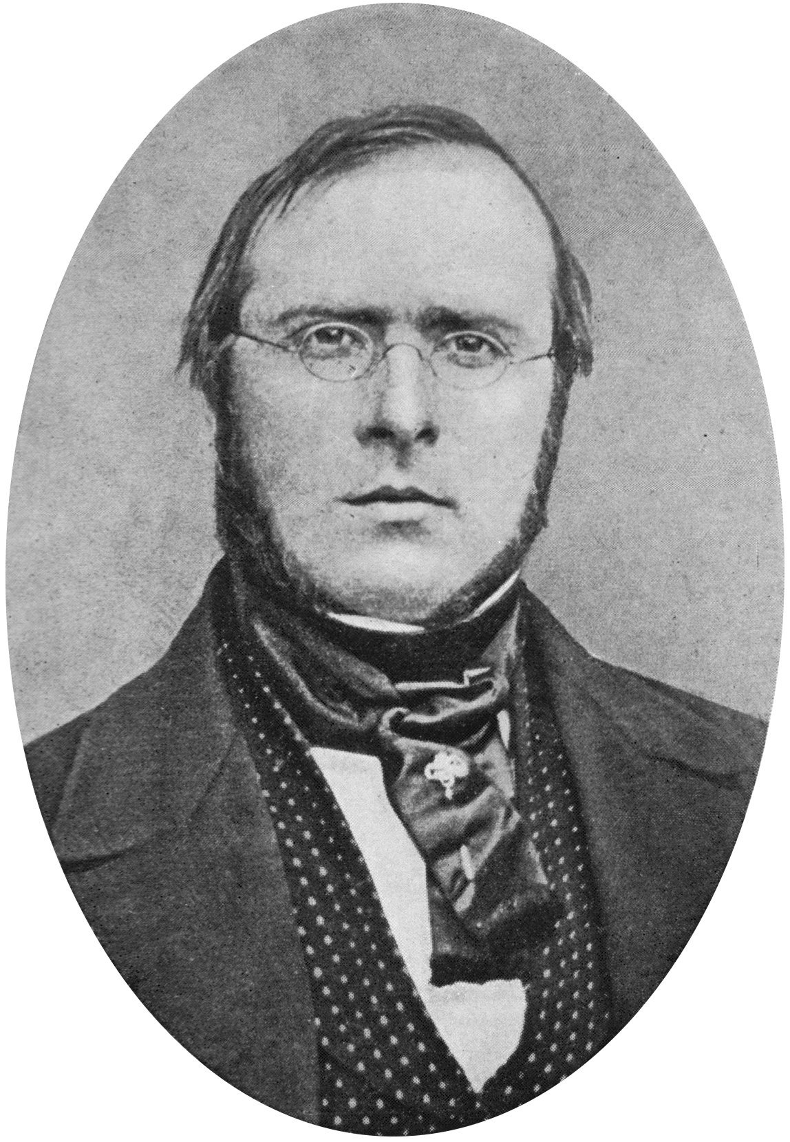
The ciliospinal center (in Latin: ''centrum ciliospinale'') is a structure which receives input from the pretectum, and has output to the superior cervical ganglion. It is located in the intermediolateral cell columns (IMLCC) of the spinal cord between C8 and T2. It plays a role in the control of the iris dilator muscle. It is also known as "Budge's center", or "centre". It is associated with a reflex identified by Augustus Volney Waller and Ludwig Julius Budge in 1852. See also * Horner's syndrome Horner's syndrome, also known as oculosympathetic paresis, is a combination of symptoms that arises when a group of nerves known as the sympathetic trunk is damaged. The signs and symptoms occur on the same side (ipsilateral) as it is a lesion ... References Spinal cord {{anatomy-stub ... [...More Info...] [...Related Items...] OR: [Wikipedia] [Google] [Baidu] |
Superior Cervical Ganglion
The superior cervical ganglion (SCG) is part of the autonomic nervous system (ANS); more specifically, it is part of the sympathetic nervous system, a division of the ANS most commonly associated with the fight or flight response. The ANS is composed of pathways that lead to and from ganglia, groups of nerve cells. A ganglion allows a large amount of divergence in a neuronal pathway and also enables a more localized circuitry for control of the innervated targets. The SCG is the only ganglion in the sympathetic nervous system that innervates the head and neck. It is the largest and most rostral (superior) of the three cervical ganglia. The SCG innervates many organs, glands and parts of the carotid system in the head. Structure Location The SCG is located opposite the second and third cervical vertebrae. It lies deep to the sheath of the internal carotid artery and internal jugular vein, and anterior to the Longus capitis muscle. The SCG contains neurons that supply sympathetic ... [...More Info...] [...Related Items...] OR: [Wikipedia] [Google] [Baidu] |
Pretectum
In neuroanatomy, the pretectal area, or pretectum, is a midbrain structure composed of seven nuclei and comprises part of the subcortical visual system. Through reciprocal bilateral projections from the retina, it is involved primarily in mediating behavioral responses to acute changes in ambient light such as the pupillary light reflex, the optokinetic reflex, and temporary changes to the circadian rhythm. In addition to the pretectum's role in the visual system, the anterior pretectal nucleus has been found to mediate somatosensory and nociceptive information. Location and structure The pretectum is a bilateral group of highly interconnected nuclei located near the junction of the midbrain and forebrain. The pretectum is generally classified as a midbrain structure, although because of its proximity to the forebrain it is sometimes classified as part of the caudal diencephalon (forebrain). Within vertebrates, the pretectum is located directly anterior to the superior colliculus ... [...More Info...] [...Related Items...] OR: [Wikipedia] [Google] [Baidu] |
Intermediolateral Nucleus
The intermediolateral nucleus (IML) is a region of grey matter found in one of the three grey columns of the spinal cord, the lateral grey column. This is Rexed lamina VII. The intermediolateral cell column exists at vertebral levels T1 – L3. It mediates the entire sympathetic innervation of the body, but the nucleus resides in the grey matter of the spinal cord. Rexed Lamina VII contains several well defined nuclei including the nucleus dorsalis (Clarke's column), the intermediolateral nucleus, and the sacral autonomic nucleus. It extends from T1 to L3, and contains the autonomic motor neurons that give rise to the preganglionic fibers of the sympathetic nervous system, (preganglionic sympathetic general visceral efferent General visceral efferent fibers (GVE) or visceral efferents or autonomic efferents, are the efferent nerve fibers of the autonomic nervous system (also known as the ''visceral efferent nervous system'' that provide motor innervation to smooth m ...s). ... [...More Info...] [...Related Items...] OR: [Wikipedia] [Google] [Baidu] |
Iris Dilator Muscle
The iris dilator muscle (pupil dilator muscle, pupillary dilator, radial muscle of iris, radiating fibers), is a smooth muscle of the eye, running radially in the iris and therefore fit as a dilator. The pupillary dilator consists of a spokelike arrangement of modified contractile cells called myoepithelial cells. These cells are stimulated by the sympathetic nervous system. When stimulated, the cells contract, widening the pupil and allowing more light to enter the eye. Structure Innervation It is innervated by the sympathetic system, which acts by releasing noradrenaline, which acts on α1-receptors. page 163 Thus, when presented with a threatening stimulus that activates the fight-or-flight response, this innervation contracts the muscle and dilates the pupil, thus temporarily letting more light reach the retina. The dilator muscle is innervated more specifically by postganglionic sympathetic nerves arising from the superior cervical ganglion as the sympathetic root of cil ... [...More Info...] [...Related Items...] OR: [Wikipedia] [Google] [Baidu] |
Augustus Volney Waller
Augustus Volney Waller FRS (21 December 1816 – 18 September 1870) was a British neurophysiologist. He was the first to describe the degeneration of severed nerve fibers, now known as Wallerian degeneration. Life The son of William Waller of Elverton Farm, near Faversham, Kent, was born on 21 December 1816. His youth was spent at Nice, where his father died in 1830. Waller was then sent back to England, where he lived, first with Dr. Lacon Lambe of Tewkesbury, and afterwards with William Lambe the vegetarian. His father sharing Lambe's views, Augustus was brought up until the age of eighteen on a vegetarian diet. Waller studied in Paris, where he obtained the degree of M.D. in 1840, and in the following year he was admitted a licentiate of the Society of Apothecaries in London. He then entered general medical practice at St. Mary Abbott's Terrace, Kensington. He soon acquired a considerable practice, but after the publication of two papers in the ''Philosophical Transactio ... [...More Info...] [...Related Items...] OR: [Wikipedia] [Google] [Baidu] |
Ludwig Julius Budge
Ludwig Julius Budge (11 September 1811, in Wetzlar – 14 July 1888, in Greifswald) was a German physiologist Physiology (; ) is the scientific study of functions and mechanisms in a living system. As a sub-discipline of biology, physiology focuses on how organisms, organ systems, individual organs, cells, and biomolecules carry out the chemical a .... He studied medicine at the Universities of University of Marburg, Marburg, University of Berlin, Berlin and University of Würzburg, Würzburg, and following graduation worked as a general practitioner in Wetzlar and Altenkirchen. In 1843 he was privat-docent to the medical faculty at University of Bonn, Bonn, where he became an associate professor in 1847. In 1856 he was appointed professor of anatomy and physiology at the University of Greifswald.ADB:Budge, Lud ... [...More Info...] [...Related Items...] OR: [Wikipedia] [Google] [Baidu] |
Horner's Syndrome
Horner's syndrome, also known as oculosympathetic paresis, is a combination of symptoms that arises when a group of nerves known as the sympathetic trunk is damaged. The signs and symptoms occur on the same side (ipsilateral) as it is a lesion of the sympathetic trunk. It is characterized by miosis (a constricted pupil), partial ptosis (a weak, droopy eyelid), apparent anhidrosis (decreased sweating), with apparent enophthalmos (inset eyeball). The nerves of the sympathetic trunk arise from the spinal cord in the chest, and from there ascend to the neck and face. The nerves are part of the sympathetic nervous system, a division of the autonomic (or involuntary) nervous system. Once the syndrome has been recognized, medical imaging and response to particular eye drops may be required to identify the location of the problem and the underlying cause. Signs and symptoms Signs that are found in people with Horner's syndrome on the affected side of the face include the following: * ... [...More Info...] [...Related Items...] OR: [Wikipedia] [Google] [Baidu] |

