|
Cardiac Auscultation
Heart sounds are the noises generated by the beating heart and the resultant flow of blood through it. Specifically, the sounds reflect the turbulence created when the heart valves snap shut. In cardiac auscultation, an examiner may use a stethoscope to listen for these unique and distinct sounds that provide important auditory data regarding the condition of the heart. In healthy adults, there are two normal heart sounds, often described as a ''lub'' and a ''dub'' that occur in sequence with each heartbeat. These are the first heart sound (S1) and second heart sound (S2), produced by the closing of the atrioventricular valves and semilunar valves, respectively. In addition to these normal sounds, a variety of other sounds may be present including heart murmurs, adventitious sounds, and gallop rhythms S3 and S4. Heart murmurs are generated by turbulent flow of blood and a murmur to be heard as turbulent flow must require pressure difference of at least 30 mm of Hg between ... [...More Info...] [...Related Items...] OR: [Wikipedia] [Google] [Baidu] |
Stethoscope
The stethoscope is a medical device for auscultation, or listening to internal sounds of an animal or human body. It typically has a small disc-shaped resonator that is placed against the skin, and one or two tubes connected to two earpieces. A stethoscope can be used to listen to the sounds made by the heart, lungs or intestines, as well as blood flow in arteries and veins. In combination with a manual sphygmomanometer, it is commonly used when measuring blood pressure. Less commonly, "mechanic's stethoscopes", equipped with rod shaped chestpieces, are used to listen to internal sounds made by machines (for example, sounds and vibrations emitted by worn ball bearings), such as diagnosing a malfunctioning automobile engine by listening to the sounds of its internal parts. Stethoscopes can also be used to check scientific vacuum chambers for leaks and for various other small-scale acoustic monitoring tasks. A stethoscope that intensifies auscultatory sounds is called a phonen ... [...More Info...] [...Related Items...] OR: [Wikipedia] [Google] [Baidu] |
Stenosis
A stenosis (from Ancient Greek στενός, "narrow") is an abnormal narrowing in a blood vessel or other tubular organ or structure such as foramina and canals. It is also sometimes called a stricture (as in urethral stricture). ''Stricture'' as a term is usually used when narrowing is caused by contraction of smooth muscle (e.g. achalasia, prinzmetal angina); ''stenosis'' is usually used when narrowing is caused by lesion that reduces the space of lumen (e.g. atherosclerosis). The term coarctation is another synonym, but is commonly used only in the context of aortic coarctation. Restenosis is the recurrence of stenosis after a procedure. Types The resulting syndrome depends on the structure affected. Examples of vascular stenotic lesions include: * Intermittent claudication (peripheral artery stenosis) * Angina ( coronary artery stenosis) * Carotid artery stenosis which predispose to (strokes and transient ischaemic episodes) * Renal artery stenosis The types of sten ... [...More Info...] [...Related Items...] OR: [Wikipedia] [Google] [Baidu] |
Diastole
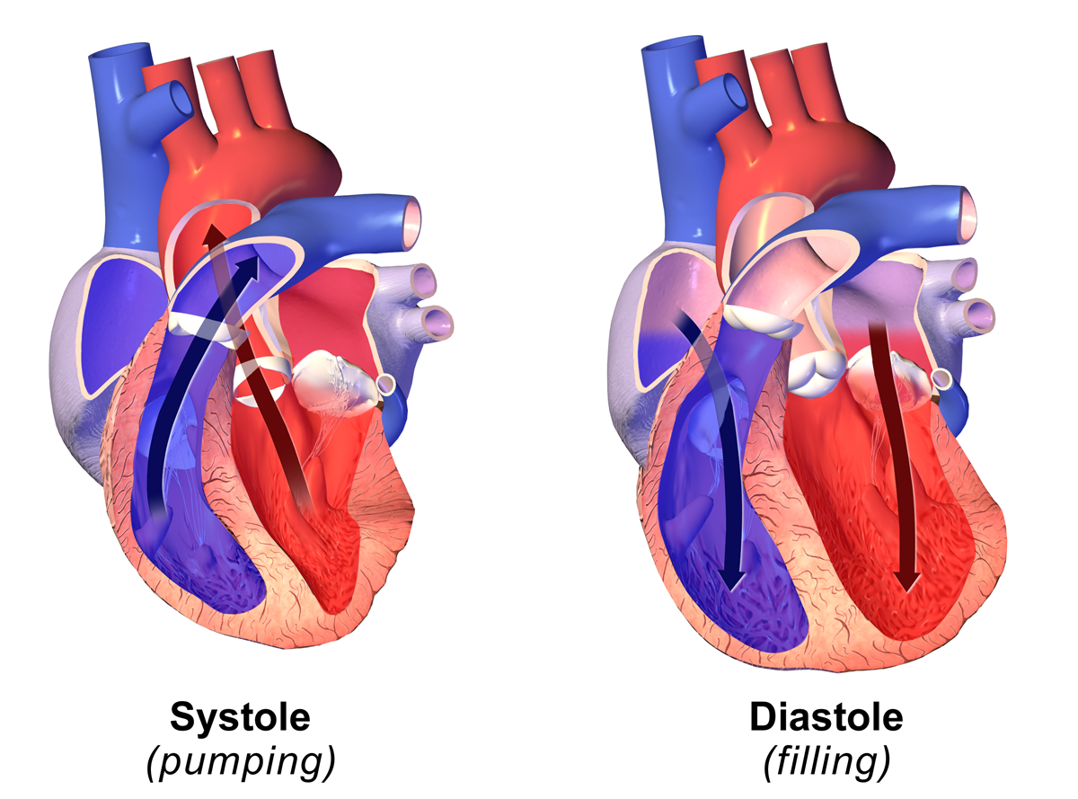
Diastole ( ) is the relaxed phase of the cardiac cycle when the chambers of the heart are re-filling with blood. The contrasting phase is systole when the heart chambers are contracting. Atrial diastole is the relaxing of the atria, and ventricular diastole the relaxing of the ventricles. The term originates from the Greek word (''diastolē''), meaning "dilation", from (''diá'', "apart") + (''stéllein'', "to send"). Role in cardiac cycle A typical heart rate is 75 beats per minute (bpm), which means that the cardiac cycle that produces one heartbeat, lasts for less than one second. The cycle requires 0.3 sec in ventricular systole (contraction)—pumping blood to all body systems from the two ventricles; and 0.5 sec in diastole (dilation), re-filling the four chambers of the heart, for a total of 0.8 sec to complete the cycle. Early ventricular diastole During early ventricular diastole, pressure in the two ventricles begins to drop from the peak reached during systo ... [...More Info...] [...Related Items...] OR: [Wikipedia] [Google] [Baidu] |
Pulmonary Valve
The pulmonary valve (sometimes referred to as the pulmonic valve) is a valve of the heart that lies between the right ventricle and the pulmonary artery and has three cusps. It is one of the four valves of the heart and one of the two semilunar valves, the other being the aortic valve. Similar to the aortic valve, the pulmonary valve opens in ventricular systole, when the pressure in the right ventricle rises above the pressure in the pulmonary artery. At the end of ventricular systole, when the pressure in the right ventricle falls rapidly, the pressure in the pulmonary artery will close the pulmonary valve. The closure of the pulmonary valve contributes the P2 component of the second heart sound (S2). The right heart is a low-pressure system, so the P2 component of the second heart sound is usually softer than the A2 component of the second heart sound. However, it is physiologically normal in some young people to hear both components separated during inhalation. Description * ... [...More Info...] [...Related Items...] OR: [Wikipedia] [Google] [Baidu] |
Aortic Valve
The aortic valve is a valve in the heart of humans and most other animals, located between the left ventricle and the aorta. It is one of the four valves of the heart and one of the two semilunar valves, the other being the pulmonary valve. The aortic valve normally has three cusps or leaflets, although in 1–2% of the population it is found to congenitally have two leaflets. The aortic valve is the last structure in the heart the blood travels through before stopping the flow through the systemic circulation. Structure The aortic valve normally has three cusps however there is some discrepancy in their naming. They may be called the left coronary, right coronary and non-coronary cusp. Some sources also advocate they be named as a left, right and posterior cusp. Anatomists have traditionally named them the left posterior (origin of left coronary), anterior (origin of the right coronary) and right posterior. The three cusps, when the valve is closed, contain a sinus called an a ... [...More Info...] [...Related Items...] OR: [Wikipedia] [Google] [Baidu] |
Semilunar Valves
A heart valve is a one-way valve that allows blood to flow in one direction through the chambers of the heart. Four valves are usually present in a mammalian heart and together they determine the pathway of blood flow through the heart. A heart valve opens or closes according to differential blood pressure on each side. The four valves in the mammalian heart are two atrioventricular valves separating the upper atria from the lower ventricles – the mitral valve in the left heart, and the tricuspid valve in the right heart. The other two valves are at the entrance to the arteries leaving the heart these are the semilunar valves – the aortic valve at the aorta, and the pulmonary valve at the pulmonary artery. The heart also has a coronary sinus valve, and an inferior vena cava valve, not discussed here. Structure The heart valves and the chambers are lined with endocardium. Heart valves separate the atria from the ventricles, or the ventricles from a blood vessel. Heart va ... [...More Info...] [...Related Items...] OR: [Wikipedia] [Google] [Baidu] |
Right Bundle Branch Block
A right bundle branch block (RBBB) is a heart block in the right bundle branch of the electrical conduction system. During a right bundle branch block, the right ventricle is not directly activated by impulses travelling through the right bundle branch. The left ventricle, however, is still normally activated by the left bundle branch. These impulses are then able to travel through the myocardium of the left ventricle to the right ventricle and depolarize the right ventricle this way. As conduction through the myocardium is slower than conduction through the Bundle of His-Purkinje fibres, the QRS complex is seen to be widened. The QRS complex often shows an extra deflection that reflects the rapid depolarisation of the left ventricle followed by the slower depolarisation of the right ventricle. Incomplete right bundle branch block Incomplete right bundle branch block (IRBBB) is an conduction abnormality in the right bundle branch block. While a complete RBBB has a QRS dura ... [...More Info...] [...Related Items...] OR: [Wikipedia] [Google] [Baidu] |
Chordae Tendineae
The chordae tendineae (tendinous cords), colloquially known as the heart strings, are inelastic cords of fibrous connective tissue that connect the papillary muscles to the tricuspid valve and the mitral valve in the heart. Structure The chordae tendineae connect the atrioventricular valves (tricuspid and mitral), to the papillary muscles within the ventricles. Multiple chordae tendineae attach to each leaflet or cusp of the valves. Chordae tendineae contain elastin in a delicate structure notably at their periphery. Tendon of Todaro The ''tendon of Todaro'' is a continuation of the Eustachian valve of the inferior vena cava and the valve of the coronary sinus. Along with the opening of the coronary sinus and the septal cusp of the tricuspid valve, it makes up Koch's triangle. The apex of Koch's triangle is the location of the atrioventricular node. Function During atrial systole, blood flows from the atria to the ventricles down the pressure gradient. Chordae tendineae are ... [...More Info...] [...Related Items...] OR: [Wikipedia] [Google] [Baidu] |
Systole (medicine)
Systole ( ) is the part of the cardiac cycle during which some chambers of the heart contract after refilling with blood. The term originates, via New Latin, from Ancient Greek (''sustolē''), from (''sustéllein'' 'to contract'; from ''sun'' 'together' + ''stéllein'' 'to send'), and is similar to the use of the English term ''to squeeze''. The mammalian heart has four chambers: the left atrium above the left ventricle (lighter pink, see graphic), which two are connected through the mitral (or bicuspid) valve; and the right atrium above the right ventricle (lighter blue), connected through the tricuspid valve. The atria are the receiving blood chambers for the circulation of blood and the ventricles are the discharging chambers. In late ventricular diastole, the atrial chambers contract and send blood to the larger, lower ventricle chambers. This flow fills the ventricles with blood, and the resulting pressure closes the valves to the atria. The ventricles now perform i ... [...More Info...] [...Related Items...] OR: [Wikipedia] [Google] [Baidu] |
Mitral Valve
The mitral valve (), also known as the bicuspid valve or left atrioventricular valve, is one of the four heart valves. It has two cusps or flaps and lies between the left atrium and the left ventricle of the heart. The heart valves are all one-way valves allowing blood flow in just one direction. The mitral valve and the tricuspid valve are known as the atrioventricular valves because they lie between the atria and the ventricles. In normal conditions, blood flows through an open mitral valve during diastole with contraction of the left atrium, and the mitral valve closes during systole with contraction of the left ventricle. The valve opens and closes because of pressure differences, opening when there is greater pressure in the left atrium than ventricle and closing when there is greater pressure in the left ventricle than atrium. In abnormal conditions, blood may flow backward through the valve ( mitral regurgitation) or the mitral valve may be narrowed (mitral stenosis). Rh ... [...More Info...] [...Related Items...] OR: [Wikipedia] [Google] [Baidu] |
Tricuspid Valve
The tricuspid valve, or right atrioventricular valve, is on the right dorsal side of the mammalian heart, at the superior portion of the right ventricle. The function of the valve is to allow blood to flow from the right atrium to the right ventricle during diastole, and to close to prevent backflow ( regurgitation) from the right ventricle into the right atrium during right ventricular contraction ( systole). Structure The tricuspid valve usually has three cusps or leaflets, named the anterior, posterior, and septal cusps. Each leaflet is connected via chordae tendineae to the anterior, posterior, and septal papillary muscles of the right ventricle, respectively. Tricuspid valves may also occur with two or four leaflets; the number may change over a lifetime. Function The tricuspid valve functions as a one-way valve that closes during ventricular systole to prevent regurgitation of blood from the right ventricle back into the right atrium. It opens during ventricular diastol ... [...More Info...] [...Related Items...] OR: [Wikipedia] [Google] [Baidu] |
Heart Valves
A heart valve is a one-way valve that allows blood to flow in one direction through the chambers of the heart. Four valves are usually present in a mammalian heart and together they determine the pathway of blood flow through the heart. A heart valve opens or closes according to differential blood pressure on each side. The four valves in the mammalian heart are two atrioventricular valves separating the upper atria from the lower ventricles – the mitral valve in the left heart, and the tricuspid valve in the right heart. The other two valves are at the entrance to the arteries leaving the heart these are the semilunar valves – the aortic valve at the aorta, and the pulmonary valve at the pulmonary artery. The heart also has a coronary sinus valve, and an inferior vena cava valve, not discussed here. Structure The heart valves and the chambers are lined with endocardium. Heart valves separate the atria from the ventricles, or the ventricles from a blood vessel. Heart va ... [...More Info...] [...Related Items...] OR: [Wikipedia] [Google] [Baidu] |


