|
Buttock Cells
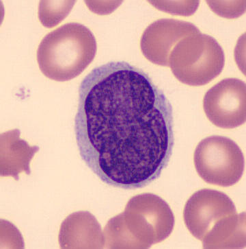
Buttock cells are cells having a notched appearance that are found in certain malignancies, such as non-Hodgkin's lymphoma (including follicular lymphoma), mycosis fungoides, and Sézary syndrome. See also * Clue cell * Koilocyte * Large cell Large cell is a term used in oncology. It does not refer to a particular type of cell; rather it refers to cells that are larger than would be normally expected for that type. It is frequently used when describing lymphoma and lung cancer. It was ... References Blood cells Lymphoma {{oncology-stub ... [...More Info...] [...Related Items...] OR: [Wikipedia] [Google] [Baidu] |
Non-Hodgkin's Lymphoma
Non-Hodgkin lymphoma (NHL), also known as non-Hodgkin's lymphoma, is a group of blood cancers that includes all types of lymphomas except Hodgkin lymphomas. Symptoms include lymphadenopathy, enlarged lymph nodes, fever, night sweats, weight loss, and tiredness. Other symptoms may include bone pain, chest pain, or itchiness. Some forms are slow-growing while others are fast-growing. Lymphomas are types of cancer that develop from lymphocytes, a type of white blood cell. Risk factors include immunodeficiency, poor immune function, autoimmune diseases, Helicobacter pylori infection, ''Helicobacter pylori'' infection, hepatitis C, obesity, and Epstein–Barr virus infection. The World Health Organization classifies lymphomas into five major groups, including one for Hodgkin lymphoma. Within the four groups for NHL are over 60 specific types of lymphoma. Diagnosis is by bone marrow biopsy, examination of a bone marrow or lymph node biopsy. Medical imaging is done to help with cance ... [...More Info...] [...Related Items...] OR: [Wikipedia] [Google] [Baidu] |
Follicular Lymphoma
Follicular lymphoma (FL) is a cancer that involves certain types of white blood cells known as lymphocytes. The cancer originates from the uncontrolled division of specific types of B-cells known as centrocytes and centroblasts. These cells normally occupy the follicles (nodular swirls of various types of lymphocytes) in the germinal centers of lymphoid tissues such as lymph nodes. The cancerous cells in FL typically form follicular or follicle-like structures (see adjacent Figure) in the tissues they invade. These structures are usually the dominant histological feature of this cancer. There are several synonymous and obsolete terms for FL such as CB/CC lymphoma (centroblastic and centrocytic lymphoma), nodular lymphoma, Brill-Symmers Disease, and the subtype designation, follicular large-cell lymphoma. In the US and Europe, this disease is the second most common form of non-Hodgkin's lymphomas, exceeded only by diffuse large B-cell lymphoma. FL accounts for 10-20% of non-Hodgkin ... [...More Info...] [...Related Items...] OR: [Wikipedia] [Google] [Baidu] |
Mycosis Fungoides
Mycosis fungoides, also known as Alibert-Bazin syndrome or granuloma fungoides, is the most common form of cutaneous T-cell lymphoma. It generally affects the skin, but may progress internally over time. Symptoms include rash, tumors, skin lesions, and itchy skin. While the cause remains unclear, most cases are not hereditary. Most cases are in people over 20 years of age, and it is more common in men than women. Treatment options include sunlight exposure, ultraviolet light, topical corticosteroids, chemotherapy, and radiotherapy. Signs and symptoms The symptoms of mycosis fungoides are categorized into three clinical stages: the patch stage, the plaque stage, and the tumour stage. The patch stage is defined by flat, reddish patches of varying sizes that may have a wrinkled appearance. They can also look yellowish in people with darker skin. The plaque stage follows the patch stage of mycosis fungoides. It is characterized by the presence of raised lesions that appear reddis ... [...More Info...] [...Related Items...] OR: [Wikipedia] [Google] [Baidu] |
Clue Cell
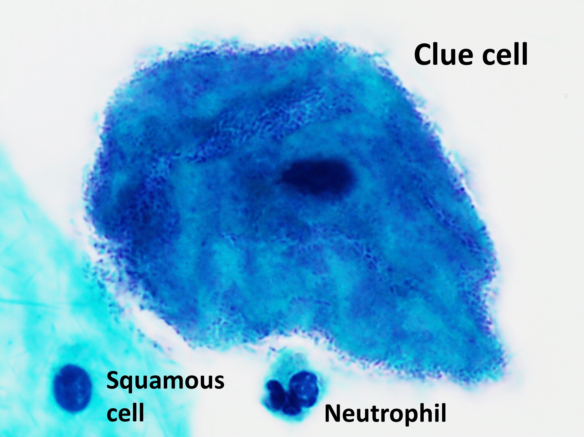
Clue cells are epithelial cells of the vagina that get their distinctive stippled appearance by being covered with bacteria. The etymology behind the term "clue" cell derives from the original research article from Gardner and Dukes describing the characteristic cells. The name was chosen for its brevity in describing the sine qua non of bacterial vaginosis. They are a medical sign of bacterial vaginosis, particularly that caused by ''Gardnerella vaginalis'', a group of Gram-variable bacteria. This bacterial infection is characterized by a foul, fishy smelling, thin gray vaginal discharge, and an increase in vaginal pH from around 4.5 to over 5.5. References External links Overviewat WebMD WebMD is an American corporation known primarily as an online publisher of news and information pertaining to human health and well-being. The site includes information pertaining to drugs. It is one of the top healthcare websites. It was foun ... Clue cell in Gram stain aClue cell i ... [...More Info...] [...Related Items...] OR: [Wikipedia] [Google] [Baidu] |
Koilocyte
A koilocyte is a squamous epithelial cell that has undergone a number of structural changes, which occur as a result of infection of the cell by human papillomavirus (HPV). Identification of these cells by pathologists can be useful in diagnosing various HPV-associated lesions. Koilocytosis Koilocytosis or koilocytic atypia or koilocytotic atypia are terms used in histology and cytology to describe the presence of koilocytes in a specimen. Koilocytes may have the following cellular changes: * Nuclear enlargement (two to three times normal size). * Irregularity of the nuclear membrane contour, creating a wrinkled or raisinoid appearance. * A darker than normal staining pattern in the nucleus, known as hyperchromasia. * A clear area around the nucleus, known as a perinuclear halo or perinuclear cytoplasmic vacuolization. Collectively, these types of changes are called a cytopathic effect; various types of cytopathic effect can be seen in many different cell types infected by many ... [...More Info...] [...Related Items...] OR: [Wikipedia] [Google] [Baidu] |
Large Cell
Large cell is a term used in oncology. It does not refer to a particular type of cell; rather it refers to cells that are larger than would be normally expected for that type. It is frequently used when describing lymphoma and lung cancer. It was more frequently used in the past than it is used today, when doctors often could tell little about a cell other than its size, and it was used for classification systems such as the "Working Formulation" for lymphoma. As such, the term lives on in the names of many conditions, even when the size of the cell is no longer one of the most important diagnostic criteria. The phrase giant cell is also frequently used, especially with carcinoma. Giant cell tumors include giant-cell tumor of bone, giant-cell tumor of the tendon sheath, and giant cell fibroblastoma. See also * Anaplastic large-cell lymphoma * Buttock cell * Nosology References External links Overviewat Mayo Clinic The Mayo Clinic () is a nonprofit American academic ... [...More Info...] [...Related Items...] OR: [Wikipedia] [Google] [Baidu] |
Blood Cells
A blood cell, also called a hematopoietic cell, hemocyte, or hematocyte, is a cell produced through hematopoiesis and found mainly in the blood. Major types of blood cells include red blood cells (erythrocytes), white blood cells (leukocytes), and platelets (thrombocytes). Together, these three kinds of blood cells add up to a total 45% of the blood tissue by volume, with the remaining 55% of the volume composed of plasma, the liquid component of blood. Red blood cells Red blood cells or ''erythrocytes'', primarily carry oxygen and collect carbon dioxide through the use of hemoglobin. Hemoglobin is an iron-containing protein that gives red blood cells their color and facilitates transportation of oxygen from the lungs to tissues and carbon dioxide from tissues to the lungs to be exhaled. Red blood cells are the most abundant cell in the blood, accounting for about 40-45% of its volume. Red blood cells are circular, biconcave, disk-shaped and deformable to allow them to squeeze ... [...More Info...] [...Related Items...] OR: [Wikipedia] [Google] [Baidu] |


.jpg)
