|
Buccinator Crest
The buccinator crest (Latin ''crista buccinatoria'') is a bony crest of the human mandible, that passes from the base of the coronoid process to the area of the third molar.https://medical-dictionary.thefreedictionary.com/buccinator+crest The free dictionary's medical dictionary defenition of buccinator crest The alveolar border of the buccinator muscle The buccinator () is a thin quadrilateral muscle occupying the interval between the maxilla and the mandible at the side of the face. It forms the anterior part of the cheek or the lateral wall of the oral cavity.Illustrated Anatomy of the Head ... attaches upon it. References Bones of the head and neck {{musculoskeletal-stub ... [...More Info...] [...Related Items...] OR: [Wikipedia] [Google] [Baidu] |
Sobo 1909 93
{{disambiguation, geo, surname ...
Sobo may refer to: Places * Sobo, La Brea, Trinidad and Tobago * Mount Sobo, Japan * SoBo or South Mumbai Other * Sobo (deity) * Sobo language (other) * Alexandra Sobo (born 1987), Romanian volleyball player * Sobo, an app for recording and distributing sound snippets, developed by Alan Braverman Alan Michael Braverman is an American businessman. He is co-founder and initial CTO of Xoom Corporation/Eventbrite with Kevin Hartz and Geni.com/Yammer with David O. Sacks. In 2014 Braverman worked on Sobo, "an audio version of Twitter" Bra ... [...More Info...] [...Related Items...] OR: [Wikipedia] [Google] [Baidu] |
Crest (anatomy)
A crest is any of various anatomical features appearing as a raised point or ridge, most prominently those on the head or back of an animal. *A part of a bone: **Sagittal crest **Cnemial crest **Iliac crest **Frontal crest ** Infratemporal crest ** Anterior lacrimal crest ** Posterior lacrimal crest **Buccinator crest *A feature on various animals: **Crest (feathers) ** Display feature or thermoregulatory feature in various reptiles ** Sail (anatomy), also known as ''crest'' in some animals **The point of a horse's neck where the mane grows from *Neural crest Neural crest cells are a temporary group of cells unique to vertebrates that arise from the embryonic ectoderm germ layer, and in turn give rise to a diverse cell lineage—including melanocytes, craniofacial cartilage and bone, smooth muscle, per ..., a temporary group of cells unique to vertebrates that arise from the embryonic ectoderm cell layer {{SIA ... [...More Info...] [...Related Items...] OR: [Wikipedia] [Google] [Baidu] |
Mandible
In anatomy, the mandible, lower jaw or jawbone is the largest, strongest and lowest bone in the human facial skeleton. It forms the lower jaw and holds the lower tooth, teeth in place. The mandible sits beneath the maxilla. It is the only movable bone of the skull (discounting the ossicles of the middle ear). It is connected to the temporal bones by the temporomandibular joints. The bone is formed prenatal development, in the fetus from a fusion of the left and right mandibular prominences, and the point where these sides join, the mandibular symphysis, is still visible as a faint ridge in the midline. Like other symphyses in the body, this is a midline articulation where the bones are joined by fibrocartilage, but this articulation fuses together in early childhood.Illustrated Anatomy of the Head and Neck, Fehrenbach and Herring, Elsevier, 2012, p. 59 The word "mandible" derives from the Latin word ''mandibula'', "jawbone" (literally "one used for chewing"), from ''wikt:mandere ... [...More Info...] [...Related Items...] OR: [Wikipedia] [Google] [Baidu] |
Coronoid Process Of The Mandible
In human anatomy, the mandible's coronoid process (from Greek ''korōnē'', denoting something hooked) is a thin, triangular eminence, which is flattened from side to side and varies in shape and size. Its anterior border is convex and is continuous below with the anterior border of the ramus. Its ''posterior border'' is concave and forms the anterior boundary of the mandibular notch. The ''lateral surface'' is smooth, and affords insertion to the temporalis and masseter muscles. Its ''medial surface'' gives insertion to the temporalis, and presents a ridge which begins near the apex of the process and runs downward and forward to the inner side of the last molar tooth. Between this ridge and the anterior border is a grooved triangular area, the upper part of which gives attachment to the temporalis, the lower part to some fibers of the buccinator. Clinical significance Fractures of the mandible are common. However, coronoid process fractures are very rare. Isolated fractures of th ... [...More Info...] [...Related Items...] OR: [Wikipedia] [Google] [Baidu] |
Third Molar
A third molar, commonly called wisdom tooth, is one of the three molars per quadrant of the human dentition. It is the most posterior of the three. The age at which wisdom teeth come through ( erupt) is variable, but this generally occurs between late teens and early twenties. Most adults have four wisdom teeth, one in each of the four quadrants, but it is possible to have none, fewer, or more, in which case the extras are called supernumerary teeth. Wisdom teeth may get stuck ( impacted) against other teeth if there is not enough space for them to come through normally. Impacted wisdom teeth are still sometimes removed for orthodontic treatment, believing that they move the other teeth and cause crowding, though this is not held anymore as true. Impacted wisdom teeth may suffer from tooth decay if oral hygiene becomes more difficult. Wisdom teeth which are partially erupted through the gum may also cause inflammation and infection in the surrounding gum tissues, termed pericoron ... [...More Info...] [...Related Items...] OR: [Wikipedia] [Google] [Baidu] |
Dental Alveolus
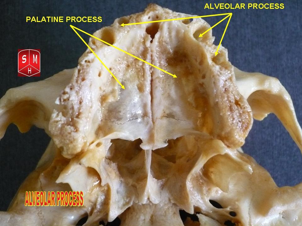
Dental alveoli (singular ''alveolus'') are sockets in the jaws in which the roots of teeth are held in the alveolar process with the periodontal ligament. The lay term for dental alveoli is tooth sockets. A joint that connects the roots of the teeth and the alveolus is called ''gomphosis'' (plural ''gomphoses''). Alveolar bone is the bone that surrounds the roots of the teeth forming bone sockets. In mammals, tooth sockets are found in the maxilla, the premaxilla, and the mandible. Etymology 1706, "a hollow," especially "the socket of a tooth," from Latin alveolus "a tray, trough, basin; bed of a small river; small hollow or cavity," diminutive of alvus "belly, stomach, paunch, bowels; hold of a ship," from PIE root *aulo- "hole, cavity" (source also of Greek aulos "flute, tube, pipe;" Serbo-Croatian, Polish, Russian ulica "street," originally "narrow opening;" Old Church Slavonic uliji, Lithuanian aulys "beehive" (hollow trunk), Armenian yli "pregnant"). The word was extended in ... [...More Info...] [...Related Items...] OR: [Wikipedia] [Google] [Baidu] |
Buccinator Muscle
The buccinator () is a thin quadrilateral muscle occupying the interval between the maxilla and the mandible at the side of the face. It forms the anterior part of the cheek or the lateral wall of the oral cavity.Illustrated Anatomy of the Head and Neck, Fehrenbach and Herring, Elsevier, 2012, page 91 Structure It arises from the outer surfaces of the alveolar processes of the maxilla and mandible, corresponding to the three pairs of molar teeth and in the mandible, it is attached upon the buccinator crest posterior to the third molar; and behind, from the anterior border of the pterygomandibular raphe which separates it from the constrictor pharyngis superior. The fibers converge toward the angle of the mouth, where the central fibers intersect each other, those from below being continuous with the upper segment of the orbicularis oris, and those from above with the lower segment; the upper and lower fibers are continued forward into the corresponding lip without decussation ... [...More Info...] [...Related Items...] OR: [Wikipedia] [Google] [Baidu] |


