|
Bronchoscope
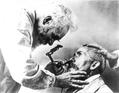
Bronchoscopy is an endoscopic technique of visualizing the inside of the airways for diagnostic and therapeutic purposes. An instrument (bronchoscope) is inserted into the airways, usually through the nose or mouth, or occasionally through a tracheostomy. This allows the practitioner to examine the patient's airways for abnormalities such as foreign bodies, bleeding, tumors, or inflammation. Specimens may be taken from inside the lungs. The construction of bronchoscopes ranges from rigid metal tubes with attached lighting devices to flexible optical fiber instruments with realtime video equipment. History The German laryngologist Gustav Killian is attributed with performing the first bronchoscopy in 1897. Killian used a rigid bronchoscope to remove a pork bone. The procedure was done in an awake patient using topical cocaine as a local anesthetic. From this time until the 1970s, rigid bronchoscopes were used exclusively. Chevalier Jackson, refined the rigid bronchoscope in the 1 ... [...More Info...] [...Related Items...] OR: [Wikipedia] [Google] [Baidu] |
Bronchoalveolar Lavage
Bronchoalveolar lavage (BAL) (also known as bronchoalveolar washing) is a diagnostic method of the lower respiratory system in which a bronchoscope is passed through the mouth or nose into an appropriate airway in the lungs, with a measured amount of fluid introduced and then collected for examination. This method is typically performed to diagnose pathogenic infections of the lower respiratory airways (leading to, for example pneumonia and COVID-19), though it also has been shown to have utility in diagnosing interstitial lung disease. Bronchoalveolar lavage can be a more sensitive method of detection than nasal swabs in respiratory molecular diagnostics, as has been the case with SARS-CoV-2 where bronchoalveolar lavage samples detect copies of viral RNA after negative nasal swab testing. In particular, bronchoalveolar lavage is commonly used to diagnose infections in people with immune system problems, pneumonia in people on ventilators, and acute respiratory distress syndrome ... [...More Info...] [...Related Items...] OR: [Wikipedia] [Google] [Baidu] |
Shigeto Ikeda
was a Japanese people, Japanese physician, regarded as the "father" of fiberoptic bronchoscopy. He graduated from Keio University School of Medicine in 1952 and joined the Thoracic Surgery department. In 1966, he developed the first flexible bronchoscope in conjunction with Machida Endoscope Co. Ltd (later taken over by Pentax) and Olympus Corporation, Olympus Optical Co., Ltd. This allowed better visualisation of the upper lobe bronchus, bronchi than is possible with the rigid bronchoscope. Successive improvements under his supervision included the development of video-bronchoscopy. His motto was "there is more hope with the bronchoscope". References 1925 births 2001 deaths Japanese thoracic surgeons 20th-century surgeons {{japan-med-bio-stub ... [...More Info...] [...Related Items...] OR: [Wikipedia] [Google] [Baidu] |
Victor Negus
Sir Victor Ewings Negus, MS, FRCS (6 February 1887 – 15 July 1974) was a British surgeon who specialised in laryngology and also made fundamental contributions to comparative anatomy with his work on the structure and evolution of the larynx. He was born and educated in London, studying at King's College School, then King's College London, followed by King's College Hospital. The final years of his medical training were interrupted by the First World War, during which he served with the Royal Army Medical Corps. After the war, he qualified as a surgeon and studied with laryngologists in France and the USA before resuming his career at King's College Hospital where he became a junior surgeon in 1924. In the 1920s, Negus worked on aspects of both throat surgery and the anatomy of the larynx, the latter work contributing to his degree of Master of Surgery (1924). His surgical innovations included designs for laryngoscopes, bronchoscopes, oesophagoscopes, an operating table, and t ... [...More Info...] [...Related Items...] OR: [Wikipedia] [Google] [Baidu] |
Chevalier Jackson
Chevalier Quixote Jackson (November 4, 1865 – August 16, 1958) was an American pioneer in laryngology. He is sometimes known as the "father of endoscopy", although Philipp Bozzini (1773–1809) is also often given this sobriquet. Chevalier Q. Jackson extracted over 2000 swallowed foreign bodies from patients. The collection is currently on display at the Mütter Museum in Philadelphia. Biography Jackson was born in Pittsburgh, Pennsylvania. He went to school at the Western University of Pennsylvania (now the University of Pittsburgh) from 1879 to 1883, and received his MD from Jefferson Medical College in Philadelphia. He also studied laryngology in England. His work reduced the risks involved in a tracheotomy. He essentially invented the modern science of endoscopy of the upper airway and esophagus, using hollow tubes with illumination (esophagoscopes and bronchoscopes). He developed methods for removing foreign bodies from the esophagus and the airway with great safety — a h ... [...More Info...] [...Related Items...] OR: [Wikipedia] [Google] [Baidu] |
Tracheostomy
Tracheotomy (, ), or tracheostomy, is a surgical airway management procedure which consists of making an incision (cut) on the anterior aspect (front) of the neck and opening a direct airway through an incision in the Vertebrate trachea, trachea (windpipe). The resulting Stoma (medicine), stoma (hole) can serve independently as an airway or as a site for a tracheal tube or tracheostomy tube to be inserted; this tube allows a person to breathe without the use of the nose or mouth. Etymology and terminology The etymology of the word ''tracheotomy'' comes from two Greek language, Greek words: the root (linguistics), root ''tom-'' (from Ancient Greek, Greek τομή ''tomḗ'') meaning "to cut", and the word ''trachea'' (from Greek τραχεία ''tracheía''). The word ''tracheostomy'', including the root ''stom-'' (from Greek στόμα ''stóma'') meaning "mouth," refers to the making of a semi-permanent or permanent opening, and to the opening itself. Some sources offer differ ... [...More Info...] [...Related Items...] OR: [Wikipedia] [Google] [Baidu] |
Gustav Killian
Gustav Killian (2 June 1860 – 24 February 1921) was a German laryngologist and founder of the bronchoscopy. Life and death His father Johann Baptist Caesar Killian (1820–1889), the son of a ''städtischen Wegeaufsehers'' an urban way overseer, was a ''doctor of philosophiae'' and high school teacher born in Mainz and later living in Bensheim. His mother Apollonia (1833–1865), née Höpfel died early, at age 31 of cholera. He was also born in Mainz, and educated at the University of Freiburg-im-Breisgau. He made revolutionary advances in the diagnosis and treatment of affections of the infralaryngeal passages, especially in the diagnosis and removal of foreign bodies in the bronchial tubes, by means of his new art of bronchoscopic control. His first college appointment was as assistant to Professor Hack of the chair of otolaryngology in Mainz. The sudden death of Wilhelm Hack (1851–1887) led to his succession by Killian, although he was not made professor at the time. H ... [...More Info...] [...Related Items...] OR: [Wikipedia] [Google] [Baidu] |
Electrocautery
Cauterization (or cauterisation, or cautery) is a medical practice or technique of burning a part of a body to remove or close off a part of it. It destroys some tissue in an attempt to mitigate bleeding and damage, remove an undesired growth, or minimize other potential medical harm, such as infections when antibiotics are unavailable. The practice was once widespread for treatment of wounds. Its utility before the advent of antibiotics was said to be effective at more than one level: *To prevent exsanguination *To close amputations Cautery was historically believed to prevent infection, but current research shows that cautery actually increases the risk for infection by causing more tissue damage and providing a more hospitable environment for bacterial growth. Actual cautery refers to the metal device, generally heated to a dull red glow, that a physician applies to produce blisters, to stop bleeding of a blood vessel, and for other similar purposes., page 16. The main forms ... [...More Info...] [...Related Items...] OR: [Wikipedia] [Google] [Baidu] |
Eyepiece
An eyepiece, or ocular lens, is a type of lens that is attached to a variety of optical devices such as telescopes and microscopes. It is named because it is usually the lens that is closest to the eye when someone looks through the device. The objective lens or mirror collects light and brings it to focus creating an image. The eyepiece is placed near the focal point of the objective to magnify this image. The amount of magnification depends on the focal length of the eyepiece. An eyepiece consists of several "lens elements" in a housing, with a "barrel" on one end. The barrel is shaped to fit in a special opening of the instrument to which it is attached. The image can be focused by moving the eyepiece nearer and further from the objective. Most instruments have a focusing mechanism to allow movement of the shaft in which the eyepiece is mounted, without needing to manipulate the eyepiece directly. The eyepieces of binoculars are usually permanently mounted in the binocula ... [...More Info...] [...Related Items...] OR: [Wikipedia] [Google] [Baidu] |
Bowden Cable
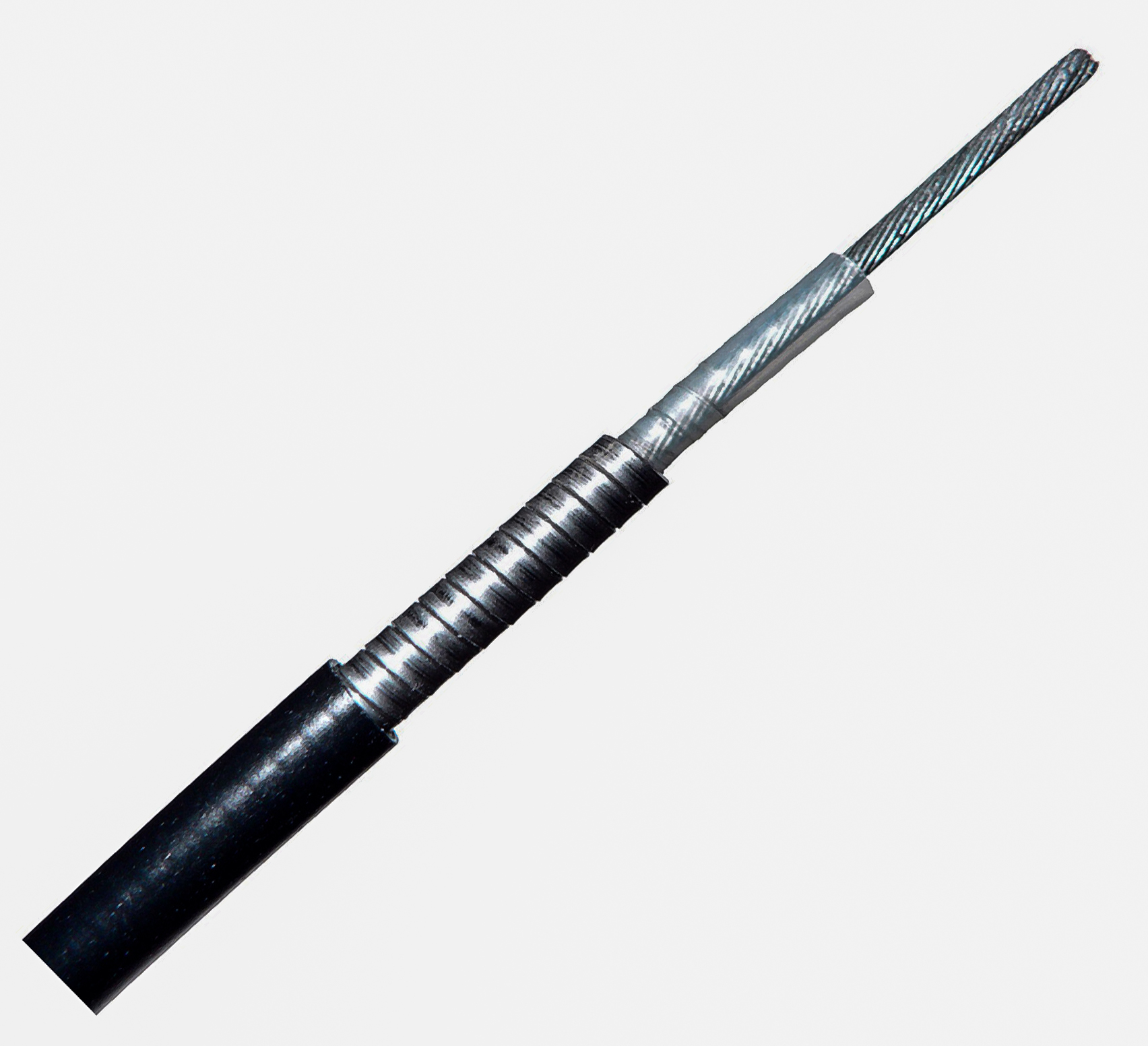
A Bowden cable ( ) is a type of flexible cable used to transmit mechanical force or energy by the movement of an inner cable relative to a hollow outer cable housing. The housing is generally of composite construction, consisting of an inner lining, a longitudinally incompressible layer such as a helical winding or a sheaf of steel wire, and a protective outer covering. The linear movement of the inner cable is most often used to transmit a pulling force, although push/pull cables have gained popularity in recent years e.g. as gear shift cables. Many light aircraft use a push/pull Bowden cable for the throttle control, and here it is normal for the inner element to be a solid wire, rather than a multi-strand cable. Usually, provision is made for adjusting the cable tension using an inline hollow bolt (often called a "barrel adjuster"), which lengthens or shortens the cable housing relative to a fixed anchor point. Lengthening the housing (turning the barrel adjuster out) ti ... [...More Info...] [...Related Items...] OR: [Wikipedia] [Google] [Baidu] |
Segmental Bronchus
A bronchus is a passage or airway in the lower respiratory tract that conducts air into the lungs. The first or primary bronchi pronounced (BRAN-KAI) to branch from the trachea at the carina are the right main bronchus and the left main bronchus. These are the widest bronchi, and enter the right lung, and the left lung at each hilum. The main bronchi branch into narrower secondary bronchi or lobar bronchi, and these branch into narrower tertiary bronchi or segmental bronchi. Further divisions of the segmental bronchi are known as 4th order, 5th order, and 6th order segmental bronchi, or grouped together as subsegmental bronchi. The bronchi, when too narrow to be supported by cartilage, are known as bronchioles. No gas exchange takes place in the bronchi. Structure The trachea (windpipe) divides at the carina into two main or primary bronchi, the left bronchus and the right bronchus. The carina of the trachea is located at the level of the sternal angle and the fifth thoracic verte ... [...More Info...] [...Related Items...] OR: [Wikipedia] [Google] [Baidu] |
Lobar Bronchus
A bronchus is a passage or airway in the lower respiratory tract that conducts air into the lungs. The first or primary bronchi pronounced (BRAN-KAI) to branch from the trachea at the carina are the right main bronchus and the left main bronchus. These are the widest bronchi, and enter the right lung, and the left lung at each hilum. The main bronchi branch into narrower secondary bronchi or lobar bronchi, and these branch into narrower tertiary bronchi or segmental bronchi. Further divisions of the segmental bronchi are known as 4th order, 5th order, and 6th order segmental bronchi, or grouped together as subsegmental bronchi. The bronchi, when too narrow to be supported by cartilage, are known as bronchioles. No gas exchange takes place in the bronchi. Structure The trachea (windpipe) divides at the carina into two main or primary bronchi, the left bronchus and the right bronchus. The carina of the trachea is located at the level of the sternal angle and the fifth thoracic verte ... [...More Info...] [...Related Items...] OR: [Wikipedia] [Google] [Baidu] |


