|
Bronchopleural Fistula
A bronchopleural fistula (BPF) is a fistula between the pleural space and the lung. It can develop following pneumonectomy, lung ablation, post-traumatically, or with certain types of infection. It may also develop when large airways are in communication with the pleural space following a large pneumothorax or other loss of pleural negative pressure, especially during positive pressure mechanical ventilation. On imaging, the diagnosis is suspected indirectly on radiograph. Increased gas in the pneumonectomy operative bed, or new gas within a loculated effusion are highly suggestive of the diagnosis. Infectious causes include tuberculosis, ''Actinomyces israelii ''Actinomyces israelii'' is a species of Gram-positive, rod-shaped bacteria within the genus ''Actinomyces''. Known to live commensally on and within humans, ''A. israelii'' is an opportunistic pathogen and a cause of actinomycosis. Many physi ...'', '' Nocardia'', and '' Blastomyces dermatitidis''. Malignancy and trauma ... [...More Info...] [...Related Items...] OR: [Wikipedia] [Google] [Baidu] |
Fistula
A fistula (plural: fistulas or fistulae ; from Latin ''fistula'', "tube, pipe") in anatomy is an abnormal connection between two hollow spaces (technically, two epithelialized surfaces), such as blood vessels, intestines, or other hollow organs. Types of fistula can be described by their location. Anal fistulas connect between the anal canal and the perianal skin. Anovaginal or rectovaginal fistulas occur when a hole develops between the anus or rectum and the vagina. Colovaginal fistulas occur between the colon and the vagina. Urinary tract fistulas are abnormal openings within the urinary tract or an abnormal connection between the urinary tract and another organ such as between the bladder and the uterus in a vesicouterine fistula, between the bladder and the vagina in a vesicovaginal fistula, and between the urethra and the vagina in urethrovaginal fistula. When occurring between two parts of the intestine, it is known as an enteroenteral fistula, between the small intest ... [...More Info...] [...Related Items...] OR: [Wikipedia] [Google] [Baidu] |
Pleural Space
The pleural cavity, pleural space, or interpleural space is the potential space between the pleurae of the pleural sac that surrounds each lung. A small amount of serous pleural fluid is maintained in the pleural cavity to enable lubrication between the membranes, and also to create a pressure gradient. The serous membrane that covers the surface of the lung is the visceral pleura and is separated from the outer membrane the parietal pleura by just the film of pleural fluid in the pleural cavity. The visceral pleura follows the fissures of the lung and the root of the lung structures. The parietal pleura is attached to the mediastinum, the upper surface of the diaphragm, and to the inside of the ribcage. Structure In humans, the left and right lungs are completely separated by the mediastinum, and there is no communication between their pleural cavities. Therefore, in cases of a unilateral pneumothorax, the contralateral lung will remain functioning normally unless there is a ... [...More Info...] [...Related Items...] OR: [Wikipedia] [Google] [Baidu] |
Lung
The lungs are the primary organs of the respiratory system in humans and most other animals, including some snails and a small number of fish. In mammals and most other vertebrates, two lungs are located near the backbone on either side of the heart. Their function in the respiratory system is to extract oxygen from the air and transfer it into the bloodstream, and to release carbon dioxide from the bloodstream into the atmosphere, in a process of gas exchange. Respiration is driven by different muscular systems in different species. Mammals, reptiles and birds use their different muscles to support and foster breathing. In earlier tetrapods, air was driven into the lungs by the pharyngeal muscles via buccal pumping, a mechanism still seen in amphibians. In humans, the main muscle of respiration that drives breathing is the diaphragm. The lungs also provide airflow that makes vocal sounds including human speech possible. Humans have two lungs, one on the left and on ... [...More Info...] [...Related Items...] OR: [Wikipedia] [Google] [Baidu] |
Pneumonectomy
A pneumonectomy (or pneumectomy) is a surgical procedure to remove a lung first successfully done in 1933 by Dr. Evarts Graham. This is not to be confused with a lobectomy or segmentectomy, which only removes one part of the lung. There are two types of pneumonectomy, simple and extra pleural. A simple pneumonectomy removes just the lung. An extra pleural pneumonectomy take away part of the diaphragm, the parietal pleura, and the pericardium on that side as well. Indications The most common reason for a pneumonectomy is to remove tumourous tissue arising from lung cancer. Other reasons can arise are a traumatic lung injury, bronchiectasis, tuberculosis, a congenital defect, and fungal infections. Contraindications Tests The operation will reduce the respiratory capacity of the patient and before conducting a pneumonectomy, survivability after the removal has to be assessed. If at all possible, a pulmonary function test (PFT) should be done prior. It has been found that fo ... [...More Info...] [...Related Items...] OR: [Wikipedia] [Google] [Baidu] |
Pneumothorax
A pneumothorax is an abnormal collection of air in the pleural space between the lung and the chest wall. Symptoms typically include sudden onset of sharp, one-sided chest pain and shortness of breath. In a minority of cases, a one-way valve is formed by an area of damaged tissue, and the amount of air in the space between chest wall and lungs increases; this is called a tension pneumothorax. This can cause a steadily worsening oxygen shortage and low blood pressure. This leads to a type of shock called obstructive shock, which can be fatal unless reversed. Very rarely, both lungs may be affected by a pneumothorax. It is often called a "collapsed lung", although that term may also refer to atelectasis. A primary spontaneous pneumothorax is one that occurs without an apparent cause and in the absence of significant lung disease. A secondary spontaneous pneumothorax occurs in the presence of existing lung disease. Smoking increases the risk of primary spontaneous pneumothora ... [...More Info...] [...Related Items...] OR: [Wikipedia] [Google] [Baidu] |
Mechanical Ventilation
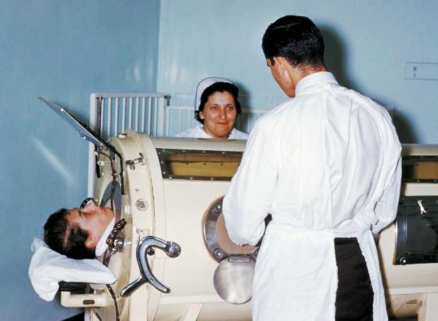
Mechanical ventilation, assisted ventilation or intermittent mandatory ventilation (IMV), is the medical term for using a machine called a ventilator to fully or partially provide artificial ventilation. Mechanical ventilation helps move air into and out of the lungs, with the main goal of helping the delivery of oxygen and removal of carbon dioxide. Mechanical ventilation is used for many reasons, including to protect the airway due to mechanical or neurologic cause, to ensure adequate oxygenation, or to remove excess carbon dioxide from the lungs. Various healthcare providers are involved with the use of mechanical ventilation and people who require ventilators are typically monitored in an intensive care unit. Mechanical ventilation is termed invasive if it involves an instrument to create an airway that is placed inside the trachea. This is done through an endotracheal tube or nasotracheal tube. For non-invasive ventilation in people who are conscious, face or nasal mask ... [...More Info...] [...Related Items...] OR: [Wikipedia] [Google] [Baidu] |
Tuberculosis
Tuberculosis (TB) is an infectious disease usually caused by '' Mycobacterium tuberculosis'' (MTB) bacteria. Tuberculosis generally affects the lungs, but it can also affect other parts of the body. Most infections show no symptoms, in which case it is known as latent tuberculosis. Around 10% of latent infections progress to active disease which, if left untreated, kill about half of those affected. Typical symptoms of active TB are chronic cough with blood-containing mucus, fever, night sweats, and weight loss. It was historically referred to as consumption due to the weight loss associated with the disease. Infection of other organs can cause a wide range of symptoms. Tuberculosis is spread from one person to the next through the air when people who have active TB in their lungs cough, spit, speak, or sneeze. People with Latent TB do not spread the disease. Active infection occurs more often in people with HIV/AIDS and in those who smoke. Diagnosis of active TB is ... [...More Info...] [...Related Items...] OR: [Wikipedia] [Google] [Baidu] |
Actinomyces Israelii
''Actinomyces israelii'' is a species of Gram-positive, rod-shaped bacteria within the genus ''Actinomyces''. Known to live commensally on and within humans, ''A. israelii'' is an opportunistic pathogen and a cause of actinomycosis. Many physiologically diverse strains of the species are known to exist, though not all are strict anaerobes. It was named after the German surgeon James Adolf Israel (1848–1926), who studied the organism for the first time in 1878. Pathogenesis Actinomycosis is most frequently caused by ''A. israelii''. It is a normal colonizer of the vagina, colon, and mouth. Infection is established first by a breach of the mucosal barrier during various procedures (dental, gastrointestinal), aspiration, or pathologies such as diverticulitis Diverticulitis, specifically colonic diverticulitis, is a gastrointestinal disease characterized by inflammation of abnormal pouches—diverticula—which can develop in the wall of the large intestine. Symptoms t ... [...More Info...] [...Related Items...] OR: [Wikipedia] [Google] [Baidu] |
Nocardia
''Nocardia'' is a genus of weakly staining Gram-positive, catalase-positive, rod-shaped bacteria. It forms partially acid-fast beaded branching filaments (acting as fungi, but being truly bacteria). It contains a total of 85 species. Some species are nonpathogenic, while others are responsible for nocardiosis. ''Nocardia'' species are found worldwide in soil rich in organic matter. In addition, they are oral microflora found in healthy gingiva, as well as periodontal pockets. Most ''Nocardia'' infections are acquired by inhalation of the bacteria or through traumatic introduction. Culture and staining ''Nocardia'' colonies have a variable appearance, but most species appear to have aerial hyphae when viewed with a dissecting microscope, particularly when they have been grown on nutritionally limiting media. ''Nocardia'' grow slowly on nonselective culture media, and are strict aerobes with the ability to grow in a wide temperature range. Some species are partially acid-fast ... [...More Info...] [...Related Items...] OR: [Wikipedia] [Google] [Baidu] |
Blastomyces Dermatitidis
''Blastomyces dermatitidis'' is a dimorphic fungus that causes blastomycosis, an invasive and often serious fungal infection found occasionally in humans and other animals. It lives in soil and wet, decaying wood, often in an area close to a waterway such as a lake, river or stream. Indoor growth may also occur, for example, in accumulated debris in damp sheds or shacks. The fungus is endemic to parts of eastern North America, particularly boreal northern Ontario, southeastern Manitoba, Quebec south of the St. Lawrence River, parts of the U.S. Appalachian mountains and interconnected eastern mountain chains, the west bank of Lake Michigan, the state of Wisconsin, and the entire Mississippi Valley including the valleys of some major tributaries such as the Ohio River. In addition, it occurs rarely in Africa both north and south of the Sahara Desert, as well as in the Arabian Peninsula and the Indian subcontinent. Though it has never been directly observed growing in nature, it is ... [...More Info...] [...Related Items...] OR: [Wikipedia] [Google] [Baidu] |


