|
Bone Scintigraphy
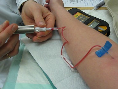
A bone scan or bone scintigraphy is a nuclear medicine imaging technique of the bone. It can help diagnose a number of bone conditions, including cancer of the bone or metastasis, location of bone inflammation and fractures (that may not be visible in traditional X-ray images), and bone infection (osteomyelitis). Nuclear medicine provides functional imaging and allows visualisation of bone metabolism or bone remodeling, which most other imaging techniques (such as X-ray computed tomography, CT) cannot. Bone scintigraphy competes with positron emission tomography (PET) for imaging of abnormal metabolism in bones, but is considerably less expensive. Bone scintigraphy has higher sensitivity but lower specificity than CT or MRI for diagnosis of scaphoid fractures following negative plain radiography. History Some of the earliest investigations into skeletal metabolism were carried out by George de Hevesy in the 1930s, using phosphorus-32 and by Charles Pecher in the 1940s. In t ... [...More Info...] [...Related Items...] OR: [Wikipedia] [Google] [Baidu] |
Nuclear Medicine
Nuclear medicine or nucleology is a medical specialty involving the application of radioactive substances in the diagnosis and treatment of disease. Nuclear imaging, in a sense, is "radiology done inside out" because it records radiation emitting from within the body rather than radiation that is generated by external sources like X-rays. In addition, nuclear medicine scans differ from radiology, as the emphasis is not on imaging anatomy, but on the function. For such reason, it is called a physiological imaging modality. Single photon emission computed tomography (SPECT) and positron emission tomography (PET) scans are the two most common imaging modalities in nuclear medicine. Diagnostic medical imaging Diagnostic In nuclear medicine imaging, radiopharmaceuticals are taken internally, for example, through inhalation, intravenously or orally. Then, external detectors (gamma cameras) capture and form images from the radiation emitted by the radiopharmaceuticals. This process ... [...More Info...] [...Related Items...] OR: [Wikipedia] [Google] [Baidu] |
Charles Pecher
Charles Pecher (26 November 1913 – 28 August 1941) was a Belgian pioneer in nuclear medicine. He discovered and introduced strontium-89 in medical therapeutic procedures in 1939. He was the first to report a possible therapeutic role for the beta emitting radionuclide strontium-89 in the palliation of bone pain associated with metastatic bone disease. His autoradiographies of animals or organs after administration of strontium-89 or phosphorus-32 started the development of bone scintigraphy. The groundbreaking work of Pecher was forgotten for decades due to the classification of information linked to the Manhattan project. The therapeutic use of 89Sr was only approved in 1993 for the palliative treatment of breast and prostate cancers metastatic to the bones for use in the US and became the first bone-seeking radiopharmaceutical that came into widespread use. Biography Born in Antwerp on 26 November 1913, Pecher was the son of the liberal politician Édouard Pecher and Emili ... [...More Info...] [...Related Items...] OR: [Wikipedia] [Google] [Baidu] |
Hydroxyapatite
Hydroxyapatite, also called hydroxylapatite (HA), is a naturally occurring mineral form of calcium apatite with the formula Ca5(PO4)3(OH), but it is usually written Ca10(PO4)6(OH)2 to denote that the crystal unit cell comprises two entities. Hydroxyapatite is the hydroxyl endmember of the complex apatite group. The OH− ion can be replaced by fluoride, chloride or carbonate, producing fluorapatite or chlorapatite. It crystallizes in the hexagonal crystal system. Pure hydroxyapatite powder is white. Naturally occurring apatites can, however, also have brown, yellow, or green colorations, comparable to the discolorations of dental fluorosis. Up to 50% by volume and 70% by weight of human bone is a modified form of hydroxyapatite, known as bone mineral. Carbonated calcium-deficient hydroxyapatite is the main mineral of which dental enamel and dentin are composed. Hydroxyapatite crystals are also found in pathological calcifications such as those found in breast tumors, as w ... [...More Info...] [...Related Items...] OR: [Wikipedia] [Google] [Baidu] |
Adsorb
Adsorption is the adhesion of atoms, ions or molecules from a gas, liquid or dissolved solid to a surface. This process creates a film of the ''adsorbate'' on the surface of the ''adsorbent''. This process differs from absorption, in which a fluid (the ''absorbate'') is dissolved by or permeates a liquid or solid (the ''absorbent''). Adsorption is a '' surface phenomenon'', while absorption involves the whole volume of the material, although adsorption does often precede absorption. The term ''sorption'' encompasses both processes, while ''desorption'' is the reverse of it. Like surface tension, adsorption is a consequence of surface energy. In a bulk material, all the bonding requirements (be they ionic, covalent or metallic) of the constituent atoms of the material are fulfilled by other atoms in the material. However, atoms on the surface of the adsorbent are not wholly surrounded by other adsorbent atoms and therefore can attract adsorbates. The exact nature of the bond ... [...More Info...] [...Related Items...] OR: [Wikipedia] [Google] [Baidu] |
Methylene Diphosphonate
Medronic acid (conjugate base, medronate), also known as methylene diphosphonate, is the smallest bisphosphonate. Its complex with radioactive technetium, 99mTc medronic acid, is used in nuclear medicine to detect bone abnormalities, including metastases Metastasis is a pathogenic agent's spread from an initial or primary site to a different or secondary site within the host's body; the term is typically used when referring to metastasis by a cancerous tumor. The newly pathological sites, then .... References External links * Bisphosphonates {{organic-compound-stub ... [...More Info...] [...Related Items...] OR: [Wikipedia] [Google] [Baidu] |
Radiopharmaceutical
Radiopharmaceuticals, or medicinal radiocompounds, are a group of pharmaceutical drugs containing radioactive isotopes. Radiopharmaceuticals can be used as diagnostic and therapeutic agents. Radiopharmaceuticals emit radiation themselves, which is different from contrast media which absorb or alter external electromagnetism or ultrasound. Radiopharmacology is the branch of pharmacology that specializes in these agents. The main group of these compounds are the radiotracers used to diagnose dysfunction in body tissues. While not all medical isotopes are radioactive, radiopharmaceuticals are the oldest and still most common such drugs. Drug nomenclature As with other pharmaceutical drugs, there is standardization of the drug nomenclature for radiopharmaceuticals, although various standards coexist. The International Nonproprietary Names (INNs), United States Pharmacopeia (USP) names, and IUPAC names for these agents are usually similar other than trivial style differences. The d ... [...More Info...] [...Related Items...] OR: [Wikipedia] [Google] [Baidu] |
Diphosphonates
Bisphosphonates are a class of drugs that prevent the loss of bone density, used to treat osteoporosis and similar diseases. They are the most commonly prescribed drugs used to treat osteoporosis. They are called bisphosphonates because they have two phosphonate () groups. They are thus also called diphosphonates ('' bis-'' or '' di-'' + ''phosphonate''). Evidence shows that they reduce the risk of fracture in post-menopausal women with osteoporosis. Bone tissue undergoes constant remodeling and is kept in balance (homeostasis) by osteoblasts creating bone and osteoclasts destroying bone. Bisphosphonates inhibit the digestion of bone by encouraging osteoclasts to undergo apoptosis, or cell death, thereby slowing bone loss. The uses of bisphosphonates include the prevention and treatment of osteoporosis, Paget's disease of bone, bone metastasis (with or without hypercalcemia), multiple myeloma, primary hyperparathyroidism, osteogenesis imperfecta, fibrous dysplasia, and o ... [...More Info...] [...Related Items...] OR: [Wikipedia] [Google] [Baidu] |
Phosphate
In chemistry, a phosphate is an anion, salt, functional group or ester derived from a phosphoric acid. It most commonly means orthophosphate, a derivative of orthophosphoric acid . The phosphate or orthophosphate ion is derived from phosphoric acid by the removal of three protons . Removal of one or two protons gives the dihydrogen phosphate ion and the hydrogen phosphate ion ion, respectively. These names are also used for salts of those anions, such as ammonium dihydrogen phosphate and trisodium phosphate. File:3-phosphoric-acid-3D-balls.png, Phosphoricacid File:2-dihydrogenphosphate-3D-balls.png, Dihydrogenphosphate File:1-hydrogenphosphate-3D-balls.png, Hydrogenphosphate File:0-phosphate-3D-balls.png, Phosphate In organic chemistry, phosphate or orthophosphate is an organophosphate, an ester of orthophosphoric acid of the form where one or more hydrogen atoms are replaced by organic groups. An example is trimethyl phosphate, . The term also refers to the triv ... [...More Info...] [...Related Items...] OR: [Wikipedia] [Google] [Baidu] |
Technetium-99m
Technetium-99m (99mTc) is a metastable nuclear isomer of technetium-99 (itself an isotope of technetium), symbolized as 99mTc, that is used in tens of millions of medical diagnostic procedures annually, making it the most commonly used medical radioisotope in the world. Technetium-99m is used as a radioactive tracer and can be detected in the body by medical equipment (gamma cameras). It is well suited to the role, because it emits readily detectable gamma rays with a photon energy of 140 keV (these 8.8 pm photons are about the same wavelength as emitted by conventional X-ray diagnostic equipment) and its half-life for gamma emission is 6.0058 hours (meaning 93.7% of it decays to 99Tc in 24 hours). The relatively "short" physical half-life of the isotope and its biological half-life of 1 day (in terms of human activity and metabolism) allows for scanning procedures which collect data rapidly but keep total patient radiation exposure low. The same characteristics make the ... [...More Info...] [...Related Items...] OR: [Wikipedia] [Google] [Baidu] |
Rectilinear Scanner
A rectilinear scanner is an imaging device, used to capture emission from radiopharmaceuticals in nuclear medicine. The image is created by physically moving a radiation detector over the surface of a radioactive patient. It has become obsolete in medical imaging, largely replaced by the gamma camera since the late 1960s. History One of the first rectilinear scanners was developed by Benedict Cassen in 1950. Before then hand-held detectors had been used to locate radioactive materials in patients, but the Cassen system (designed for Iodine-131) combined a motor driven photomultiplier tube and printing mechanism. Subsequent developments improved the detection systems, movement, display and printing of images. Components Cassen's original rectilinear scanner used calcium tungstate (CaWo4) crystal as the radiation detector. Later systems used a Sodium iodide (NaI) scintillator, as in a gamma camera. The detector must be connected by mechanical or electronic means to an output sys ... [...More Info...] [...Related Items...] OR: [Wikipedia] [Google] [Baidu] |
Isotopes Of Strontium
The alkaline earth metal strontium (38Sr) has four stable, naturally occurring isotopes: 84Sr (0.56%), 86Sr (9.86%), 87Sr (7.0%) and 88Sr (82.58%). Its standard atomic weight is 87.62(1). Only 87Sr is radiogenic; it is produced by decay from the radioactive alkali metal 87 Rb, which has a half-life of 4.88 × 10¹⁰ years (i.e. more than three times longer than the current age of the universe). Thus, there are two sources of 87Sr in any material: primordial, formed during nucleosynthesis along with 84Sr, 86Sr and 88Sr; and that formed by radioactive decay of 87Rb. The ratio 87Sr/86Sr is the parameter typically reported in geologic investigations; ratios in minerals and rocks have values ranging from about 0.7 to greater than 4.0 (see rubidium–strontium dating). Because strontium has an electron configuration similar to that of calcium, it readily substitutes for calcium in minerals. In addition to the four stable isotopes, thirty-two unstable isotopes of strontium are known to e ... [...More Info...] [...Related Items...] OR: [Wikipedia] [Google] [Baidu] |
Fluorine-18
Fluorine-18 (18F) is a fluorine radioisotope which is an important source of positrons. It has a mass of 18.0009380(6) u and its half-life is 109.771(20) minutes. It decays by positron emission 96% of the time and electron capture 4% of the time. Both modes of decay yield stable oxygen-18. Natural occurrence is a natural trace radioisotope produced by cosmic ray spallation of atmospheric argon as well as by reaction of protons with natural oxygen: 18O + p → 18F + n.18O">sup>18Oater with high energy protons (typically ~18 MeV). The fluorine produced is in the form of a water solution of 18F.html" ;"title="sup>18F">sup>18F luoride, which is then used in a rapid chemical synthesis of various radio pharmaceuticals. The organic oxygen-18 pharmaceutical molecule is not made before the production of the radiopharmaceutical, as high energy protons destroy such molecules ( radiolysis). Radiopharmaceuticals using fluorine must therefore be synthesized after the fluorine-18 has been p ... [...More Info...] [...Related Items...] OR: [Wikipedia] [Google] [Baidu] |


