|
Bone Graft
Bone grafting is a surgical procedure that replaces missing bone in order to repair bone fractures that are extremely complex, pose a significant health risk to the patient, or fail to heal properly. Some small or acute fractures can be cured without bone grafting, but the risk is greater for large fractures like compound fractures. Bone generally has the ability to regenerate completely but requires a very small fracture space or some sort of scaffold to do so. Bone grafts may be autologous (bone harvested from the patient's own body, often from the iliac crest), allograft (cadaveric bone usually obtained from a bone bank), or synthetic (often made of hydroxyapatite or other naturally occurring and biocompatible substances) with similar mechanical properties to bone. Most bone grafts are expected to be resorbed and replaced as the natural bone heals over a few months' time. The principles involved in successful bone grafts include osteoconduction (guiding the reparative growth ... [...More Info...] [...Related Items...] OR: [Wikipedia] [Google] [Baidu] |
Bone Fractures
A bone fracture (abbreviated FRX or Fx, Fx, or #) is a medical condition in which there is a partial or complete break in the continuity of any bone in the body. In more severe cases, the bone may be broken into several fragments, known as a ''comminuted fracture''. A bone fracture may be the result of high force impact or stress, or a minimal trauma injury as a result of certain medical conditions that weaken the bones, such as osteoporosis, osteopenia, bone cancer, or osteogenesis imperfecta, where the fracture is then properly termed a pathologic fracture. Signs and symptoms Although bone tissue contains no pain receptors, a bone fracture is painful for several reasons: * Breaking in the continuity of the periosteum, with or without similar discontinuity in endosteum, as both contain multiple pain receptors. * Edema and hematoma of nearby soft tissues caused by ruptured bone marrow evokes pressure pain. * Involuntary muscle spasms trying to hold bone fragments i ... [...More Info...] [...Related Items...] OR: [Wikipedia] [Google] [Baidu] |
Implantology
A dental implant (also known as an endosseous implant or fixture) is a prosthesis that interfaces with the bone of the jaw or skull to support a dental prosthesis such as a crown, bridge, denture, or facial prosthesis or to act as an orthodontic anchor. The basis for modern dental implants is a biologic process called osseointegration, in which materials such as titanium or zirconia form an intimate bond to bone. The implant fixture is first placed so that it is likely to osseointegrate, then a dental prosthetic is added. A variable amount of healing time is required for osseointegration before either the dental prosthetic (a tooth, bridge or denture) is attached to the implant or an abutment is placed which will hold a dental prosthetic/crown. Success or failure of implants depends on the health of the person receiving the treatment, drugs which affect the chances of osseointegration, and the health of the tissues in the mouth. The amount of stress that will be put on the impl ... [...More Info...] [...Related Items...] OR: [Wikipedia] [Google] [Baidu] |
Allograft
Allotransplant (''allo-'' meaning "other" in Greek) is the transplantation of cells, tissues, or organs to a recipient from a genetically non-identical donor of the same species. The transplant is called an allograft, allogeneic transplant, or homograft. Most human tissue and organ transplants are allografts. It is contrasted with autotransplantation (from one part of the body to another in the same person), syngenic transplantation of isografts (grafts transplanted between two genetically identical individuals) and xenotransplantation (from other species). Allografts can be referred to as "homostatic" if they are biologically inert when transplanted, such as bone and cartilage. An immune response against an allograft or xenograft is termed rejection. An allogenic bone marrow transplant can result in an immune attack on the recipient, called graft-versus-host disease. Procedure Material is obtained from a donor who is a living person, or a deceased person's body recei ... [...More Info...] [...Related Items...] OR: [Wikipedia] [Google] [Baidu] |
Dentin
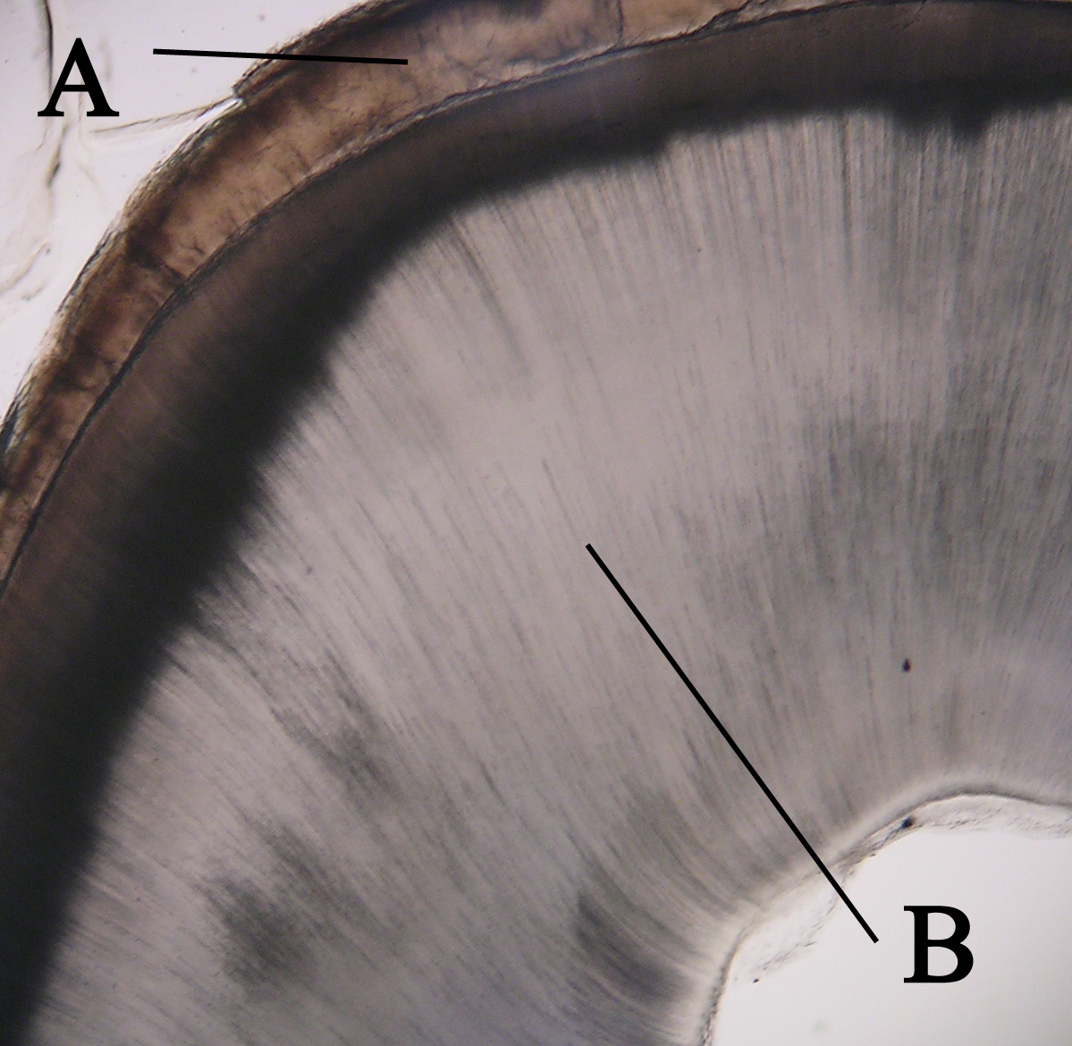
Dentin () (American English) or dentine ( or ) (British English) ( la, substantia eburnea) is a calcified tissue of the body and, along with enamel, cementum, and pulp, is one of the four major components of teeth. It is usually covered by enamel on the crown and cementum on the root and surrounds the entire pulp. By volume, 45% of dentin consists of the mineral hydroxyapatite, 33% is organic material, and 22% is water. Yellow in appearance, it greatly affects the color of a tooth due to the translucency of enamel. Dentin, which is less mineralized and less brittle than enamel, is necessary for the support of enamel. Dentin rates approximately 3 on the Mohs scale of mineral hardness. There are two main characteristics which distinguish dentin from enamel: firstly, dentin forms throughout life; secondly, dentin is sensitive and can become hypersensitive to changes in temperature due to the sensory function of odontoblasts, especially when enamel recedes and dentin channels b ... [...More Info...] [...Related Items...] OR: [Wikipedia] [Google] [Baidu] |
Anterior Superior Iliac Spine
The anterior superior iliac spine ( abbreviated: ASIS) is a bony projection of the iliac bone, and an important landmark of surface anatomy. It refers to the anterior extremity of the iliac crest of the pelvis. It provides attachment for the inguinal ligament, and the sartorius muscle. The tensor fasciae latae muscle attaches to the lateral aspect of the superior anterior iliac spine, and also about 5 cm away at the iliac tubercle. Structure The anterior superior iliac spine refers to the anterior extremity of the iliac crest of the pelvis. This is a key surface landmark, and easily palpated. It provides attachment for the inguinal ligament, the sartorius muscle, and the tensor fasciae latae muscle. A variety of structures lie close to the anterior superior iliac spine, including the subcostal nerve, the femoral artery (which passes between it and the pubic symphysis), and the iliohypogastric nerve. Clinical significance The anterior superior iliac sp ... [...More Info...] [...Related Items...] OR: [Wikipedia] [Google] [Baidu] |
Vital Bone Graft
Vital or Vitals may refer to: Places * Vital Creek, a creek located in the Omineca Country region of British Columbia * Vital Range, a subrange in the Omineca Mountains in British Columbia People * Vital (given name) *Vital (surname) Arts, entertainment, and media Music * ''Vital'' (Anberlin album), 2012 * ''Vital'' (Fernando Otero album), a 2010 album by Fernando Otero * ''Vital'' (Van der Graaf Generator album), 1978 * ''Vital'', a 2009 studio album by Norman Bedard * ''Vitals'' (Mutemath album), 2015 Other uses in arts, entertainment, and media * ''Vital'' (film), a 2004 Japanese movie directed by Shinya Tsukamoto * ''Vitals'' (novel), a 2002 science fiction/techno-thriller novel by Greg Bear Other uses * Vital (grape), a Portuguese wine grape grown in the Alcobaça wine region * USS ''Vital'', two US warships * Vital currents, the concept of currents within the body found in Yoga * VITAL for Children, a charitable organisation * Vital Forsikring, a Norwegian insu ... [...More Info...] [...Related Items...] OR: [Wikipedia] [Google] [Baidu] |
Periosteum
The periosteum is a membrane that covers the outer surface of all bones, except at the articular surfaces (i.e. the parts within a joint space) of long bones. Endosteum lines the inner surface of the medullary cavity of all long bones. Structure The periosteum consists of an outer fibrous layer, and an inner cambium layer (or osteogenic layer). The fibrous layer is of dense irregular connective tissue, containing fibroblasts, while the cambium layer is highly cellular containing progenitor cells that develop into osteoblasts. These osteoblasts are responsible for increasing the width of a long bone and the overall size of the other bone types. After a bone fracture, the progenitor cells develop into osteoblasts and chondroblasts, which are essential to the healing process. The outer fibrous layer and the inner cambium layer is differentiated under electron micrography. As opposed to osseous tissue, the periosteum has nociceptors, sensory neurons that make it very sensit ... [...More Info...] [...Related Items...] OR: [Wikipedia] [Google] [Baidu] |
Human Skull
The skull is a bone protective cavity for the brain. The skull is composed of four types of bone i.e., cranial bones, facial bones, ear ossicles and hyoid bone. However two parts are more prominent: the cranium and the mandible. In humans, these two parts are the neurocranium and the viscerocranium ( facial skeleton) that includes the mandible as its largest bone. The skull forms the anterior-most portion of the skeleton and is a product of cephalisation—housing the brain, and several sensory structures such as the eyes, ears, nose, and mouth. In humans these sensory structures are part of the facial skeleton. Functions of the skull include protection of the brain, fixing the distance between the eyes to allow stereoscopic vision, and fixing the position of the ears to enable sound localisation of the direction and distance of sounds. In some animals, such as horned ungulates (mammals with hooves), the skull also has a defensive function by providing the mount (on the f ... [...More Info...] [...Related Items...] OR: [Wikipedia] [Google] [Baidu] |
Human Mandible
In anatomy, the mandible, lower jaw or jawbone is the largest, strongest and lowest bone in the human facial skeleton. It forms the lower jaw and holds the lower teeth in place. The mandible sits beneath the maxilla. It is the only movable bone of the skull (discounting the ossicles of the middle ear). It is connected to the temporal bones by the temporomandibular joints. The bone is formed in the fetus from a fusion of the left and right mandibular prominences, and the point where these sides join, the mandibular symphysis, is still visible as a faint ridge in the midline. Like other symphyses in the body, this is a midline articulation where the bones are joined by fibrocartilage, but this articulation fuses together in early childhood.Illustrated Anatomy of the Head and Neck, Fehrenbach and Herring, Elsevier, 2012, p. 59 The word "mandible" derives from the Latin word ''mandibula'', "jawbone" (literally "one used for chewing"), from '' mandere'' "to chew" and ''-bula' ... [...More Info...] [...Related Items...] OR: [Wikipedia] [Google] [Baidu] |
Ribs
The rib cage, as an enclosure that comprises the ribs, vertebral column and sternum in the thorax of most vertebrates, protects vital organs such as the heart, lungs and great vessels. The sternum, together known as the thoracic cage, is a semi-rigid bony and cartilaginous structure which surrounds the thoracic cavity and supports the shoulder girdle to form the core part of the human skeleton. A typical human thoracic cage consists of 12 pairs of ribs and the adjoining costal cartilages, the sternum (along with the manubrium and xiphoid process), and the 12 thoracic vertebrae articulating with the ribs. Together with the skin and associated fascia and muscles, the thoracic cage makes up the thoracic wall and provides attachments for extrinsic skeletal muscles of the neck, upper limbs, upper abdomen and back. The rib cage intrinsically holds the muscles of respiration (diaphragm, intercostal muscles, etc.) that are crucial for active inhalation and forced exhalatio ... [...More Info...] [...Related Items...] OR: [Wikipedia] [Google] [Baidu] |
Fibula
The fibula or calf bone is a human leg, leg bone on the Lateral (anatomy), lateral side of the tibia, to which it is connected above and below. It is the smaller of the two bones and, in proportion to its length, the most slender of all the long bones. Its upper extremity is small, placed toward the back of the Upper extremity of tibia, head of the tibia, below the knee, knee joint and excluded from the formation of this joint. Its lower extremity inclines a little forward, so as to be on a plane anterior to that of the upper end; it projects below the tibia and forms the lateral part of the ankle, ankle joint. Structure The bone has the following components: * Lateral malleolus * Interosseous membrane connecting the fibula to the tibia, forming a syndesmosis joint * The superior tibiofibular articulation is an arthrodial joint between the lateral condyle of tibia, lateral condyle of the tibia and the head of the fibula. * The inferior tibiofibular articulation (tibiofibular synd ... [...More Info...] [...Related Items...] OR: [Wikipedia] [Google] [Baidu] |


