|
Binasal Hemianopsia
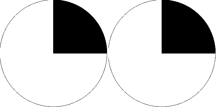
Binasal hemianopsia is the medical description of a type of partial blindness where vision is missing in the inner half of both the right and left visual field. It is associated with certain lesions of the eye and of the central nervous system, such as congenital hydrocephalus. Causes In binasal hemianopsia, vision is missing in the inner (nasal or medial) half of both the right and left visual fields. Information from the nasal visual field falls on the temporal (lateral) retina. Those lateral retinal nerve fibers do not cross in the optic chiasm. Calcification of the internal carotid arteries can impinge the uncrossed, lateral retinal fibers, leading to loss of vision in the nasal field. Clinical testing of visual fields (by confrontation) can produce a false positive result, particularly in inferior nasal quadrants. Management Etymology The absence of vision in half of a visual field is described as ''hemianopsia''. The absence of visual perception in one quarter of a vis ... [...More Info...] [...Related Items...] OR: [Wikipedia] [Google] [Baidu] |
Blindness
Visual impairment, also known as vision impairment, is a medical definition primarily measured based on an individual's better eye visual acuity; in the absence of treatment such as correctable eyewear, assistive devices, and medical treatment– visual impairment may cause the individual difficulties with normal daily tasks including reading and walking. Low vision is a functional definition of visual impairment that is chronic, uncorrectable with treatment or correctable lenses, and impacts daily living. As such low vision can be used as a disability metric and varies based on an individual's experience, environmental demands, accommodations, and access to services. The American Academy of Ophthalmology defines visual impairment as the best-corrected visual acuity of less than 20/40 in the better eye, and the World Health Organization defines it as a presenting acuity of less than 6/12 in the better eye. The term blindness is used for complete or nearly complete vision loss. In ... [...More Info...] [...Related Items...] OR: [Wikipedia] [Google] [Baidu] |
Central Nervous System
The central nervous system (CNS) is the part of the nervous system consisting primarily of the brain and spinal cord. The CNS is so named because the brain integrates the received information and coordinates and influences the activity of all parts of the bodies of bilaterally symmetric and triploblastic animals—that is, all multicellular animals except sponges and diploblasts. It is a structure composed of nervous tissue positioned along the rostral (nose end) to caudal (tail end) axis of the body and may have an enlarged section at the rostral end which is a brain. Only arthropods, cephalopods and vertebrates have a true brain (precursor structures exist in onychophorans, gastropods and lancelets). The rest of this article exclusively discusses the vertebrate central nervous system, which is radically distinct from all other animals. Overview In vertebrates, the brain and spinal cord are both enclosed in the meninges. The meninges provide a barrier to chemicals dissolv ... [...More Info...] [...Related Items...] OR: [Wikipedia] [Google] [Baidu] |
Hydrocephalus
Hydrocephalus is a condition in which an accumulation of cerebrospinal fluid (CSF) occurs within the brain. This typically causes increased intracranial pressure, pressure inside the skull. Older people may have headaches, double vision, poor balance, urinary incontinence, personality changes, or mental impairment. In babies, it may be seen as a rapid increase in head size. Other symptoms may include vomiting, sleepiness, seizures, and Parinaud's syndrome, downward pointing of the eyes. Hydrocephalus can occur due to birth defects or be acquired later in life. Associated birth defects include neural tube defects and those that result in aqueductal stenosis. Other causes include meningitis, brain tumors, traumatic brain injury, intraventricular hemorrhage, and subarachnoid hemorrhage. The four types of hydrocephalus are communicating, noncommunicating, ''ex vacuo'', and normal pressure hydrocephalus, normal pressure. Diagnosis is typically made by physical examination and medic ... [...More Info...] [...Related Items...] OR: [Wikipedia] [Google] [Baidu] |
Visual Perception
Visual perception is the ability to interpret the surrounding environment through photopic vision (daytime vision), color vision, scotopic vision (night vision), and mesopic vision (twilight vision), using light in the visible spectrum reflected by objects in the environment. This is different from visual acuity, which refers to how clearly a person sees (for example "20/20 vision"). A person can have problems with visual perceptual processing even if they have 20/20 vision. The resulting perception is also known as vision, sight, or eyesight (adjectives ''visual'', ''optical'', and ''ocular'', respectively). The various physiological components involved in vision are referred to collectively as the visual system, and are the focus of much research in linguistics, psychology, cognitive science, neuroscience, and molecular biology, collectively referred to as vision science. Visual system In humans and a number of other mammals, light enters the eye through the cornea and is ... [...More Info...] [...Related Items...] OR: [Wikipedia] [Google] [Baidu] |
Visual Field
The visual field is the "spatial array of visual sensations available to observation in introspectionist psychological experiments". Or simply, visual field can be defined as the entire area that can be seen when an eye is fixed straight at a point. The equivalent concept for optical instruments and image sensors is the field of view (FOV). In optometry, ophthalmology, and neurology, a visual field test is used to determine whether the visual field is affected by diseases that cause local scotoma or a more extensive loss of vision or a reduction in sensitivity (increase in threshold). Normal limits The normal (monocular) human visual field extends to approximately 60 degrees nasally (toward the nose, or inward) from the vertical meridian in each eye, to 107 degrees temporally (away from the nose, or outwards) from the vertical meridian, and approximately 70 degrees above and 80 below the horizontal meridian. The binocular visual field is the superimposition of the two monocular ... [...More Info...] [...Related Items...] OR: [Wikipedia] [Google] [Baidu] |
Hemianopsia
Hemianopsia, or hemianopia, is a loss of vision or blindness (anopsia) in half the visual field, usually on one side of the vertical midline. The most common causes of this damage are stroke, brain tumor, and trauma. This article deals only with permanent hemianopsia, and not with transitory or temporary hemianopsia, as identified by William Wollaston PRS in 1824. Temporary hemianopsia can occur in the aura phase of migraine. Etymology The word ''hemianopsia'' is from Greek origins, where: * ''hemi'' means "half", * ''an'' means "without", and * ''opsia'' means "seeing". Types When the pathology involves both eyes, it is either homonymous or heteronymous. Homonymous hemianopsia Paris as seen with left homonymous hemianopsia. A homonymous hemianopsia is the loss of half of the visual field on the same side in both eyes. The visual images that we see to the right side travel from both eyes to the left side of the brain, while the visual images we see to the left sid ... [...More Info...] [...Related Items...] OR: [Wikipedia] [Google] [Baidu] |
Quadrantanopsia
Quadrantanopia, quadrantanopsia, refers to an anopia (loss of vision) affecting a quarter of the visual field. It can be associated with a lesion of an optic radiation In neuroanatomy, the optic radiation (also known as the geniculocalcarine tract, the geniculostriate pathway, and posterior thalamic radiation) are axons from the neurons in the lateral geniculate nucleus to the primary visual cortex. The optic .... While quadrantanopia can be caused by lesions in the temporal and parietal lobes of the brain, it is most commonly associated with lesions in the occipital lobe.Kolb, B & Whishaw, I.Q. Human Neuropsychology, Sixth Edition, p.361; Worth Publishers (2008) Presentation An interesting aspect of quadrantanopia is that there exists a distinct and sharp border between the intact and damaged visual fields, due to an anatomical separation of the quadrants of the visual field. For example, information in the left half of visual field is processed in the right occip ... [...More Info...] [...Related Items...] OR: [Wikipedia] [Google] [Baidu] |
Anopsia
An anopsia () is a defect in the visual field. If the defect is only partial, then the portion of the field with the defect can be used to isolate the underlying cause. Types of partial anopsia: *Hemianopsia **Homonymous hemianopsia **Heteronymous hemianopsia ***Binasal hemianopsia ***Bitemporal hemianopsia **Superior hemianopia **Inferior hemianopia *Quadrantanopia Quadrantanopia, quadrantanopsia, refers to an anopia (loss of vision) affecting a quarter of the visual field. It can be associated with a lesion of an optic radiation. While quadrantanopia can be caused by lesions in the temporal and parieta ... References External links Visual disturbances and blindness {{eye-stub ... [...More Info...] [...Related Items...] OR: [Wikipedia] [Google] [Baidu] |
Bitemporal Hemianopsia
Bitemporal hemianopsia, is the medical description of a type of partial blindness where vision is missing in the outer half of both the right and left visual field. It is usually associated with lesions of the optic chiasm, the area where the optic nerves from the right and left eyes cross near the pituitary gland. Causes In bitemporal hemianopsia, vision is missing in the outer (temporal or lateral) half of both the right and left visual fields. Information from the temporal visual field falls on the nasal (medial) retina. The nasal retina is responsible for carrying the information along the optic nerve, and crosses to the other side at the optic chiasm. When there is compression at optic chiasm, the visual impulse from both nasal retina are affected, leading to inability to view the temporal, or peripheral, vision. This phenomenon is known as bitemporal hemianopsia. Knowing the neurocircuitry of visual signal flow through the optic tract is very important in understanding bitem ... [...More Info...] [...Related Items...] OR: [Wikipedia] [Google] [Baidu] |
Homonymous Hemianopsia
Hemianopsia, or hemianopia, is a visual field loss on the left or right side of the vertical midline. It can affect one eye but usually affects both eyes. Homonymous hemianopsia (or homonymous hemianopia) is hemianopic visual field loss on the same side of both eyes. Homonymous hemianopsia occurs because the right half of the brain has visual pathways for the left hemifield of both eyes, and the left half of the brain has visual pathways for the right hemifield of both eyes. When one of these pathways is damaged, the corresponding visual field is lost. Signs and symptoms Paris as seen with right homonymous hemianopsia Mobility can be difficult for people with homonymous hemianopsia. "Patients frequently complain of bumping into obstacles on the side of the field loss, thereby bruising their arms and legs." People with homonymous hemianopsia often experience discomfort in crowds. "A patient with this condition may be unaware of what he or she cannot see and frequently bumps ... [...More Info...] [...Related Items...] OR: [Wikipedia] [Google] [Baidu] |
Blindness
Visual impairment, also known as vision impairment, is a medical definition primarily measured based on an individual's better eye visual acuity; in the absence of treatment such as correctable eyewear, assistive devices, and medical treatment– visual impairment may cause the individual difficulties with normal daily tasks including reading and walking. Low vision is a functional definition of visual impairment that is chronic, uncorrectable with treatment or correctable lenses, and impacts daily living. As such low vision can be used as a disability metric and varies based on an individual's experience, environmental demands, accommodations, and access to services. The American Academy of Ophthalmology defines visual impairment as the best-corrected visual acuity of less than 20/40 in the better eye, and the World Health Organization defines it as a presenting acuity of less than 6/12 in the better eye. The term blindness is used for complete or nearly complete vision loss. In ... [...More Info...] [...Related Items...] OR: [Wikipedia] [Google] [Baidu] |


.jpg)