|
Balloon Aortic Valvuloplasty
Aortic valvuloplasty, also known as balloon aortic valvuloplasty (BAV), is a procedure used to improve blood flow through the aortic valve in conditions that cause aortic stenosis, or narrowing of the aortic valve. It can be performed in various patient populations including fetuses, newborns, children, adults, and pregnant women. The procedure involves using a balloon catheter to dilate the narrowed aortic valve by inflating the balloon. Medical Uses Guidelines and indications are specific to different patient populations. For adults with aortic stenosis, guidelines suggest that balloon aortic valvuloplasty (BAV) is to be used as a temporary procedure to improve blood flow through the aortic valve to alleviate symptoms and stabilize clinically before having more invasive procedures done, including aortic valve replacement (AVR) or transcatheter aortic valve implantation (TAVI). The most common conditions that deem patients too unstable for AVR or TAVI are pulmonary edema and card ... [...More Info...] [...Related Items...] OR: [Wikipedia] [Google] [Baidu] |
Aortic Valve
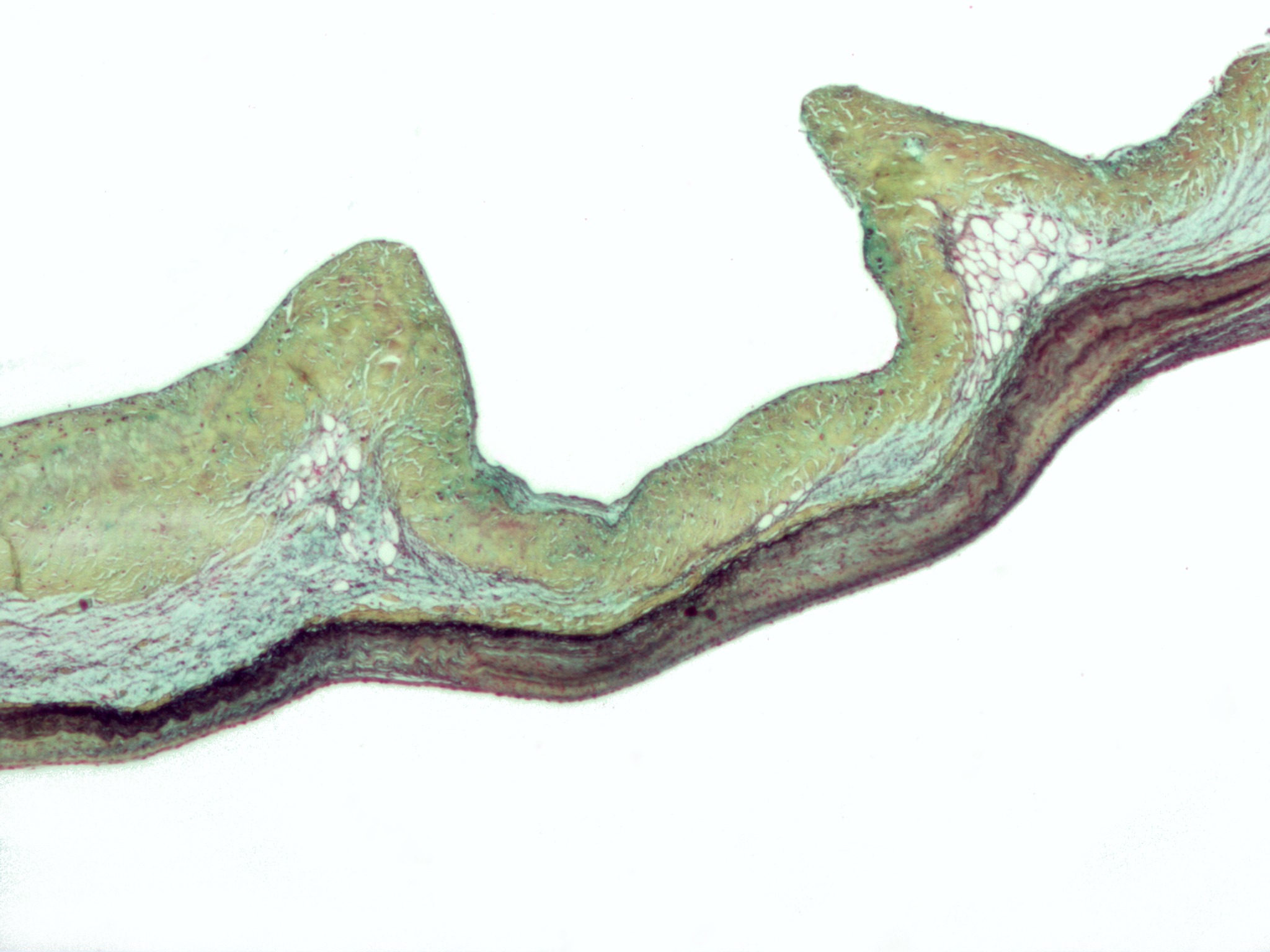
The aortic valve is a valve in the heart of humans and most other animals, located between the left ventricle and the aorta. It is one of the four valves of the heart and one of the two semilunar valves, the other being the pulmonary valve. The aortic valve normally has three cusps or leaflets, although in 1–2% of the population it is found to congenitally have two leaflets. The aortic valve is the last structure in the heart the blood travels through before stopping the flow through the systemic circulation. Structure The aortic valve normally has three cusps however there is some discrepancy in their naming. They may be called the left coronary, right coronary and non-coronary cusp. Some sources also advocate they be named as a left, right and posterior cusp. Anatomists have traditionally named them the left posterior (origin of left coronary), anterior (origin of the right coronary) and right posterior. The three cusps, when the valve is closed, contain a sinus called an a ... [...More Info...] [...Related Items...] OR: [Wikipedia] [Google] [Baidu] |
Carotid Artery , an artery on each side of the head and neck supplying blood to the brain
{{SIA ...
Carotid artery may refer to: * Common carotid artery, often "carotids" or "carotid", an artery on each side of the neck which divides into the external carotid artery and internal carotid artery * External carotid artery, an artery on each side of the head and neck supplying blood to the face, scalp, skull, neck and meninges * Internal carotid artery The internal carotid artery (Latin: arteria carotis interna) is an artery in the neck which supplies the anterior circulation of the brain. In human anatomy, the internal and external carotids arise from the common carotid arteries, where these b ... [...More Info...] [...Related Items...] OR: [Wikipedia] [Google] [Baidu] |
Kidney Function
Assessment of kidney function occurs in different ways, using the presence of symptoms and signs, as well as measurements using urine tests, blood tests, and medical imaging. Functions of a healthy kidney include maintaining a person's fluid balance, maintaining an acid-base balance; regulating electrolytes including sodium, potassium, and other electrolytes; clearing toxins; regulating blood pressure; and regulating hormones, such as erythropoietin; and activation of vitamin D. Introduction The functions of the kidney include maintenance of acid-base balance; regulation of fluid balance; regulation of sodium, potassium, and other electrolytes; clearance of toxins; absorption of glucose, amino acids, and other small molecules; regulation of blood pressure; production of various hormones, such as erythropoietin; and activation of vitamin D. Much of renal physiology is studied at the level of the nephron, the smallest functional unit of the kidney. Each nephron b ... [...More Info...] [...Related Items...] OR: [Wikipedia] [Google] [Baidu] |
Arrhythmia
Arrhythmias, also known as cardiac arrhythmias, heart arrhythmias, or dysrhythmias, are irregularities in the heartbeat, including when it is too fast or too slow. A resting heart rate that is too fast – above 100 beats per minute in adults – is called tachycardia, and a resting heart rate that is too slow – below 60 beats per minute – is called bradycardia. Some types of arrhythmias have no symptoms. Symptoms, when present, may include palpitations or feeling a pause between heartbeats. In more serious cases, there may be lightheadedness, passing out, shortness of breath or chest pain. While most cases of arrhythmia are not serious, some predispose a person to complications such as stroke or heart failure. Others may result in sudden death. Arrhythmias are often categorized into four groups: extra beats, supraventricular tachycardias, ventricular arrhythmias and bradyarrhythmias. Extra beats include premature atrial contractions, premature ventricular contract ... [...More Info...] [...Related Items...] OR: [Wikipedia] [Google] [Baidu] |
Infection
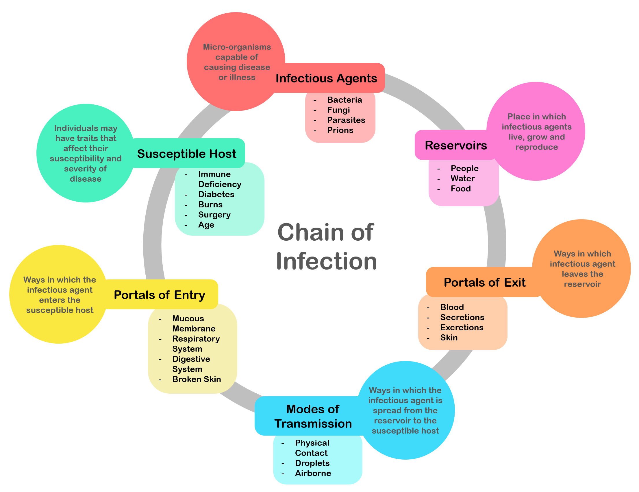
An infection is the invasion of tissues by pathogens, their multiplication, and the reaction of host tissues to the infectious agent and the toxins they produce. An infectious disease, also known as a transmissible disease or communicable disease, is an illness resulting from an infection. Infections can be caused by a wide range of pathogens, most prominently bacteria and viruses. Hosts can fight infections using their immune system. Mammalian hosts react to infections with an innate response, often involving inflammation, followed by an adaptive response. Specific medications used to treat infections include antibiotics, antivirals, antifungals, antiprotozoals, and antihelminthics. Infectious diseases resulted in 9.2 million deaths in 2013 (about 17% of all deaths). The branch of medicine that focuses on infections is referred to as infectious disease. Types Infections are caused by infectious agents (pathogens) including: * Bacteria (e.g. ''Mycobacterium tuberculosis'', ... [...More Info...] [...Related Items...] OR: [Wikipedia] [Google] [Baidu] |
Aortic Regurgitation
Aortic regurgitation (AR), also known as aortic insufficiency (AI), is the leaking of the aortic valve of the heart that causes blood to flow in the reverse direction during ventricular diastole, from the aorta into the left ventricle. As a consequence, the cardiac muscle is forced to work harder than normal. Signs and symptoms Symptoms of aortic regurgitation are similar to those of heart failure and include the following: * Dyspnea on exertion * Orthopnea * Paroxysmal nocturnal dyspnea * Palpitations * Angina pectoris * Cyanosis (in acute cases) Causes In terms of the cause of aortic regurgitation, is often due to the aortic root dilation ('' annuloaortic ectasia''), which is idiopathic in over 80% of cases, but otherwise may result from aging, syphilitic aortitis, osteogenesis imperfecta, aortic dissection, Behçet's disease, reactive arthritis and systemic hypertension.Chapter 1: Diseases of the Cardiovascular system > Section: Valvular Heart Disease in: Aortic root dilation ... [...More Info...] [...Related Items...] OR: [Wikipedia] [Google] [Baidu] |
Stroke
A stroke is a medical condition in which poor blood flow to the brain causes cell death. There are two main types of stroke: ischemic, due to lack of blood flow, and hemorrhagic, due to bleeding. Both cause parts of the brain to stop functioning properly. Signs and symptoms of a stroke may include an inability to move or feel on one side of the body, problems understanding or speaking, dizziness, or loss of vision to one side. Signs and symptoms often appear soon after the stroke has occurred. If symptoms last less than one or two hours, the stroke is a transient ischemic attack (TIA), also called a mini-stroke. A hemorrhagic stroke may also be associated with a severe headache. The symptoms of a stroke can be permanent. Long-term complications may include pneumonia and loss of bladder control. The main risk factor for stroke is high blood pressure. Other risk factors include high blood cholesterol, tobacco smoking, obesity, diabetes mellitus, a previous TIA, end-st ... [...More Info...] [...Related Items...] OR: [Wikipedia] [Google] [Baidu] |
New York Heart Association Functional Classification
The New York Heart Association (NYHA) Functional Classification provides a simple way of classifying the extent of heart failure. It places patients in one of four categories based on how much they are limited during physical activity; the limitations/symptoms are in regard to normal breathing and varying degrees in shortness of breath and/or angina. It originated in 1928, when no measurements of cardiac function were possible, to provide a common language for physicians to communicate. Despite difficulties in applying it, such as the challenge of consistently classifying patients in class II or III, because functional capacity is such a powerful determinant of outcome it remains arguably the most important prognostic marker in routine clinical use in heart failure today. With time the classification system evolved and updated multiple times. Presently, the ninth edition of the NYHA classification is being used in the clinical practice released in the year 1994 by the Criteria Comm ... [...More Info...] [...Related Items...] OR: [Wikipedia] [Google] [Baidu] |
Left Ventricle
A ventricle is one of two large chambers toward the bottom of the heart that collect and expel blood towards the peripheral beds within the body and lungs. The blood pumped by a ventricle is supplied by an atrium, an adjacent chamber in the upper heart that is smaller than a ventricle. Interventricular means between the ventricles (for example the interventricular septum), while intraventricular means within one ventricle (for example an intraventricular block). In a four-chambered heart, such as that in humans, there are two ventricles that operate in a double circulatory system: the right ventricle pumps blood into the pulmonary circulation to the lungs, and the left ventricle pumps blood into the systemic circulation through the aorta. Structure Ventricles have thicker walls than atria and generate higher blood pressures. The physiological load on the ventricles requiring pumping of blood throughout the body and lungs is much greater than the pressure generated by the atria ... [...More Info...] [...Related Items...] OR: [Wikipedia] [Google] [Baidu] |
Pacing Technology
An artificial cardiac pacemaker (or artificial pacemaker, so as not to be confused with the natural cardiac pacemaker) or pacemaker is a medical device that generates electrical impulses delivered by electrodes to the chambers of the heart either the upper atria, or lower ventricles to cause the targeted chambers to contract and pump blood. By doing so, the pacemaker regulates the function of the electrical conduction system of the heart. The primary purpose of a pacemaker is to maintain an adequate heart rate, either because the heart's natural pacemaker is not fast enough, or because there is a block in the heart's electrical conduction system. Modern pacemakers are externally programmable and allow a cardiologist, particularly a cardiac electrophysiologist, to select the optimal pacing modes for individual patients. Most pacemakers are on demand, in which the stimulation of the heart is based on the dynamic demand of the circulatory system. Others send out a fixed rate of i ... [...More Info...] [...Related Items...] OR: [Wikipedia] [Google] [Baidu] |
Seldinger Technique
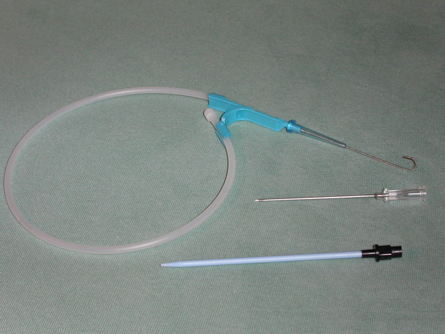
The Seldinger technique, also known as Seldinger wire technique, is a medical procedure to obtain safe access to blood vessels and other hollow organs. It is named after Sven Ivar Seldinger (1921–1998), a Swedish radiologist who introduced the procedure in 1953. Uses The Seldinger technique is used for angiography, insertion of chest drains and central venous catheters, insertion of PEG tubes using the push technique, insertion of the leads for an artificial pacemaker or implantable cardioverter-defibrillator, and numerous other interventional medical procedures. Complications The initial puncture is with a sharp instrument, and this may lead to hemorrhage or perforation of the organ in question. Infection is a possible complication, and hence asepsis is practiced during most Seldinger procedures. Loss of the guidewire into the cavity or blood vessel is a significant and generally preventable complication. Description The desired vessel or cavity is punctured with a shar ... [...More Info...] [...Related Items...] OR: [Wikipedia] [Google] [Baidu] |
Medical Ultrasound
Medical ultrasound includes diagnostic techniques (mainly imaging techniques) using ultrasound, as well as therapeutic applications of ultrasound. In diagnosis, it is used to create an image of internal body structures such as tendons, muscles, joints, blood vessels, and internal organs, to measure some characteristics (e.g. distances and velocities) or to generate an informative audible sound. Its aim is usually to find a source of disease or to exclude pathology. The usage of ultrasound to produce visual images for medicine is called medical ultrasonography or simply sonography. The practice of examining pregnant women using ultrasound is called obstetric ultrasonography, and was an early development of clinical ultrasonography. Ultrasound is composed of sound waves with frequencies which are significantly higher than the range of human hearing (>20,000 Hz). Ultrasonic images, also known as sonograms, are created by sending pulses of ultrasound into tissue using a pr ... [...More Info...] [...Related Items...] OR: [Wikipedia] [Google] [Baidu] |


.png)


