 |
Buccal Membrane
The oral mucosa is the mucous membrane lining the inside of the mouth. It comprises stratified squamous epithelium, termed "oral epithelium", and an underlying connective tissue termed ''lamina propria''. The oral cavity has sometimes been described as a mirror that reflects the health of the individual. Changes indicative of disease are seen as alterations in the oral mucosa lining the mouth, which can reveal systemic conditions, such as diabetes or vitamin deficiency, or the local effects of chronic tobacco or alcohol use. The oral mucosa tends to heal faster and with less scar formation compared to the skin. The underlying mechanism remains unknown, but research suggests that extracellular vesicles might be involved. Classification Oral mucosa can be divided into three main categories based on function and histology: *Lining mucosa, nonkeratinized stratified squamous epithelium, found almost everywhere else in the oral cavity, including the: **Alveolar mucosa, the lining betw ... [...More Info...] [...Related Items...] OR: [Wikipedia] [Google] [Baidu] |
|
Mucous Membrane
A mucous membrane or mucosa is a membrane that lines various cavities in the body of an organism and covers the surface of internal organs. It consists of one or more layers of epithelial cells overlying a layer of loose connective tissue. It is mostly of endodermal origin and is continuous with the skin at body openings such as the eyes, eyelids, ears, inside the nose, inside the mouth, lips, the genital areas, the urethral opening and the anus. Some mucous membranes secrete mucus, a thick protective fluid. The function of the membrane is to stop pathogens and dirt from entering the body and to prevent bodily tissues from becoming dehydrated. Structure The mucosa is composed of one or more layers of epithelial cells that secrete mucus, and an underlying lamina propria of loose connective tissue. The type of cells and type of mucus secreted vary from organ to organ and each can differ along a given tract. Mucous membranes line the digestive, respiratory and reproduct ... [...More Info...] [...Related Items...] OR: [Wikipedia] [Google] [Baidu] |
|
 |
Lingual Papillae
Lingual papillae (singular papilla) are small structures on the upper surface of the tongue that give it its characteristic rough texture. The four types of papillae on the human tongue have different structures and are accordingly classified as circumvallate (or vallate), fungiform, filiform, and foliate. All except the filiform papillae are associated with taste buds. Structure In living subjects, lingual papillae are more readily seen when the tongue is dry. There are four types of papillae present on the tongue: Filiform papillae Filiform papillae are the most numerous of the lingual papillae. They are fine, small, cone-shaped papillae covering most of the dorsum of the tongue. They are responsible for giving the tongue its texture and are responsible for the sensation of touch. Unlike the other kinds of papillae, filiform papillae do not contain taste buds. They cover most of the front two-thirds of the tongue's surface. They appear as very small, conical or cylindrical ... [...More Info...] [...Related Items...] OR: [Wikipedia] [Google] [Baidu] |
 |
Nicotinic Stomatitis
Nicotinic acetylcholine receptors, or nAChRs, are receptor polypeptides that respond to the neurotransmitter acetylcholine. Nicotinic receptors also respond to drugs such as the agonist nicotine. They are found in the central and peripheral nervous system, muscle, and many other tissues of many organisms. At the neuromuscular junction they are the primary receptor in muscle for motor nerve-muscle communication that controls muscle contraction. In the peripheral nervous system: (1) they transmit outgoing signals from the presynaptic to the postsynaptic cells within the sympathetic and parasympathetic nervous system, and (2) they are the receptors found on skeletal muscle that receive acetylcholine released to signal for muscular contraction. In the immune system, nAChRs regulate inflammatory processes and signal through distinct intracellular pathways. In insects, the cholinergic system is limited to the central nervous system. The nicotinic receptors are considered cholinerg ... [...More Info...] [...Related Items...] OR: [Wikipedia] [Google] [Baidu] |
 |
Biopsy
A biopsy is a medical test commonly performed by a surgeon, interventional radiologist, or an interventional cardiologist. The process involves extraction of sample cells or tissues for examination to determine the presence or extent of a disease. The tissue is then fixed, dehydrated, embedded, sectioned, stained and mounted before it is generally examined under a microscope by a pathologist; it may also be analyzed chemically. When an entire lump or suspicious area is removed, the procedure is called an excisional biopsy. An incisional biopsy or core biopsy samples a portion of the abnormal tissue without attempting to remove the entire lesion or tumor. When a sample of tissue or fluid is removed with a needle in such a way that cells are removed without preserving the histological architecture of the tissue cells, the procedure is called a needle aspiration biopsy. Biopsies are most commonly performed for insight into possible cancerous or inflammatory conditions. H ... [...More Info...] [...Related Items...] OR: [Wikipedia] [Google] [Baidu] |
 |
Bruxism
Bruxism is excessive teeth grinding or jaw clenching. It is an oral parafunctional activity; i.e., it is unrelated to normal function such as eating or talking. Bruxism is a common behavior; reports of prevalence range from 8% to 31% in the general population. Several symptoms are commonly associated with bruxism, including aching jaw muscles, headaches, hypersensitive teeth, tooth wear, and damage to dental restorations (e.g. crowns and fillings). Symptoms may be minimal, without patient awareness of the condition. If nothing is done, after a while many teeth start wearing down until the whole tooth is gone. There are two main types of bruxism: one occurs during sleep (nocturnal bruxism) and one during wakefulness (awake bruxism). Dental damage may be similar in both types, but the symptoms of sleep bruxism tend to be worse on waking and improve during the course of the day, and the symptoms of awake bruxism may not be present at all on waking, and then worsen over the day. ... [...More Info...] [...Related Items...] OR: [Wikipedia] [Google] [Baidu] |
|
Linea Alba (cheek)
The linea alba (Latin for ''white line''), in dentistry, is a horizontal streak on the buccal mucosa (inner surface of the cheek), level with the occlusion (biting plane). It usually extends from the commissure to the posterior teeth, and can extend to the inner lip mucosa and corners of the mouth. The linea alba is a common finding and most likely associated with pressure, frictional irritation, or sucking trauma from the facial surfaces of the teeth. It may be mistaken for a lesion requiring treatment and may be found in individuals who chew tobacco. Clinical considerations *The linea alba is usually present bilaterally *It is restricted to dentulous areas (i.e., in areas where there are missing teeth the line will be absent - unless a denture is worn) *It presents an asymptomatic, linear elevation, with a whitish colour, at the level of the occlusal line of the teeth Treatment Treatment is not required. See also * Crenated tongue * Morsicatio buccarum * Linea alba (abdomen) ... [...More Info...] [...Related Items...] OR: [Wikipedia] [Google] [Baidu] |
|
|
Progenitor Cell
A progenitor cell is a biological cell that can differentiate into a specific cell type. Stem cells and progenitor cells have this ability in common. However, stem cells are less specified than progenitor cells. Progenitor cells can only differentiate into their "target" cell type. The most important difference between stem cells and progenitor cells is that stem cells can replicate indefinitely, whereas progenitor cells can divide only a limited number of times. Controversy about the exact definition remains and the concept is still evolving. The terms "progenitor cell" and "stem cell" are sometimes equated. Properties Most progenitors are identified as oligopotent. In this point of view, they can compare to adult stem cells, but progenitors are said to be in a further stage of cell differentiation. They are in the "center" between stem cells and fully differentiated cells. The kind of potency they have depends on the type of their "parent" stem cell and also on their niche. ... [...More Info...] [...Related Items...] OR: [Wikipedia] [Google] [Baidu] |
|
 |
Keratinocyte
Keratinocytes are the primary type of cell found in the epidermis, the outermost layer of the skin. In humans, they constitute 90% of epidermal skin cells. Basal cells in the basal layer (''stratum basale'') of the skin are sometimes referred to as basal keratinocytes. Keratinocytes form a barrier against environmental damage by heat, UV radiation, water loss, pathogenic bacteria, fungi, parasites, and viruses. A number of structural proteins, enzymes, lipids, and antimicrobial peptides contribute to maintain the important barrier function of the skin. Keratinocytes differentiate from epidermal stem cells in the lower part of the epidermis and migrate towards the surface, finally becoming corneocytes and eventually be shed off, which happens every 40 to 56 days in humans. Function The primary function of keratinocytes is the formation of a barrier against environmental damage by heat, UV radiation, water loss, pathogenic bacteria, fungi, parasites, and viruses. Pathogens i ... [...More Info...] [...Related Items...] OR: [Wikipedia] [Google] [Baidu] |
|
Cell Differentiation
Cellular differentiation is the process in which a stem cell alters from one type to a differentiated one. Usually, the cell changes to a more specialized type. Differentiation happens multiple times during the development of a multicellular organism as it changes from a simple zygote to a complex system of tissues and cell types. Differentiation continues in adulthood as adult stem cells divide and create fully differentiated daughter cells during tissue repair and during normal cell turnover. Some differentiation occurs in response to antigen exposure. Differentiation dramatically changes a cell's size, shape, membrane potential, metabolic activity, and responsiveness to signals. These changes are largely due to highly controlled modifications in gene expression and are the study of epigenetics. With a few exceptions, cellular differentiation almost never involves a change in the DNA sequence itself. Although metabolic composition does get altered quite dramatical ... [...More Info...] [...Related Items...] OR: [Wikipedia] [Google] [Baidu] |
|
 |
Soft Palate
The soft palate (also known as the velum, palatal velum, or muscular palate) is, in mammals, the soft tissue constituting the back of the roof of the mouth. The soft palate is part of the palate of the mouth; the other part is the hard palate. The soft palate is distinguished from the hard palate at the front of the mouth in that it does not contain bone. Structure Muscles The five muscles of the soft palate play important roles in swallowing and breathing. The muscles are: # Tensor veli palatini, which is involved in swallowing # Palatoglossus, involved in swallowing # Palatopharyngeus, involved in breathing # Levator veli palatini, involved in swallowing # Musculus uvulae, which moves the uvula These muscles are innervated by the pharyngeal plexus via the vagus nerve, with the exception of the tensor veli palatini. The tensor veli palatini is innervated by the mandibular division of the trigeminal nerve (V3). Function The soft palate is moveable, consisting of mu ... [...More Info...] [...Related Items...] OR: [Wikipedia] [Google] [Baidu] |
 |
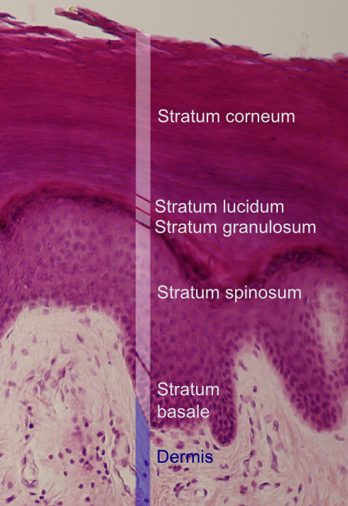
Stratum Corneum
The stratum corneum (Latin for 'horny layer') is the outermost layer of the epidermis. The human stratum corneum comprises several levels of flattened corneocytes that are divided into two layers: the ''stratum disjunctum'' and ''stratum compactum''. The skin's protective acid mantle and lipid barrier sit on top of the stratum disjunctum. The stratum disjunctum is the uppermost and loosest layer of skin. The stratum compactum is the comparatively deeper, more compacted and more cohesive part of the stratum corneum. The corneocytes of the stratum disjunctum are larger, more rigid and more hydrophobic than that of the stratum compactum. The stratum corneum is the dead tissue that performs protective and adaptive physiological functions including mechanical shear, impact resistance, water flux and hydration regulation, microbial proliferation and invasion regulation, initiation of inflammation through cytokine activation and dendritic cell activity, and selective permeability to e ... [...More Info...] [...Related Items...] OR: [Wikipedia] [Google] [Baidu] |
 |
Stratum Granulosum
The stratum granulosum (or granular layer) is a thin layer of cells in the epidermis lying above the stratum spinosum and below the stratum corneum ( stratum lucidum on the soles and palms).James, William; Berger, Timothy; Elston, Dirk (2005) ''Andrews' Diseases of the Skin: Clinical Dermatology'' (10th ed.). Saunders. Page 2. . Keratinocytes migrating from the underlying stratum spinosum become known as granular cells in this layer. These cells contain keratohyalin granules, which are filled with histidine- and cysteine-rich proteins that appear to bind the keratin filaments together. Therefore, the main function of keratohyalin granules is to bind intermediate keratin filaments together.Marks, James G; Miller, Jeffery (2006). ''Lookingbill and Marks' Principles of Dermatology'' (4th ed.). Elsevier Inc. Page 7. . At the transition between this layer and the stratum corneum, cells secrete lamellar bodies (containing lipids and proteins) into the extracellular space. This resu ... [...More Info...] [...Related Items...] OR: [Wikipedia] [Google] [Baidu] |