|
Brachiocephalic Trunk
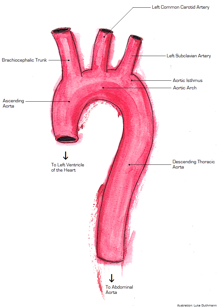
The brachiocephalic artery (or brachiocephalic trunk or innominate artery) is an artery of the mediastinum that supplies blood to the right arm and the head and neck. It is the first branch of the aortic arch. Soon after it emerges, the brachiocephalic artery divides into the right common carotid artery and the right subclavian artery. There is no brachiocephalic artery for the left side of the body. The left common carotid, and the left subclavian artery, come directly off the aortic arch. However, there are two brachiocephalic veins. Structure The brachiocephalic artery arises, on a level with the upper border of the second right costal cartilage, from the start of the aortic arch, on a plane anterior to the origin of the left carotid artery. It ascends obliquely upward, backward, and to the right to the level of the upper border of the right sternoclavicular articulation, where it divides into the right common carotid artery and right subclavian arteries. The artery then ... [...More Info...] [...Related Items...] OR: [Wikipedia] [Google] [Baidu] |
Heart
The heart is a muscular organ found in most animals. This organ pumps blood through the blood vessels of the circulatory system. The pumped blood carries oxygen and nutrients to the body, while carrying metabolic waste such as carbon dioxide to the lungs. In humans, the heart is approximately the size of a closed fist and is located between the lungs, in the middle compartment of the chest, called the mediastinum. In humans, other mammals, and birds, the heart is divided into four chambers: upper left and right atria and lower left and right ventricles. Commonly, the right atrium and ventricle are referred together as the right heart and their left counterparts as the left heart. Fish, in contrast, have two chambers, an atrium and a ventricle, while most reptiles have three chambers. In a healthy heart, blood flows one way through the heart due to heart valves, which prevent backflow. The heart is enclosed in a protective sac, the pericardium, which also contains ... [...More Info...] [...Related Items...] OR: [Wikipedia] [Google] [Baidu] |
Superior Vena Cava
The superior vena cava (SVC) is the anatomical terms of location#Superior and inferior, superior of the two venae cavae, the great vein, venous trunks that return deoxygenated blood from the circulatory system, systemic circulation to the atrium (heart), right atrium of the heart. It is a large-diameter (24 mm) short length vein that receives venous return from the upper half of the body, above the thoracic diaphragm, diaphragm. Venous return from the lower half, below the diaphragm, flows through the inferior vena cava. The SVC is located in the anterior right superior mediastinum. It is the typical site of central venous access via a central venous catheter or a peripherally inserted central catheter. Mentions of "the cava" without further specification usually refer to the SVC. Structure The superior vena cava is formed by the left and right brachiocephalic veins, which receive blood from the upper limbs, head and neck, behind the lower border of the first right costal ca ... [...More Info...] [...Related Items...] OR: [Wikipedia] [Google] [Baidu] |
Ascending Aorta
The ascending aorta (AAo) is a portion of the aorta commencing at the upper part of the base of the left ventricle, on a level with the lower border of the third costal cartilage behind the left half of the sternum. Structure It passes obliquely upward, forward, and to the right, in the direction of the heart's axis, as high as the upper border of the second right costal cartilage, describing a slight curve in its course, and being situated, about behind the posterior surface of the sternum. The total length is about . Components The aortic root is the portion of the aorta beginning at the aortic annulus and extending to the sinotubular junction. It is sometimes regarded as a part of the ascending aorta, and sometimes regarded as a separate entity from the rest of the ascending aorta. Between each commissure of the aortic valve and opposite the cusps of the aortic valve, three small dilatations called the aortic sinuses. The sinotubular junction is the point in the ascend ... [...More Info...] [...Related Items...] OR: [Wikipedia] [Google] [Baidu] |
Ventral Aorta
The aorta ( ) is the main and largest artery in the human body, originating from the left ventricle of the heart and extending down to the abdomen, where it splits into two smaller arteries (the common iliac arteries). The aorta distributes oxygenated blood to all parts of the body through the systemic circulation. Structure Sections In anatomical sources, the aorta is usually divided into sections. One way of classifying a part of the aorta is by anatomical compartment, where the thoracic aorta (or thoracic portion of the aorta) runs from the heart to the diaphragm. The aorta then continues downward as the abdominal aorta (or abdominal portion of the aorta) from the diaphragm to the aortic bifurcation. Another system divides the aorta with respect to its course and the direction of blood flow. In this system, the aorta starts as the ascending aorta, travels superiorly from the heart, and then makes a hairpin turn known as the aortic arch. Following the aortic arch, the ... [...More Info...] [...Related Items...] OR: [Wikipedia] [Google] [Baidu] |
Aortic Arches
The aortic arches or pharyngeal arch arteries (previously referred to as branchial arches in human embryos) are a series of six paired embryological vascular structures which give rise to the great arteries of the neck and head. They are ventral to the dorsal aorta and arise from the aortic sac. The aortic arches are formed sequentially within the pharyngeal arches and initially appear symmetrical on both sides of the embryo, but then undergo a significant remodelling to form the final asymmetrical structure of the great arteries. Structure Arches 1 and 2 The ''first'' and ''second arches'' disappear early. A remnant of the 1st arch forms part of the maxillary artery, a branch of the external carotid artery. The ventral end of the second develops into the ascending pharyngeal artery, and its dorsal end gives origin to the stapedial artery, a vessel which typically atrophies in humans but persists in some mammals. The stapedial artery passes through the ring of the stapes an ... [...More Info...] [...Related Items...] OR: [Wikipedia] [Google] [Baidu] |
Truncus Arteriosus
The truncus arteriosus is a structure that is present during embryonic development. It is an arterial trunk that originates from both ventricles of the heart that later divides into the aorta and the pulmonary trunk. Structure The truncus arteriosus and bulbus cordis are divided by the aorticopulmonary septum. The truncus arteriosus gives rise to the ascending aorta and the pulmonary trunk. The caudal end of the bulbus cordis gives rise to the smooth parts (outflow tract) of the left and right ventricles (aortic vestibule & conus arteriosus respectively). The cranial end of the bulbus cordis (also known as the conus cordis) gives rise to the aorta and pulmonary trunk with the truncus arteriosus. This makes its appearance in three portions. # Two distal ridge-like thickenings project into the lumen of the tube: the truncal and bulbar ridges. These increase in size, and ultimately meet and fuse to form a septum ( aorticopulmonary septum), which takes a spiral course toward the p ... [...More Info...] [...Related Items...] OR: [Wikipedia] [Google] [Baidu] |
Aortic Sac
The aortic sac or aortic bulb is a dilated structure in mammalian embryos, lined by endothelial cells and is the most distal part of the truncus arteriosus. It is the primordial vascular channel from which the aortic arches arise (and eventually the dorsal aortae) and is homologous to the ventral aorta of gill-bearing vertebrates. The aortic sac eventually forms right and left horns, which subsequently give rise to the brachiocephalic trunk and the proximal segment of the arch of aorta, respectively. Genes ''HAND2'' (''dHAND'') and ''HAND1'' (''eHAND'') are expressed during the development of the aortic bulb and the arteries which arise from it.Richard P. Harvey, Nadia Rosenthal (ed)Heart Development Gulf Professional Publishing, 1999; page 150-151. The protein encoded by these genes belong to the basic helix-loop-helix family of transcription factor In molecular biology, a transcription factor (TF) (or sequence-specific DNA-binding factor) is a protein that controls the rate o ... [...More Info...] [...Related Items...] OR: [Wikipedia] [Google] [Baidu] |
Internal Mammary
In human anatomy, the internal thoracic artery (ITA), previously commonly known as the internal mammary artery (a name still common among surgeons), is an artery that supplies the anterior chest wall and the breasts. It is a paired artery, with one running along each side of the sternum, to continue after its bifurcation as the superior epigastric and musculophrenic arteries. Structure The internal thoracic artery arises from the anterior surface of the subclavian artery near its origin. It has a width of between 1-2 mm. It travels downward on the inside of the rib cage, approximately 1 cm from the sides of the sternum, and thus medial to the nipple. It is accompanied by the internal thoracic vein. It runs deep to the abdominal external oblique muscle, but superficial to the vagus nerve. In adults, internal thoracic artery lies closest to the sternum at the first intercoastal space. The gap between the artery and lateral border of the sternum increases when going downwards ... [...More Info...] [...Related Items...] OR: [Wikipedia] [Google] [Baidu] |
Right Common Carotid
In anatomy, the left and right common carotid arteries (carotids) ( in Merriam-Webster Online Dictionary '.) are that supply the head and neck with oxygenated blood; they divide in the neck to form the external and internal carotid arteries< ...
[...More Info...] [...Related Items...] OR: [Wikipedia] [Google] [Baidu] |
Aorta
The aorta ( ) is the main and largest artery in the human body, originating from the left ventricle of the heart and extending down to the abdomen, where it splits into two smaller arteries (the common iliac arteries). The aorta distributes oxygenated blood to all parts of the body through the systemic circulation. Structure Sections In anatomical sources, the aorta is usually divided into sections. One way of classifying a part of the aorta is by anatomical compartment, where the thoracic aorta (or thoracic portion of the aorta) runs from the heart to the diaphragm. The aorta then continues downward as the abdominal aorta (or abdominal portion of the aorta) from the diaphragm to the aortic bifurcation. Another system divides the aorta with respect to its course and the direction of blood flow. In this system, the aorta starts as the ascending aorta, travels superiorly from the heart, and then makes a hairpin turn known as the aortic arch. Following the aortic a ... [...More Info...] [...Related Items...] OR: [Wikipedia] [Google] [Baidu] |
Thyroid
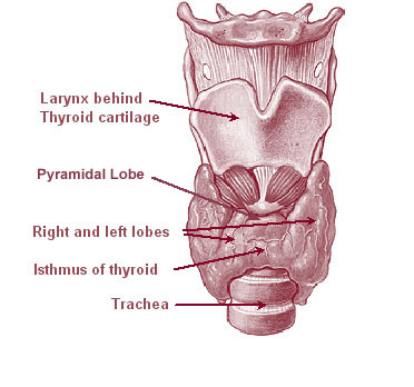
The thyroid, or thyroid gland, is an endocrine gland in vertebrates. In humans it is in the neck and consists of two connected lobes. The lower two thirds of the lobes are connected by a thin band of tissue called the thyroid isthmus. The thyroid is located at the front of the neck, below the Adam's apple. Microscopically, the functional unit of the thyroid gland is the spherical thyroid follicle, lined with follicular cells (thyrocytes), and occasional parafollicular cells that surround a lumen containing colloid. The thyroid gland secretes three hormones: the two thyroid hormones triiodothyronine (T3) and thyroxine (T4)and a peptide hormone, calcitonin. The thyroid hormones influence the metabolic rate and protein synthesis, and in children, growth and development. Calcitonin plays a role in calcium homeostasis. Secretion of the two thyroid hormones is regulated by thyroid-stimulating hormone (TSH), which is secreted from the anterior pituitary gland. TSH is reg ... [...More Info...] [...Related Items...] OR: [Wikipedia] [Google] [Baidu] |
Sternum
The sternum or breastbone is a long flat bone located in the central part of the chest. It connects to the ribs via cartilage and forms the front of the rib cage, thus helping to protect the heart, lungs, and major blood vessels from injury. Shaped roughly like a necktie, it is one of the largest and longest flat bones of the body. Its three regions are the manubrium, the body, and the xiphoid process. The word "sternum" originates from the Ancient Greek στέρνον (stérnon), meaning "chest". Structure The sternum is a narrow, flat bone, forming the middle portion of the front of the chest. The top of the sternum supports the clavicles (collarbones) and its edges join with the costal cartilages of the first two pairs of ribs. The inner surface of the sternum is also the attachment of the sternopericardial ligaments. Its top is also connected to the sternocleidomastoid muscle. The sternum consists of three main parts, listed from the top: * Manubrium * Body (gl ... [...More Info...] [...Related Items...] OR: [Wikipedia] [Google] [Baidu] |


