|
Biological Imaging
Biological imaging may refer to any imaging technique used in biology. Typical examples include: * Bioluminescence imaging, a technique for studying laboratory animals using luminescent protein * Calcium imaging, determining the calcium status of a tissue using fluorescent light * Diffuse optical imaging, using near-infrared light to generate images of the body * Diffusion-weighted imaging, a type of MRI that uses water diffusion * Fluorescence lifetime imaging, using the decay rate of a fluorescent sample * Gallium imaging, a nuclear medicine method for the detection of infections and cancers * Imaging agent, a chemical designed to allow clinicians to determine whether a mass is benign or malignant * Imaging studies, which includes many medical imaging techniques * Magnetic resonance imaging (MRI), a non-invasive method to render images of living tissues * Magneto-acousto-electrical tomography (MAET), is an imaging modality to image the electrical conductivity of biological tissu ... [...More Info...] [...Related Items...] OR: [Wikipedia] [Google] [Baidu] |
Imaging
Imaging is the representation or reproduction of an object's form; especially a visual representation (i.e., the formation of an image). Imaging technology is the application of materials and methods to create, preserve, or duplicate images. Imaging science is a multidisciplinary field concerned with the generation, collection, duplication, analysis, modification, and visualization of images,Joseph P. Hornak, ''Encyclopedia of Imaging Science and Technology'' (John Wiley & Sons, 2002) including imaging things that the human eye cannot detect. As an evolving field it includes research and researchers from physics, mathematics, electrical engineering, computer vision, computer science, and perceptual psychology. ''Imager'' are imaging sensors. Imaging chain The foundation of imaging science as a discipline is the "imaging chain" – a conceptual model describing all of the factors which must be considered when developing a system for creating visual renderings (images). In g ... [...More Info...] [...Related Items...] OR: [Wikipedia] [Google] [Baidu] |
Microscopy
Microscopy is the technical field of using microscopes to view objects and areas of objects that cannot be seen with the naked eye (objects that are not within the resolution range of the normal eye). There are three well-known branches of microscopy: optical, electron, and scanning probe microscopy, along with the emerging field of X-ray microscopy. Optical microscopy and electron microscopy involve the diffraction, reflection, or refraction of electromagnetic radiation/electron beams interacting with the specimen, and the collection of the scattered radiation or another signal in order to create an image. This process may be carried out by wide-field irradiation of the sample (for example standard light microscopy and transmission electron microscopy) or by scanning a fine beam over the sample (for example confocal laser scanning microscopy and scanning electron microscopy). Scanning probe microscopy involves the interaction of a scanning probe with the surface of the objec ... [...More Info...] [...Related Items...] OR: [Wikipedia] [Google] [Baidu] |
Ultrasound Imaging
Medical ultrasound includes diagnostic techniques (mainly medical imaging, imaging techniques) using ultrasound, as well as therapeutic ultrasound, therapeutic applications of ultrasound. In diagnosis, it is used to create an image of internal body structures such as tendons, muscles, joints, blood vessels, and internal organs, to measure some characteristics (e.g. distances and velocities) or to generate an informative audible sound. Its aim is usually to find a source of disease or to exclude pathology. The usage of ultrasound to produce visual images for medicine is called medical ultrasonography or simply sonography. The practice of examining pregnant women using ultrasound is called obstetric ultrasonography, and was an early development of clinical ultrasonography. Ultrasound is composed of sound waves with frequency, frequencies which are significantly higher than the range of human hearing (>20,000 Hz). Ultrasonic images, also known as sonograms, are created by se ... [...More Info...] [...Related Items...] OR: [Wikipedia] [Google] [Baidu] |
Photoacoustic Imaging In Biomedicine
Photoacoustic imaging or optoacoustic imaging is a biomedical imaging modality based on the photoacoustic effect. Non-ionizing laser pulses are delivered into biological tissues and part of the energy will be absorbed and converted into heat, leading to transient thermoelastic expansion and thus wideband (i.e. MHz) ultrasonic emission. The generated ultrasonic waves are detected by ultrasonic transducers and then analyzed to produce images. It is known that optical absorption is closely associated with physiological properties, such as hemoglobin concentration and oxygen saturation. As a result, the magnitude of the ultrasonic emission (i.e. photoacoustic signal), which is proportional to the local energy deposition, reveals physiologically specific optical absorption contrast. 2D or 3D images of the targeted areas can then be formed. Biomedical imaging The optical absorption in biological tissues can be due to endogenous molecules such as hemoglobin or melanin, or exogenousl ... [...More Info...] [...Related Items...] OR: [Wikipedia] [Google] [Baidu] |
Photothermal Effect
Photothermal effect is a phenomenon associated with electromagnetic radiation. It is produced by the photoexcitation of material, resulting in the production of thermal energy (heat). It is sometimes used during treatment of blood vessel lesions, laser resurfacing, laser hair removal and laser surgery. External linksQuantities, Terminology, and Symbols in Photothermal and Related SpectroscopiesResearch paper from IUPAC The International Union of Pure and Applied Chemistry (IUPAC ) is an international federation of National Adhering Organizations working for the advancement of the chemical sciences, especially by developing nomenclature and terminology. It is ...Amazing Nano Materials: Photothermal and Photoacoustic Effects Photochemistry {{chem-stub ... [...More Info...] [...Related Items...] OR: [Wikipedia] [Google] [Baidu] |
Photoacoustic Imaging
Photoacoustic imaging or optoacoustic imaging is a biomedical imaging modality based on the photoacoustic effect. Non-ionizing laser pulses are delivered into biological tissues and part of the energy will be absorbed and converted into heat, leading to transient thermoelastic expansion and thus wideband (i.e. MHz) ultrasonic emission. The generated ultrasonic waves are detected by ultrasonic transducers and then analyzed to produce images. It is known that optical absorption is closely associated with physiological properties, such as hemoglobin concentration and oxygen saturation. As a result, the magnitude of the ultrasonic emission (i.e. photoacoustic signal), which is proportional to the local energy deposition, reveals physiologically specific optical absorption contrast. 2D or 3D images of the targeted areas can then be formed. Biomedical imaging The optical absorption in biological tissues can be due to endogenous molecules such as hemoglobin or melanin, or exogenousl ... [...More Info...] [...Related Items...] OR: [Wikipedia] [Google] [Baidu] |
Optical Imaging
Medical optical imaging is the use of light as an investigational :wikt:imaging, imaging technique for medical applications. Examples include optical microscopy, spectroscopy, endoscopy, scanning laser ophthalmoscopy, laser Doppler imaging, and optical coherence tomography. Because light is an electromagnetic wave, similar phenomena occur in X-rays, microwaves, and radio waves. Optical imaging systems may be divided into diffusive and ballistic imaging systems. A model for photon migration in turbid biological media has been developed by Bonner et al. Such a model can be applied for interpretation data obtained from laser Doppler blood-flow monitors and for designing protocols for therapeutic excitation of tissue chromophores. Diffusive optical imaging Diffuse optical imaging (DOI) is a method of imaging using near-infrared spectroscopy (NIRS) or fluorescence-based methods. When used to create 3D volumetric models of the imaged material DOI is referred to as diffuse optical tomo ... [...More Info...] [...Related Items...] OR: [Wikipedia] [Google] [Baidu] |
Nuclear Medicine
Nuclear medicine or nucleology is a medical specialty involving the application of radioactive substances in the diagnosis and treatment of disease. Nuclear imaging, in a sense, is "radiology done inside out" because it records radiation emitting from within the body rather than radiation that is generated by external sources like X-rays. In addition, nuclear medicine scans differ from radiology, as the emphasis is not on imaging anatomy, but on the function. For such reason, it is called a physiological imaging modality. Single photon emission computed tomography (SPECT) and positron emission tomography (PET) scans are the two most common imaging modalities in nuclear medicine. Diagnostic medical imaging Diagnostic In nuclear medicine imaging, radiopharmaceuticals are taken internally, for example, through inhalation, intravenously or orally. Then, external detectors (gamma cameras) capture and form images from the radiation emitted by the radiopharmaceuticals. This process ... [...More Info...] [...Related Items...] OR: [Wikipedia] [Google] [Baidu] |
Thermography
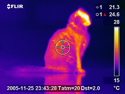
Infrared thermography (IRT), thermal video and/or thermal imaging, is a process where a Thermographic camera, thermal camera captures and creates an image of an object by using infrared radiation emitted from the object in a process, which are examples of infrared imaging science. Thermographic cameras usually detect electromagnetic radiation, radiation in the long-infrared range of the electromagnetic spectrum (roughly 9,000–14,000 nanometers or 9–14 μm) and produce images of that radiation, called thermograms. Since infrared radiation is emitted by all objects with a temperature above absolute zero according to the black body Planck's law of black-body radiation, radiation law, thermography makes it possible to see one's environment with or without optical spectrum, visible illumination. The amount of radiation emitted by an object increases with temperature; therefore, thermography allows one to see variations in temperature. When viewed through a thermal imaging camera, ... [...More Info...] [...Related Items...] OR: [Wikipedia] [Google] [Baidu] |
Non-contact Thermography
Non-contact thermography, thermographic imaging, or medical thermology is the field of thermography that uses infrared images of the human skin to assist in the diagnosis and treatment of medical conditions. Medical thermology is sometimes referred to as medical infrared imaging or tele-thermology and utilizes thermographic cameras. According to the American Academy of Thermology, Medical Thermology practitioners are licensed health care practitioners who utilize IR imaging in consistent with medically established paradigms of care. Non-medically licensed alternative practitioners who are not held to the same standard may offer thermography services but that should not be confused with the field of medical thermology. Restated, medical thermology is the use of infrared (IR) imaging to assess skin temperature as an extension of the clinician's physical exam to aid in the formation of a medical diagnosis or treatment plan. Medical Thermology does not condone those who purport t ... [...More Info...] [...Related Items...] OR: [Wikipedia] [Google] [Baidu] |
Molecular Imaging
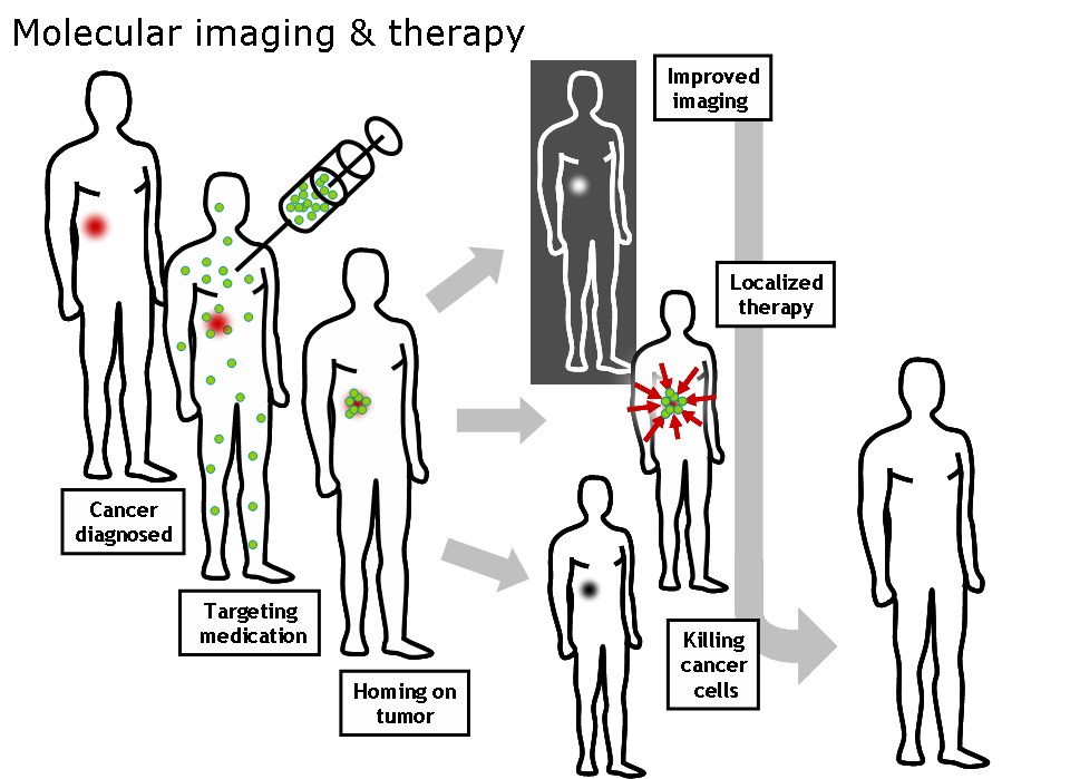
Molecular imaging is a field of medical imaging that focuses on imaging molecules of medical interest within living patients. This is in contrast to conventional methods for obtaining molecular information from preserved tissue samples, such as histology. Molecules of interest may be either ones produced naturally by the body, or synthetic molecules produced in a laboratory and injected into a patient by a doctor. The most common example of molecular imaging used clinically today is to inject a contrast agent (e.g., a microbubble, metal ion, or radioactive isotope) into a patient's bloodstream and to use an imaging modality (e.g., ultrasound, MRI, CT, PET) to track its movement in the body. Molecular imaging originated from the field of radiology from a need to better understand fundamental molecular processes inside organisms in a noninvasive manner. The ultimate goal of molecular imaging is to be able to noninvasively monitor all of the biochemical processes occurring inside ... [...More Info...] [...Related Items...] OR: [Wikipedia] [Google] [Baidu] |
Medical Imaging
Medical imaging is the technique and process of imaging the interior of a body for clinical analysis and medical intervention, as well as visual representation of the function of some organs or tissues (physiology). Medical imaging seeks to reveal internal structures hidden by the skin and bones, as well as to diagnose and treat disease. Medical imaging also establishes a database of normal anatomy and physiology to make it possible to identify abnormalities. Although imaging of removed organs and tissues can be performed for medical reasons, such procedures are usually considered part of pathology instead of medical imaging. Measurement and recording techniques that are not primarily designed to produce images, such as electroencephalography (EEG), magnetoencephalography (MEG), electrocardiography (ECG), and others, represent other technologies that produce data susceptible to representation as a parameter graph versus time or maps that contain data about the measurement loca ... [...More Info...] [...Related Items...] OR: [Wikipedia] [Google] [Baidu] |


