|
Barrel-eye
Barreleyes, also known as spook fish (a name also applied to several species of chimaera), are small deep-sea argentiniform fish comprising the family Opisthoproctidae found in tropical-to-temperate waters of the Atlantic, Pacific, and Indian Oceans. These fish are named because of their barrel-shaped, tubular eyes, which are generally directed upwards to detect the silhouettes of available prey; however, the fish are capable of directing their eyes forward, as well. The family name Opisthoproctidae is derived from the Greek words ''opisthe'' 'behind' and ''proktos'' 'anus'. Description The morphology of the Opisthoproctidae varies between three main forms: the stout, deep-bodied barreleyes of the genera ''Opisthoproctus'' and ''Macropinna'', the extremely slender and elongated spookfishes of the genera ''Dolichopteryx'' and ''Bathylychnops'', and the intermediate fusiform spookfishes of the genera ''Rhynchohyalus'' and '' Winteria''. All species have large, telescoping eyes ... [...More Info...] [...Related Items...] OR: [Wikipedia] [Google] [Baidu] |
Opisthoproctus Soleatus
''Opisthoproctus soleatus'' is a species of fish in the family Opisthoproctidae. It was first described in 1888 by Léon Vaillant. The species lives in most tropical seas, but is more common in the eastern Atlantic, from western Ireland to Mauritania and from Sierra Leone to Angola, and also in the South China Sea. ''O. soleatus'' can grow to a standard length of and usually live from about deep. Description This species is a small fish, not exceeding in length. The body of ''Opisthoproctus soleatus'' is deep and laterally compressed. Scales are large, thin, and cycloid. The ventral side of the body was described by Vaillant as a "flattened, oval, elongate sole." The sole extends forwards below the head. It is covered in large thin scales that increase in pigmentation in the distal parts. The back and sides of this fish are dark and the snout translucent, and there are several large melanophores behind and below the head. ''Opisthoprocus soleautus'' has a specialized modifica ... [...More Info...] [...Related Items...] OR: [Wikipedia] [Google] [Baidu] |
Pacific Ocean
The Pacific Ocean is the largest and deepest of Earth's five oceanic divisions. It extends from the Arctic Ocean in the north to the Southern Ocean (or, depending on definition, to Antarctica) in the south, and is bounded by the continents of Asia and Oceania in the west and the Americas in the east. At in area (as defined with a southern Antarctic border), this largest division of the World Ocean—and, in turn, the hydrosphere—covers about 46% of Earth's water surface and about 32% of its total surface area, larger than Earth's entire land area combined .Pacific Ocean . '' Britannica Concise.'' 2008: Encyclopædia Britannica, Inc. The centers of both the |
Siphonophores
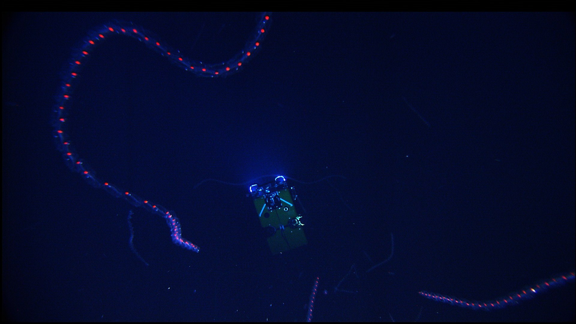
Siphonophorae (from Greek ''siphōn'' 'tube' + ''pherein'' 'to bear') is an order within Hydrozoa, which is a class of marine organisms within the phylum Cnidaria. According to the World Register of Marine Species, the order contains 175 species thus far. Although a siphonophore may appear to be an individual organism, each specimen is in fact a colonial organism composed of medusoid and polypoid zooids that are morphologically and functionally specialized. Zooids are multicellular units that develop from a single fertilized egg and combine to create functional colonies able to reproduce, digest, float, maintain body positioning, and use jet propulsion to move. Most colonies are long, thin, transparent floaters living in the pelagic zone. Like other hydrozoans, some siphonophores emit light to attract and attack prey. While many sea animals produce blue and green bioluminescence, a siphonophore in the genus ''Erenna'' was only the second life form found to produce a red ligh ... [...More Info...] [...Related Items...] OR: [Wikipedia] [Google] [Baidu] |
Nematocyst
A cnidocyte (also known as a cnidoblast or nematocyte) is an explosive cell containing one large secretory organelle called a cnidocyst (also known as a cnida () or nematocyst) that can deliver a sting to other organisms. The presence of this cell defines the phylum Cnidaria (corals, sea anemones, hydrae, jellyfish, etc.). Cnidae are used to capture prey and as a defense against predators. A cnidocyte fires a structure that contains a toxin within the cnidocyst; this is responsible for the stings delivered by a cnidarian. Structure and function Each cnidocyte contains an organelle called a cnida, cnidocyst, nematocyst, ptychocyst or spirocyst. This organelle consists of a bulb-shaped capsule containing a coiled hollow tubule structure attached to it. An immature cnidocyte is referred to as a cnidoblast or nematoblast. The externally oriented side of the cell has a hair-like trigger called a cnidocil, which is a mechano- and chemo-receptor. When the trigger is activated, the ... [...More Info...] [...Related Items...] OR: [Wikipedia] [Google] [Baidu] |
Cone Cell
Cone cells, or cones, are photoreceptor cells in the retinas of vertebrate eyes including the human eye. They respond differently to light of different wavelengths, and the combination of their responses is responsible for color vision. Cones function best in relatively bright light, called the photopic region, as opposed to rod cells, which work better in dim light, or the scotopic region. Cone cells are densely packed in the fovea centralis, a 0.3 mm diameter rod-free area with very thin, densely packed cones which quickly reduce in number towards the periphery of the retina. Conversely, they are absent from the optic disc, contributing to the blind spot. There are about six to seven million cones in a human eye (vs ~92 million rods), with the highest concentration being towards the macula. Cones are less sensitive to light than the rod cells in the retina (which support vision at low light levels), but allow the perception of color. They are also able to perceive ... [...More Info...] [...Related Items...] OR: [Wikipedia] [Google] [Baidu] |
Rhodopsin
Rhodopsin, also known as visual purple, is a protein encoded by the RHO gene and a G-protein-coupled receptor (GPCR). It is the opsin of the rod cells in the retina and a light-sensitive receptor protein that triggers visual phototransduction in rods. Rhodopsin mediates dim light vision and thus is extremely sensitive to light. When rhodopsin is exposed to light, it immediately photobleaches. In humans, it is regenerated fully in about 30 minutes, after which the rods are more sensitive. Defects in the rhodopsin gene cause eye diseases such as retinitis pigmentosa and congenital stationary night blindness. Names Rhodopsin was discovered by Franz Christian Boll in 1876. The name rhodospsin derives from Ancient Greek () for "rose", due to its pinkish color, and () for "sight". It was coined in 1878 by the German physiologist Wilhelm Friedrich Kühne (1837-1900). When George Wald discovered that rhodopsin is a holoprotein, consisting of retinal and an apoprotein, he c ... [...More Info...] [...Related Items...] OR: [Wikipedia] [Google] [Baidu] |
Rod Cell
Rod cells are photoreceptor cells in the retina of the eye that can function in lower light better than the other type of visual photoreceptor, cone cells. Rods are usually found concentrated at the outer edges of the retina and are used in peripheral vision. On average, there are approximately 92 million rod cells (vs ~6 million cones) in the human retina. Rod cells are more sensitive than cone cells and are almost entirely responsible for night vision. However, rods have little role in color vision, which is the main reason why colors are much less apparent in dim light. Structure Rods are a little longer and leaner than cones but have the same basic structure. Opsin-containing disks lie at the end of the cell adjacent to the retinal pigment epithelium, which in turn is attached to the inside of the eye. The stacked-disc structure of the detector portion of the cell allows for very high efficiency. Rods are much more common than cones, with about 120 million rod cells compar ... [...More Info...] [...Related Items...] OR: [Wikipedia] [Google] [Baidu] |
Retina
The retina (from la, rete "net") is the innermost, light-sensitive layer of tissue of the eye of most vertebrates and some molluscs. The optics of the eye create a focused two-dimensional image of the visual world on the retina, which then processes that image within the retina and sends nerve impulses along the optic nerve to the visual cortex to create visual perception. The retina serves a function which is in many ways analogous to that of the film or image sensor in a camera. The neural retina consists of several layers of neurons interconnected by synapses and is supported by an outer layer of pigmented epithelial cells. The primary light-sensing cells in the retina are the photoreceptor cells, which are of two types: rods and cones. Rods function mainly in dim light and provide monochromatic vision. Cones function in well-lit conditions and are responsible for the perception of colour through the use of a range of opsins, as well as high-acuity vision used for task ... [...More Info...] [...Related Items...] OR: [Wikipedia] [Google] [Baidu] |
Lens (anatomy)
The lens, or crystalline lens, is a transparent biconvex structure in the eye that, along with the cornea, helps to refract light to be focused on the retina. By changing shape, it functions to change the focal length of the eye so that it can focus on objects at various distances, thus allowing a sharp real image of the object of interest to be formed on the retina. This adjustment of the lens is known as '' accommodation'' (see also below). Accommodation is similar to the focusing of a photographic camera via movement of its lenses. The lens is flatter on its anterior side than on its posterior side. In humans, the refractive power of the lens in its natural environment is approximately 18 dioptres, roughly one-third of the eye's total power. Structure The lens is part of the anterior segment of the human eye. In front of the lens is the iris, which regulates the amount of light entering into the eye. The lens is suspended in place by the suspensory ligament of the lens ... [...More Info...] [...Related Items...] OR: [Wikipedia] [Google] [Baidu] |
Monterey Bay Aquarium Research Institute
The Monterey Bay Aquarium Research Institute (MBARI) is a private, non-profit oceanographic research center in Moss Landing, California. MBARI was founded in 1987 by David Packard, and is primarily funded by the David and Lucile Packard Foundation. Christopher Scholin serves as the institute's president and chief executive officer, managing a work force of approximately 220 scientists, engineers, and operations and administrative staff. At MBARI, scientists and engineers work together to develop new tools and methods for studying the ocean. Long-term funding from the David and Lucile Packard Foundation allows the institute to take on studies that traditional granting institutions may be reluctant to sponsor. Part of David Packard's charge for MBARI was to "Take risks. Ask big questions. Don't be afraid to make mistakes; if you don't make mistakes, you're not reaching far enough." MBARI's campus in Moss Landing is located near the center of Monterey Bay, at the head of the Monterey ... [...More Info...] [...Related Items...] OR: [Wikipedia] [Google] [Baidu] |
Telescope
A telescope is a device used to observe distant objects by their emission, absorption, or reflection of electromagnetic radiation. Originally meaning only an optical instrument using lenses, curved mirrors, or a combination of both to observe distant objects, the word ''telescope'' now refers to a wide range of instruments capable of detecting different regions of the electromagnetic spectrum, and in some cases other types of detectors. The first known practical telescopes were refracting telescopes with glass lenses and were invented in the Netherlands at the beginning of the 17th century. They were used for both terrestrial applications and astronomy. The reflecting telescope, which uses mirrors to collect and focus light, was invented within a few decades of the first refracting telescope. In the 20th century, many new types of telescopes were invented, including radio telescopes in the 1930s and infrared telescopes in the 1960s. Etymology The word ''telescope'' was coin ... [...More Info...] [...Related Items...] OR: [Wikipedia] [Google] [Baidu] |
Morphology (biology)
Morphology is a branch of biology dealing with the study of the form and structure of organisms and their specific structural features. This includes aspects of the outward appearance (shape, structure, colour, pattern, size), i.e. external morphology (or eidonomy), as well as the form and structure of the internal parts like bones and organs, i.e. internal morphology (or anatomy). This is in contrast to physiology, which deals primarily with function. Morphology is a branch of life science dealing with the study of gross structure of an organism or taxon and its component parts. History The etymology of the word "morphology" is from the Ancient Greek (), meaning "form", and (), meaning "word, study, research". While the concept of form in biology, opposed to function, dates back to Aristotle (see Aristotle's biology), the field of morphology was developed by Johann Wolfgang von Goethe (1790) and independently by the German anatomist and physiologist Karl Friedrich Burdach ... [...More Info...] [...Related Items...] OR: [Wikipedia] [Google] [Baidu] |

.jpg)


