|
Band Of Baillarger
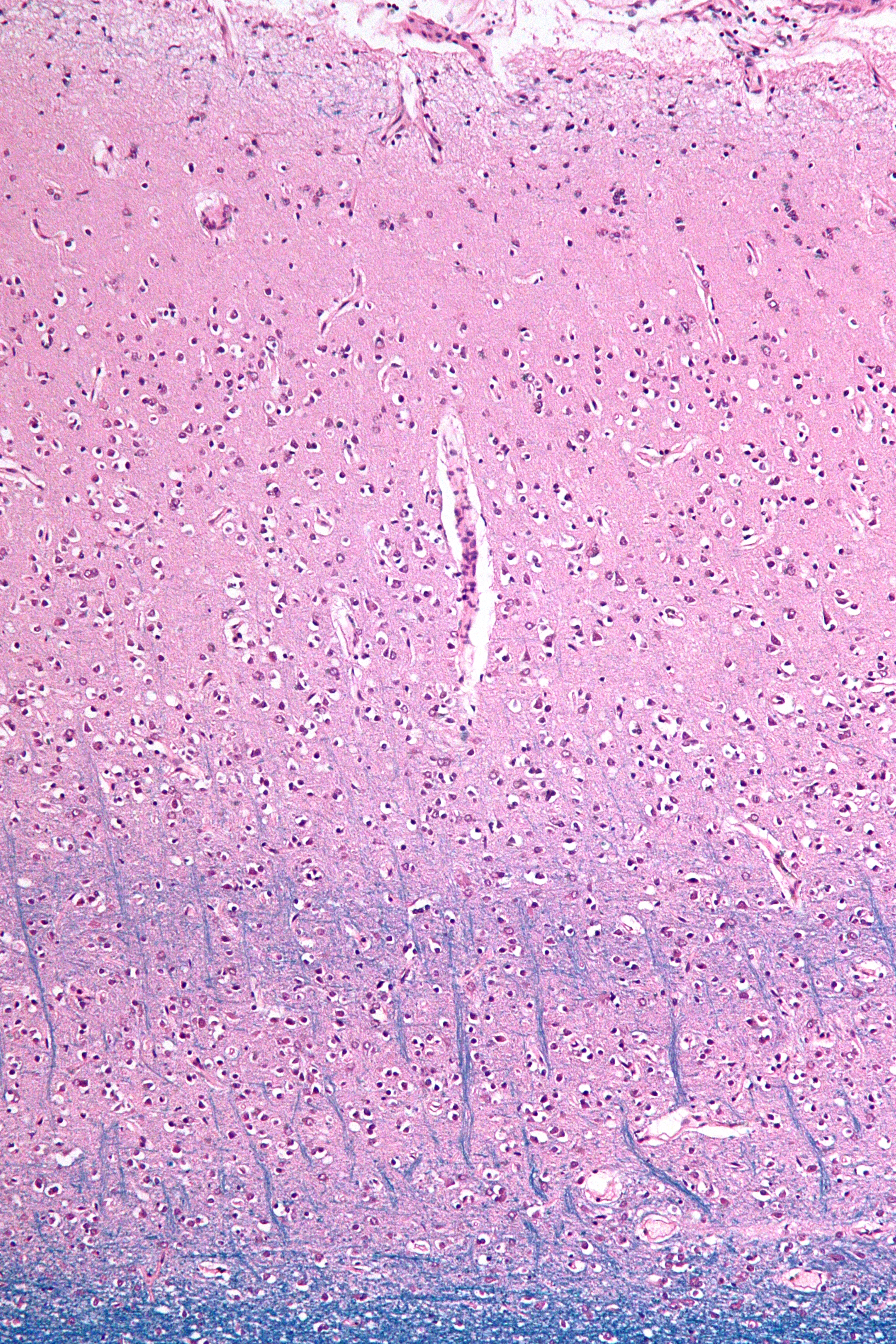
Bands of Baillarger, in neuroanatomy, are two myeloarchitectural structures, the external band of Baillarger and the internal band of Baillarger. These are myelinated fibers that course in the internal granular layer (layer IV) and internal pyramidal layer (layer V), respectively, of cerebral cortex. In the primary visual cortex (V1), the external band of Baillarger is enlarged and is called the line of Gennari. They are named after Jules Baillarger Jules Baillarger, full name Jules Gabriel François Baillarger (25 March 1809 – 31 December 1890), was a French neurologist and psychiatrist. Biography Baillarger was born in Montbazon, France. He studied medicine at the University of Paris u .... References Cerebral cortex {{neuroanatomy-stub ... [...More Info...] [...Related Items...] OR: [Wikipedia] [Google] [Baidu] |
Neuroanatomy
Neuroanatomy is the study of the structure and organization of the nervous system. In contrast to animals with radial symmetry, whose nervous system consists of a distributed network of cells, animals with bilateral symmetry have segregated, defined nervous systems. Their neuroanatomy is therefore better understood. In vertebrates, the nervous system is segregated into the internal structure of the brain and spinal cord (together called the central nervous system, or CNS) and the routes of the nerves that connect to the rest of the body (known as the peripheral nervous system, or PNS). The delineation of distinct structures and regions of the nervous system has been critical in investigating how it works. For example, much of what neuroscientists have learned comes from observing how damage or "lesions" to specific brain areas affects behavior or other neural functions. For information about the composition of non-human animal nervous systems, see nervous system. For information ab ... [...More Info...] [...Related Items...] OR: [Wikipedia] [Google] [Baidu] |
Cerebral Cortex
The cerebral cortex, also known as the cerebral mantle, is the outer layer of neural tissue of the cerebrum of the brain in humans and other mammals. The cerebral cortex mostly consists of the six-layered neocortex, with just 10% consisting of allocortex. It is separated into two cortices, by the longitudinal fissure that divides the cerebrum into the left and right cerebral hemispheres. The two hemispheres are joined beneath the cortex by the corpus callosum. The cerebral cortex is the largest site of neural integration in the central nervous system. It plays a key role in attention, perception, awareness, thought, memory, language, and consciousness. The cerebral cortex is part of the brain responsible for cognition. In most mammals, apart from small mammals that have small brains, the cerebral cortex is folded, providing a greater surface area in the confined volume of the cranium. Apart from minimising brain and cranial volume, cortical folding is crucial for the brain ... [...More Info...] [...Related Items...] OR: [Wikipedia] [Google] [Baidu] |
Visual Cortex
The visual cortex of the brain is the area of the cerebral cortex that processes visual information. It is located in the occipital lobe. Sensory input originating from the eyes travels through the lateral geniculate nucleus in the thalamus and then reaches the visual cortex. The area of the visual cortex that receives the sensory input from the lateral geniculate nucleus is the primary visual cortex, also known as visual area 1 ( V1), Brodmann area 17, or the striate cortex. The extrastriate areas consist of visual areas 2, 3, 4, and 5 (also known as V2, V3, V4, and V5, or Brodmann area 18 and all Brodmann area 19). Both hemispheres of the brain include a visual cortex; the visual cortex in the left hemisphere receives signals from the right visual field, and the visual cortex in the right hemisphere receives signals from the left visual field. Introduction The primary visual cortex (V1) is located in and around the calcarine fissure in the occipital lobe. Each hemisphere's V1 ... [...More Info...] [...Related Items...] OR: [Wikipedia] [Google] [Baidu] |
Line Of Gennari
The line of Gennari (also called the "band" or "stria" of Gennari) is a band of myelinated axons that runs parallel to the surface of the cerebral cortex on the banks of the calcarine fissure in the occipital lobe. This formation is visible to the naked eye as a white strip running through the cortical grey matter, and is the reason the V1 in primates is also referred to as the "striate cortex." The line of Gennari is due to dense axonal input from the thalamus to layer IV of visual cortex. It is the name given to the enlarged external band of Baillarger. The structure is named for its discoverer, Francesco Gennari, who first observed it in 1776 as a medical student at the University of Parma. He described it in a book which he published six years later. Although non-primate species have areas that are designated primary visual cortex, some (if not all) lack a stria of Gennari.Zilles and Wree. ''Isocortex'' in Paxinos (Ed.) ''The Rat Nervous System'', 1985. See also * Cerebral cort ... [...More Info...] [...Related Items...] OR: [Wikipedia] [Google] [Baidu] |
Jules Baillarger
Jules Baillarger, full name Jules Gabriel François Baillarger (25 March 1809 – 31 December 1890), was a French neurologist and psychiatrist. Biography Baillarger was born in Montbazon, France. He studied medicine at the University of Paris under Jean-Étienne Dominique Esquirol (1772–1840), and while a student worked as an intern at the Charenton (asylum), Charenton mental institution. In 1840 he accepted a position at the Salpêtrière, and soon after became director of a ''maison de santé'' in Ivry-sur-Seine. Among his assistants at Ivry was Louis-Victor Marcé (1828-1864). With Jacques-Joseph Moreau (1804–1884) and others, he founded the influential ''Annales médico-psychologiques'' (Medical-Psychological Annals). Contributions and theories In 1840 Baillarger was the first physician to discover that the cerebral cortex was divided into six layers of alternate white and grey laminae. His name is associated with the inner and outer Band of Baillarger, bands of Baillar ... [...More Info...] [...Related Items...] OR: [Wikipedia] [Google] [Baidu] |


