|
Ball-and-socket
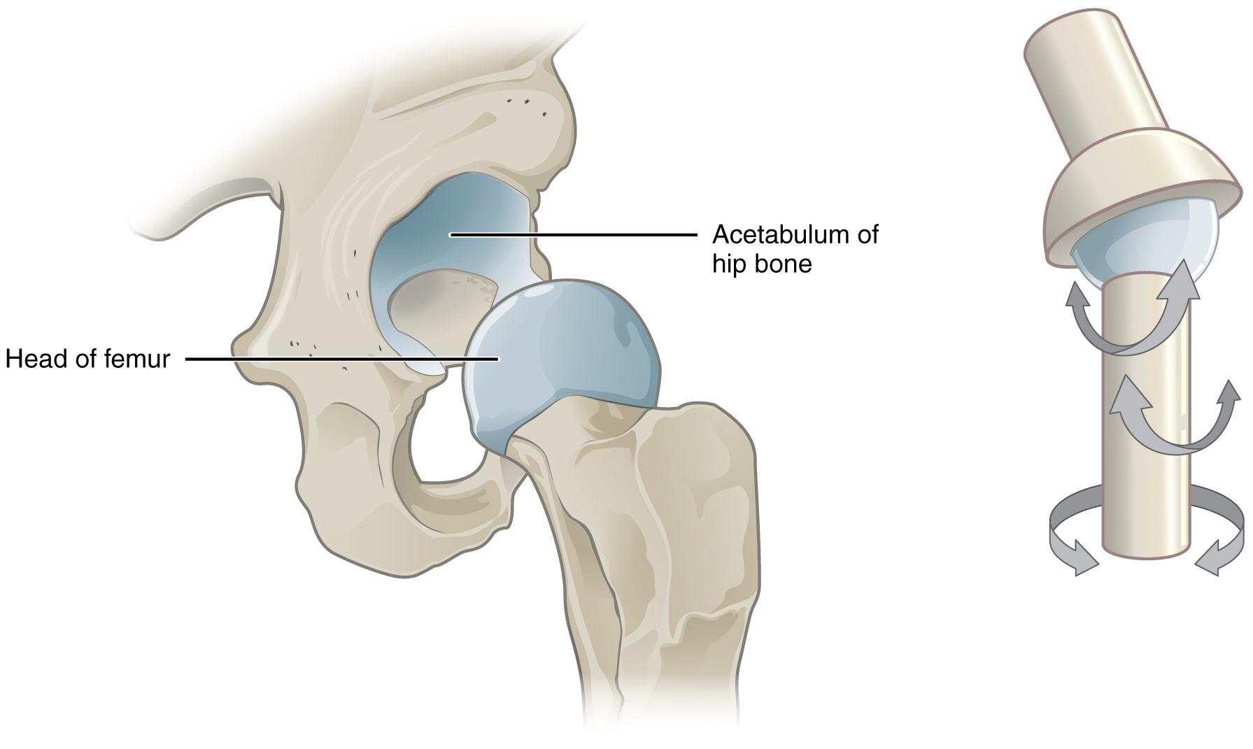
The ball-and-socket joint (or spheroid joint) is a type of synovial joint in which the ball-shaped surface of one rounded bone fits into the cup-like depression of another bone. The distal bone is capable of motion around an indefinite number of axes, which have one common center. This enables the joint to move in many directions. An enarthrosis is a special kind of spheroidal joint in which the socket covers the sphere beyond its equator.Platzer, Werner (2008) ''Color Atlas of Human Anatomy'', Volume 1p.28/ref> Examples of joints Examples of this form of articulation are found in the hip, where the round head of the femur (ball) rests in the cup-like acetabulum (socket) of the pelvis; and in the shoulder joint, where the rounded upper extremity of the humerus (ball) rests in the cup-like glenoid fossa (socket) of the shoulder blade.And the phalanges (toes, fingers)Introduction to Joints: Synovial Joints - Ball and Socket Joints (The shoulder also includes a sternoclavicular ... [...More Info...] [...Related Items...] OR: [Wikipedia] [Google] [Baidu] |
Shoulder Joint
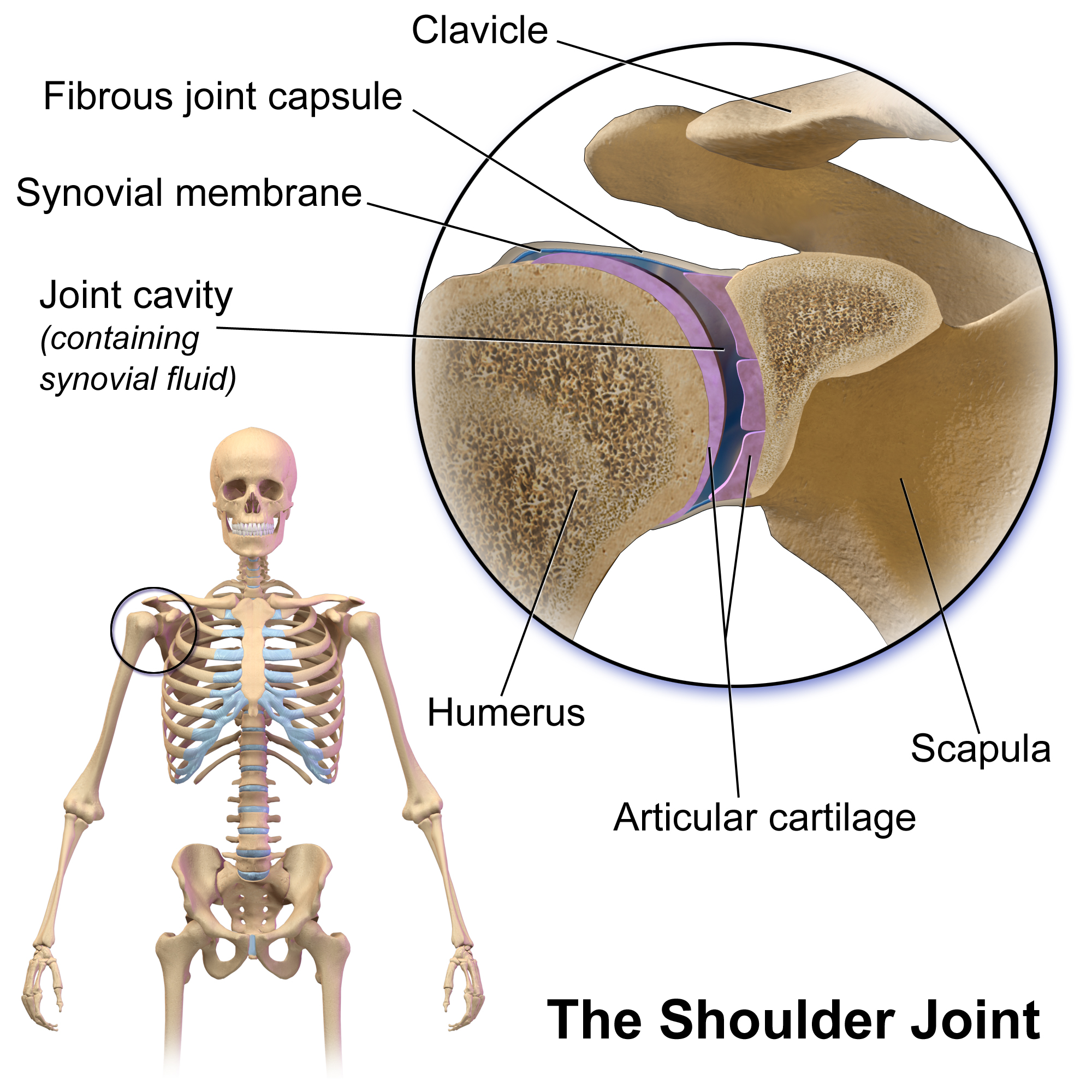
The shoulder joint (or glenohumeral joint from Greek ''glene'', eyeball, + -''oid'', 'form of', + Latin ''humerus'', shoulder) is structurally classified as a synovial joint, synovial ball-and-socket joint and functionally as a diarthrosis and multiaxial joint. It involves an articulation between the glenoid fossa of the scapula (shoulder blade) and the head of the humerus (upper arm bone). Due to the very loose joint capsule, it gives a limited interface of the humerus and scapula, it is the most mobile joint of the human body. Structure The shoulder joint is a ball-and-socket joint between the scapula and the humerus. The socket of the glenoid fossa of the scapula is itself quite shallow, but it is made deeper by the addition of the glenoid labrum. The glenoid labrum is a ring of cartilage, cartilaginous fibre attached to the circumference of the cavity. This ring is continuous with the tendon of the Biceps, biceps brachii above. Spaces Significant joint spaces are: * The ... [...More Info...] [...Related Items...] OR: [Wikipedia] [Google] [Baidu] |
Shoulder
The human shoulder is made up of three bones: the clavicle (collarbone), the scapula (shoulder blade), and the humerus (upper arm bone) as well as associated muscles, ligaments and tendons. The articulations between the bones of the shoulder make up the shoulder joints. The shoulder joint, also known as the glenohumeral joint, is the major joint of the shoulder, but can more broadly include the acromioclavicular joint. In human anatomy, the shoulder joint comprises the part of the body where the humerus attaches to the scapula, and the head sits in the glenoid cavity. The shoulder is the group of structures in the region of the joint. The shoulder joint is the main joint of the shoulder. It is a ball and socket joint that allows the arm to rotate in a circular fashion or to hinge out and up away from the body. The joint capsule is a soft tissue envelope that encircles the glenohumeral joint and attaches to the scapula, humerus, and head of the biceps. It is lined by a ... [...More Info...] [...Related Items...] OR: [Wikipedia] [Google] [Baidu] |
Glenoid Cavity
The glenoid fossa of the scapula or the glenoid cavity is a bone part of the shoulder. The word ''glenoid'' is pronounced or (both are common) and is from , "socket", reflecting the shoulder joint's ball-and-socket form. It is a shallow, pyriform articular surface, which is located on the lateral angle of the scapula. It is directed laterally and forward and articulates with the head of the humerus; it is broader below than above and its vertical diameter is the longest. This cavity forms the glenohumeral joint along with the humerus. This type of joint is classified as a synovial, ball and socket joint. The humerus is held in place within the glenoid cavity by means of the long head of the biceps tendon. This tendon originates on the superior margin of the glenoid cavity and loops over the shoulder, bracing humerus against the cavity. The rotator cuff also reinforces this joint more specifically with the supraspinatus tendon to hold the head of the humerus in the glenoid ... [...More Info...] [...Related Items...] OR: [Wikipedia] [Google] [Baidu] |
Condyloid Joint
A condyloid joint (also called condylar, ellipsoidal, or bicondylar) is an ovoid articular surface, or condyle that is received into an elliptical cavity. This permits movement in two planes, allowing flexion, extension, adduction, abduction, and circumduction. Examples Examples include: * the wrist-joint * metacarpophalangeal joints * metatarsophalangeal joints * atlanto-occipital joints These are also called ellipsoid joints. The oval-shaped condyle of one bone fits into the elliptical cavity of the other bone. These joints allow biaxial movements — i.e., forward and backward, or from side to side, but not rotation. Radiocarpal joint and metacarpophalangeal joint are examples of condyloid joints. An example of an Ellipsoid joint is the wrist; it functions similarly to the ball and socket joint The ball-and-socket joint (or spheroid joint) is a type of synovial joint in which the ball-shaped surface of one rounded bone fits into the cup-like depression of anoth ... [...More Info...] [...Related Items...] OR: [Wikipedia] [Google] [Baidu] |
Saddle Joint
A saddle joint (sellar joint, articulation by reciprocal reception) is a type of synovial joint in which the opposing surfaces are reciprocally concave and convex. It is found in the thumb, the thorax, the middle ear, and the heel. Structure In a saddle joint, one bone surface is concave while another is convex. This creates significant stability. Movements The movements of saddle joints are similar to those of the condyloid joint and include flexion, extension, adduction, abduction, and circumduction. However, axial rotation is not allowed. Saddle joints are said to be biaxial, allowing movement in the sagittal and frontal planes. Examples of saddle joints in the human body include the carpometacarpal joint of the thumb, the sternoclavicular joint of the thorax, the incudomalleolar joint of the middle ear, and the calcaneocuboid joint of the heel. Name The term "saddle" arises because the concave-convex bone interaction is compared to a horse rider riding a horse ... [...More Info...] [...Related Items...] OR: [Wikipedia] [Google] [Baidu] |
Hinge Joint
A hinge joint (ginglymus or ginglymoid) is a bone joint where the articular surfaces are molded to each other in such a manner as to permit motion only in one plane. According to one classification system they are said to be uniaxial (having one degree of freedom).Platzer, Werner (2008) ''Color Atlas of Human Anatomy', Volume 1p.28/ref> The direction which the distal bone takes in this motion is rarely in the same plane as that of the axis of the proximal bone; there is usually a certain amount of deviation from the straight line during flexion. The articular surfaces of the bones are connected by strong collateral ligaments. Examples of ginglymoid joints are the interphalangeal joints of the hand and those of the foot and the joint between the humerus and ulna. The knee joints and ankle joints are less typical, as they allow a slight degree of rotation or side-to-side movement in certain positions of the limb. The knee is the largest hinge joint in the human body ... [...More Info...] [...Related Items...] OR: [Wikipedia] [Google] [Baidu] |
Pivot Joint
In animal anatomy, a pivot joint (trochoid joint, rotary joint or lateral ginglymus) is a type of synovial joint whose movement axis is parallel to the long axis of the proximal bone, which typically has a convex articular surface. According to one classification system, a pivot joint like the other synovial joint—the hinge joint has one degree of freedom.Platzer, Werner (2008) ''Color Atlas of Human Anatomy'', Volume 1p.28/ref> Note that the degrees of freedom of a joint is not the same as a joint's range of motion. Movements Pivot joints allow rotation Rotation or rotational/rotary motion is the circular movement of an object around a central line, known as an ''axis of rotation''. A plane figure can rotate in either a clockwise or counterclockwise sense around a perpendicular axis intersect ..., which can be external (for example when rotating an arm outward), or internal (as in rotating an arm inward). When rotating the forearm, these movements are typically ca ... [...More Info...] [...Related Items...] OR: [Wikipedia] [Google] [Baidu] |
Synovial Joint
A synovial joint, also known as diarthrosis, joins bones or cartilage with a fibrous joint capsule that is continuous with the periosteum of the joined bones, constitutes the outer boundary of a synovial cavity, and surrounds the bones' articulating surfaces. This joint unites long bones and permits free bone movement and greater mobility. The synovial cavity/joint is filled with synovial fluid. The joint capsule is made up of an outer layer of fibrous membrane, which keeps the bones together structurally, and an inner layer, the synovial membrane, which seals in the synovial fluid. They are the most common and most movable type of joint in the body. As with most other joints, synovial joints achieve movement at the point of contact of the articulating bones. They originated 400 million years ago in the first jawed vertebrates. Structure Synovial joints contain the following structures: * Synovial cavity: all diarthroses have the characteristic space between the bones that is f ... [...More Info...] [...Related Items...] OR: [Wikipedia] [Google] [Baidu] |
Femoral Head
The femoral head (femur head or head of the femur) is the highest part of the thigh bone (femur The femur (; : femurs or femora ), or thigh bone is the only long bone, bone in the thigh — the region of the lower limb between the hip and the knee. In many quadrupeds, four-legged animals the femur is the upper bone of the hindleg. The Femo ...). It is supported by the femoral neck. Structure The head is globular and forms rather more than a hemisphere, is directed upward, medialward, and a little forward, the greater part of its convexity being above and in front. The femoral head's surface is smooth. It is coated with cartilage in the fresh state, except over an ovoid depression, the fovea capitis, which is situated a little below and behind the center of the femoral head, and gives attachment to the ligament of head of femur. The thickest region of the articular cartilage is at the centre of the femoral head, measuring up to 2.8 mm. The diameter of the femoral hea ... [...More Info...] [...Related Items...] OR: [Wikipedia] [Google] [Baidu] |
Acetabulum
The acetabulum (; : acetabula), also called the cotyloid cavity, is a wikt:concave, concave surface of the pelvis. The femur head, head of the femur meets with the pelvis at the acetabulum, forming the Hip#Articulation, hip joint. Structure There are three bones of the ''os coxae'' (hip bone) that come together to form the ''acetabulum''. Contributing a little more than two-fifths of the structure is the ischium, which provides lower and side boundaries to the acetabulum. The Ilium (bone), ilium forms the upper boundary, providing a little less than two-fifths of the structure of the acetabulum. The rest is formed by the Pubis (bone), pubis, near the midline. It is bounded by a prominent uneven rim, thick and strong on top, which serves as the point of attachment for the acetabular labrum. The acetabular labrum reduces the size of the opening of the acetabulum and deepens the surface of the hip joint. At the lower part of the acetabulum is the acetabular notch, which is continuo ... [...More Info...] [...Related Items...] OR: [Wikipedia] [Google] [Baidu] |
Pelvis
The pelvis (: pelves or pelvises) is the lower part of an Anatomy, anatomical Trunk (anatomy), trunk, between the human abdomen, abdomen and the thighs (sometimes also called pelvic region), together with its embedded skeleton (sometimes also called bony pelvis or pelvic skeleton). The pelvic region of the trunk includes the bony pelvis, the pelvic cavity (the space enclosed by the bony pelvis), the pelvic floor, below the pelvic cavity, and the perineum, below the pelvic floor. The pelvic skeleton is formed in the area of the back, by the sacrum and the coccyx and anteriorly and to the left and right sides, by a pair of hip bones. The two hip bones connect the spine with the lower limbs. They are attached to the sacrum posteriorly, connected to each other anteriorly, and joined with the two femurs at the hip joints. The gap enclosed by the bony pelvis, called the pelvic cavity, is the section of the body underneath the abdomen and mainly consists of the reproductive organs and ... [...More Info...] [...Related Items...] OR: [Wikipedia] [Google] [Baidu] |
Upper Extremity Of Humerus
The humerus (; : humeri) is a long bone in the arm that runs from the shoulder to the elbow. It connects the scapula and the two bones of the lower arm, the radius and ulna, and consists of three sections. The humeral upper extremity consists of a rounded head, a narrow neck, and two short processes (tubercles, sometimes called tuberosities). The body is cylindrical in its upper portion, and more prismatic below. The lower extremity consists of 2 epicondyles, 2 processes ( trochlea and capitulum), and 3 fossae ( radial fossa, coronoid fossa, and olecranon fossa). As well as its true anatomical neck, the constriction below the greater and lesser tubercles of the humerus is referred to as its surgical neck due to its tendency to fracture, thus often becoming the focus of surgeons. Etymology The word "humerus" is derived from Late Latin , from Latin , meaning upper arm, shoulder, and is linguistically related to Gothic (shoulder) and Greek . Structure Upper extremity The u ... [...More Info...] [...Related Items...] OR: [Wikipedia] [Google] [Baidu] |


