|
Autapse
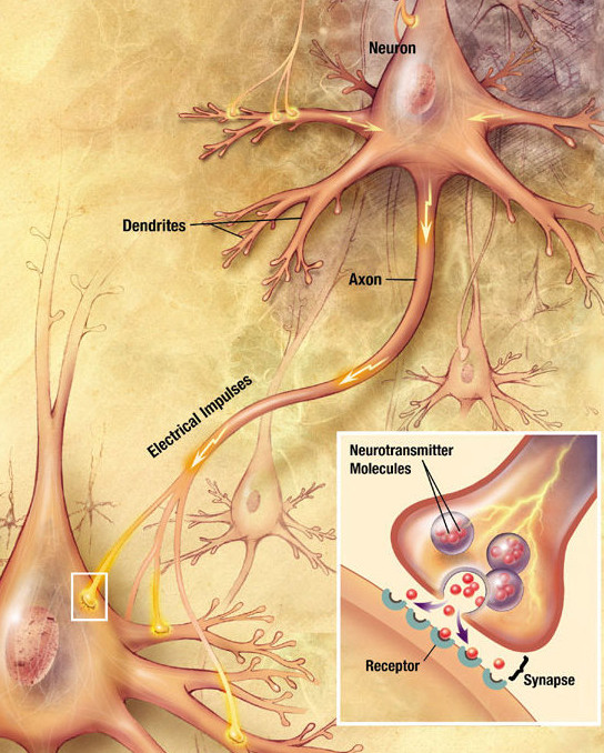
An autapse is a chemical or electrical synapse from a neuron onto itself. It can also be described as a synapse formed by the axon of a neuron on its own dendrites, '' in vivo'' or '' in vitro''. History The term "autapse" was first coined in 1972 by Van der Loos and Glaser, who observed them in Golgi preparations of the rabbit occipital cortex while originally conducting a quantitative analysis of neocortex circuitry. Also in the 1970s, autapses have been described in dog and rat cerebral cortex, monkey neostriatum, and cat spinal cord. In 2000, they were first modeled as supporting persistence in recurrent neural networks. In 2004, they were modeled as demonstrating oscillatory behavior, which was absent in the same model neuron without autapse. More specifically, the neuron oscillated between high firing rates and firing suppression, reflecting the spike bursting behavior typically found in cerebral neurons. In 2009, autapses were, for the first time, associated with ... [...More Info...] [...Related Items...] OR: [Wikipedia] [Google] [Baidu] |
Chemical Synapse
Chemical synapses are biological junctions through which neurons' signals can be sent to each other and to non-neuronal cells such as those in muscles or glands. Chemical synapses allow neurons to form circuits within the central nervous system. They are crucial to the biological computations that underlie perception and thought. They allow the nervous system to connect to and control other systems of the body. At a chemical synapse, one neuron releases neurotransmitter molecules into a small space (the synaptic cleft) that is adjacent to another neuron. The neurotransmitters are contained within small sacs called synaptic vesicles, and are released into the synaptic cleft by exocytosis. These molecules then bind to neurotransmitter receptors on the postsynaptic cell. Finally, the neurotransmitters are cleared from the synapse through one of several potential mechanisms including enzymatic degradation or re-uptake by specific transporters either on the presynaptic cell or ... [...More Info...] [...Related Items...] OR: [Wikipedia] [Google] [Baidu] |
Biological Neuron Model
Biological neuron models, also known as a spiking neuron models, are mathematical descriptions of the properties of certain cells in the nervous system that generate sharp electrical potentials across their cell membrane, roughly one millisecond in duration, called action potentials or spikes (Fig. 2). Since spikes are transmitted along the axon and synapses from the sending neuron to many other neurons, spiking neurons are considered to be a major information processing unit of the nervous system. Spiking neuron models can be divided into different categories: the most detailed mathematical models are biophysical neuron models (also called Hodgkin-Huxley models) that describe the membrane voltage as a function of the input current and the activation of ion channels. Mathematically simpler are integrate-and-fire models that describe the membrane voltage as a function of the input current and predict the spike times without a description of the biophysical processes that shape ... [...More Info...] [...Related Items...] OR: [Wikipedia] [Google] [Baidu] |
Aplysia
''Aplysia'' () is a genus of medium-sized to extremely large sea slugs, specifically sea hares, which are one clade of large sea slugs, marine gastropod mollusks. These benthic herbivorous creatures can become rather large compared with most other mollusks. They graze in tidal and subtidal zones of tropical waters, mostly in the Indo-Pacific Ocean (23 species); but they can also be found in the Atlantic Ocean (12 species), with a few species occurring in the Mediterranean. ''Aplysia'' species, when threatened, frequently release clouds of ink, it is believed in order to blind the attacker (though they are in fact considered edible by relatively few species). Following the lead of Eric R. Kandel, the genus has been studied as a model organism by neurobiologists, because its gill and siphon withdrawal reflex, as studied in ''Aplysia californica'', is mediated by electrical synapses, which allow several neurons to fire synchronously. This quick neural response is necessa ... [...More Info...] [...Related Items...] OR: [Wikipedia] [Google] [Baidu] |
Shunting Inhibition
Shunting inhibition, also known as divisive inhibition, is a form of postsynaptic potential inhibition that can be represented mathematically as reducing the excitatory potential by division, rather than linear subtraction. The term "shunting" is used because of the synaptic conductance short-circuit currents that are generated at adjacent excitatory synapses. If a shunting inhibitory synapse is activated, the input resistance is reduced locally. The amplitude of subsequent excitatory postsynaptic potential (EPSP) is reduced by this, in accordance with Ohm's Law. This simple scenario arises if the inhibitory synaptic reversal potential is identical to the resting potential. Discovery Shunting inhibition was discovered by Fatt and Katz in 1953. Mechanism Shunting inhibition is theorized to be a type of gain control Gain or GAIN may refer to: Science and technology * Gain (electronics), an electronics and signal processing term * Antenna gain * Gain (laser), the ampli ... [...More Info...] [...Related Items...] OR: [Wikipedia] [Google] [Baidu] |
Depolarization
In biology, depolarization or hypopolarization is a change within a cell, during which the cell undergoes a shift in electric charge distribution, resulting in less negative charge inside the cell compared to the outside. Depolarization is essential to the function of many cells, communication between cells, and the overall physiology of an organism. Most cells in higher organisms maintain an internal environment that is negatively charged relative to the cell's exterior. This difference in charge is called the cell's membrane potential. In the process of depolarization, the negative internal charge of the cell temporarily becomes more positive (less negative). This shift from a negative to a more positive membrane potential occurs during several processes, including an action potential. During an action potential, the depolarization is so large that the potential difference across the cell membrane briefly reverses polarity, with the inside of the cell becoming positively char ... [...More Info...] [...Related Items...] OR: [Wikipedia] [Google] [Baidu] |
Layer V
The cerebral cortex, also known as the cerebral mantle, is the outer layer of neural tissue of the cerebrum of the brain in humans and other mammals. The cerebral cortex mostly consists of the six-layered neocortex, with just 10% consisting of allocortex. It is separated into two cortices, by the longitudinal fissure that divides the cerebrum into the left and right cerebral hemispheres. The two hemispheres are joined beneath the cortex by the corpus callosum. The cerebral cortex is the largest site of neural integration in the central nervous system. It plays a key role in attention, perception, awareness, thought, memory, language, and consciousness. The cerebral cortex is part of the brain responsible for cognition. In most mammals, apart from small mammals that have small brains, the cerebral cortex is folded, providing a greater surface area in the confined volume of the cranium. Apart from minimising brain and cranial volume, cortical folding is crucial for the brain c ... [...More Info...] [...Related Items...] OR: [Wikipedia] [Google] [Baidu] |
Interneuron
Interneurons (also called internuncial neurons, relay neurons, association neurons, connector neurons, intermediate neurons or local circuit neurons) are neurons that connect two brain regions, i.e. not direct motor neurons or sensory neurons. Interneurons are the central nodes of neural circuits, enabling communication between sensory or motor neurons and the central nervous system (CNS). They play vital roles in reflexes, neuronal oscillations, and neurogenesis in the adult mammalian brain. Interneurons can be further broken down into two groups: local interneurons and relay interneurons. Local interneurons have short axons and form circuits with nearby neurons to analyze small pieces of information. Relay interneurons have long axons and connect circuits of neurons in one region of the brain with those in other regions. However, interneurons are generally considered to operate mainly within local brain areas. The interaction between interneurons allow the brain to perform ... [...More Info...] [...Related Items...] OR: [Wikipedia] [Google] [Baidu] |
Inhibitory Postsynaptic Potential
An inhibitory postsynaptic potential (IPSP) is a kind of synaptic potential that makes a postsynaptic neuron less likely to generate an action potential.Purves et al. Neuroscience. 4th ed. Sunderland (MA): Sinauer Associates, Incorporated; 2008. IPSP were first investigated in motorneurons by David P. C. Lloyd, John Eccles and Rodolfo Llinás in the 1950s and 1960s. The opposite of an inhibitory postsynaptic potential is an excitatory postsynaptic potential (EPSP), which is a synaptic potential that makes a postsynaptic neuron ''more'' likely to generate an action potential. IPSPs can take place at all chemical synapses, which use the secretion of neurotransmitters to create cell to cell signalling. Inhibitory presynaptic neurons release neurotransmitters that then bind to the postsynaptic receptors; this induces a change in the permeability of the postsynaptic neuronal membrane to particular ions. An electric current that changes the postsynaptic membrane potential to crea ... [...More Info...] [...Related Items...] OR: [Wikipedia] [Google] [Baidu] |
Hyperpolarization (biology)
Hyperpolarization is a change in a cell's membrane potential that makes it more negative. It is the opposite of a depolarization. It inhibits action potentials by increasing the stimulus required to move the membrane potential to the action potential threshold. Hyperpolarization is often caused by efflux of K+ (a cation) through K+ channels, or influx of Cl– (an anion) through Cl– channels. On the other hand, influx of cations, e.g. Na+ through Na+ channels or Ca2+ through Ca2+ channels, inhibits hyperpolarization. If a cell has Na+ or Ca2+ currents at rest, then inhibition of those currents will also result in a hyperpolarization. This voltage-gated ion channel response is how the hyperpolarization state is achieved. In neurons, the cell enters a state of hyperpolarization immediately following the generation of an action potential. While hyperpolarized, the neuron is in a refractory period that lasts roughly 2 milliseconds, during which the neuron is unabl ... [...More Info...] [...Related Items...] OR: [Wikipedia] [Google] [Baidu] |
Glutamate (neurotransmitter)
In neuroscience, glutamate refers to the anion of glutamic acid in its role as a neurotransmitter: a chemical that nerve cells use to send signals to other cells. It is by a wide margin the most abundant excitatory neurotransmitter in the vertebrate nervous system. It is used by every major excitatory function in the vertebrate brain, accounting in total for well over 90% of the synaptic connections in the human brain. It also serves as the primary neurotransmitter for some localized brain regions, such as cerebellum granule cells. Biochemical receptors for glutamate fall into three major classes, known as AMPA receptors, NMDA receptors, and metabotropic glutamate receptors. A fourth class, known as kainate receptors, are similar in many respects to AMPA receptors, but much less abundant. Many synapses use multiple types of glutamate receptors. AMPA receptors are ionotropic receptors specialized for fast excitation: in many synapses they produce excitatory electrical responses ... [...More Info...] [...Related Items...] OR: [Wikipedia] [Google] [Baidu] |


