|
Arterial Tree
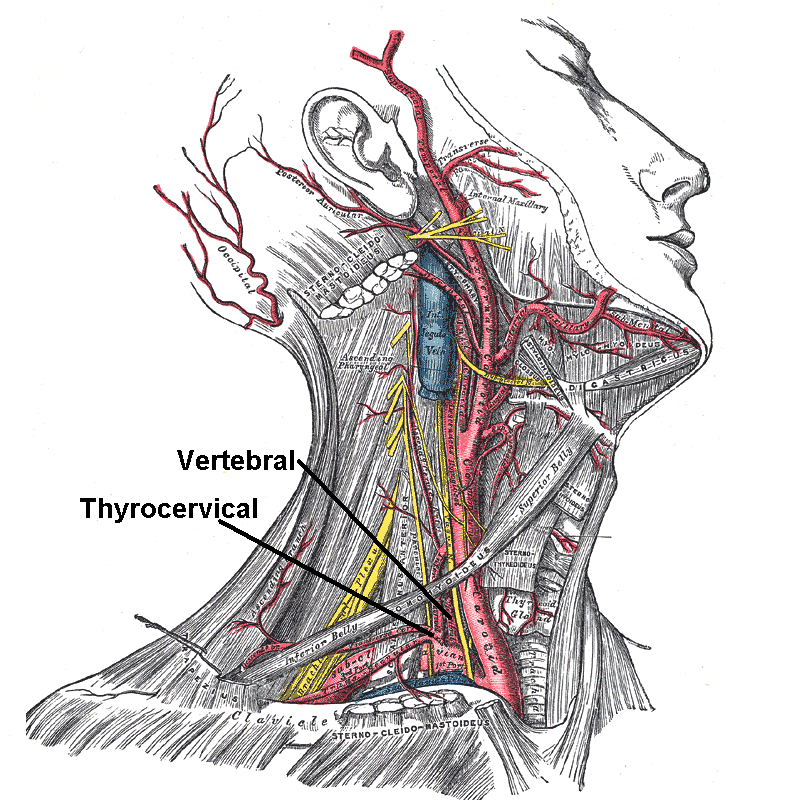
In anatomy, arterial tree is used to refer to all arteries and/or the branching pattern of the arteries. This article regards the human arterial tree. Starting from the aorta: the following are the parts Ascending aorta It is a portion of the aorta commencing at the upper part of the base of the left ventricle, on a level with the lower border of the third costal cartilage behind the left half of the sternum. Right coronary artery *posterior interventricular artery (mostly) *SA nodal artery (in 60%) *Right marginal artery Left coronary artery * anterior interventricular **septal **diagonal *circumflex **SA nodal artery (in 40%) **posterior interventricular artery (occasionally) **Left marginal arteries ** posterolateral artery * ramus intermedius (sometimes) Aortic arch brachiocephalic artery * right common carotid artery *right subclavian artery left common carotid artery (directly from arch of aorta on left mostly) internal carotid artery *ophthalmic artery * ... [...More Info...] [...Related Items...] OR: [Wikipedia] [Google] [Baidu] |
Anatomy
Anatomy () is the branch of biology concerned with the study of the structure of organisms and their parts. Anatomy is a branch of natural science that deals with the structural organization of living things. It is an old science, having its beginnings in prehistoric times. Anatomy is inherently tied to developmental biology, embryology, comparative anatomy, evolutionary biology, and phylogeny, as these are the processes by which anatomy is generated, both over immediate and long-term timescales. Anatomy and physiology, which study the structure and function (biology), function of organisms and their parts respectively, make a natural pair of related disciplines, and are often studied together. Human anatomy is one of the essential basic research, basic sciences that are applied in medicine. The discipline of anatomy is divided into macroscopic scale, macroscopic and microscopic scale, microscopic. Gross anatomy, Macroscopic anatomy, or gross anatomy, is the examination of an ... [...More Info...] [...Related Items...] OR: [Wikipedia] [Google] [Baidu] |
Right Subclavian Artery
In human anatomy, the subclavian arteries are paired major arteries of the upper thorax, below the clavicle. They receive blood from the aortic arch. The left subclavian artery supplies blood to the left arm and the right subclavian artery supplies blood to the right arm, with some branches supplying the head and thorax. On the left side of the body, the subclavian comes directly off the aortic arch, while on the right side it arises from the relatively short brachiocephalic artery when it bifurcates into the subclavian and the right common carotid artery. The usual branches of the subclavian on both sides of the body are the vertebral artery, the internal thoracic artery, the thyrocervical trunk, the costocervical trunk and the dorsal scapular artery, which may branch off the transverse cervical artery, which is a branch of the thyrocervical trunk. The subclavian becomes the axillary artery at the lateral border of the first rib. Structure From its origin, the subclavian artery t ... [...More Info...] [...Related Items...] OR: [Wikipedia] [Google] [Baidu] |
Short Posterior Ciliary Arteries
The short posterior ciliary arteries, around twenty in number, arise from the medial posterior ciliary artery and lateral posterior ciliary artery, which are branches of the ophthalmic artery as it crosses the optic nerve. Course and target They pass forward around the optic nerve to the posterior part of the eyeball, pierce the sclera around the entrance of the optic nerve, and supply the choroid (up to the equator of the eye) and ciliary processes. Some branches of the short posterior ciliary arteries also supply the optic disc via an anastomotic ring, the circle of Zinn-Haller or ''circle of Zinn'', which is associated with the fibrous extension of the ocular tendons (common tendinous ring (also annulus of Zinn)). Additional images File:Gray880.png, The terminal portion of the optic nerve and its entrance into the eyeball, in horizontal section See also * Long posterior ciliary arteries *Anterior ciliary arteries The anterior ciliary arteries are seven small arteries in ea ... [...More Info...] [...Related Items...] OR: [Wikipedia] [Google] [Baidu] |
Long Posterior Ciliary Arteries
The long posterior ciliary arteries are arteries of the head arising, together with the other ciliary arteries, from the ophthalmic artery. There are two in each eye. Course They pierce the posterior part of the sclera at some little distance from the optic nerve, and run forward, along either side of the eyeball, between the sclera and choroid, to the ciliary muscle, where they divide into two branches. These form an arterial circle, the circulus arteriosus major, around the circumference of the iris, from which numerous converging branches run, in the substance of the iris, to its pupillary margin, where they form a second (incomplete) arterial circle, the circulus arteriosus minor. Target The long posterior ciliary arteries supply the iris, ciliary body and choroid. See also * Short posterior ciliary arteries The short posterior ciliary arteries, around twenty in number, arise from the medial posterior ciliary artery and lateral posterior ciliary artery, which are branches ... [...More Info...] [...Related Items...] OR: [Wikipedia] [Google] [Baidu] |
Lacrimal Sac
The lacrimal sac or lachrymal sac is the upper dilated end of the nasolacrimal duct, and is lodged in a deep groove formed by the lacrimal bone and frontal process of the maxilla. It connects the lacrimal canaliculi, which drain tears from the eye's surface, and the nasolacrimal duct, which conveys this fluid into the nasal cavity. Lacrimal sac occlusion leads to dacryocystitis. Structure It is oval in form and measures from 12 to 15 mm. in length; its upper end is closed and rounded; its lower is continued into the nasolacrimal duct. Its superficial surface is covered by a fibrous expansion derived from the medial palpebral ligament, and its deep surface is crossed by the lacrimal part of the orbicularis oculi, which is attached to the crest on the lacrimal bone. Histology Like the nasolacrimal duct, the sac is lined by stratified columnar epithelium with mucus-secreting goblet cells, with surrounding connective tissue. The Lacrimal Sac also drains the eye of debris and ... [...More Info...] [...Related Items...] OR: [Wikipedia] [Google] [Baidu] |
Dorsal Nasal Artery
The dorsal nasal artery is an artery of the face. It is one of the two terminal branches of the ophthalmic artery. It contributes arterial supply to the lacrimal sac, and outer surface of the nose. Structure Origin The dorsal nasal artery is one of the two terminal branches of the ophthalmic artery (the other being the supratrochlear artery). It arises in the superomedial orbit. Course and relations It passes anteriorly to exit the orbit between the trochlea (superiorly), the medial palpebral ligament (inferiorly). It gives a branch to the lacrimal sac before bifurcating into two branches: one branch anastomoses with the terminal (angular) part of the facial artery and is important for the blood supply of the face; the other travels along the dorsum of the nose to supply the outer surface of the nose, and forms anastomoses with its contralateral fellow, and with the lateral nasal branch of the facial artery. Distribution The dorsal nasal artery contributes arterial supply ... [...More Info...] [...Related Items...] OR: [Wikipedia] [Google] [Baidu] |
Supratrochlear Artery
The supratrochlear artery (or frontal artery) is one of the terminal branches of the ophthalmic artery. It arises within the orbit. It exits the orbit alongside the supratrochlear nerve. It contributes arterial supply to the skin, muscles and pericranium of the forehead. Anatomy It branches from the ophthalmic artery near the trochlea of the superior oblique muscle in the orbit. Origin The supratrochlear artery branches from the ophthalmic artery in the orbit near the trochlea of the superior oblique muscle. Course After branching from the ophthalmic artery, it passes anteriorly through the superomedial orbit. It travels medial to the trochlear nerve. With the supratrochlear nerve, the supratrochlear artery exits the orbit through the supratrochlear notch (variably present), medial to the supraorbital foramen. It then ascends on the forehead. Anastomoses The supratrochlear artery anastomoses with the contralateral supratrochlear artery, and the ipsilateral supraorbital ar ... [...More Info...] [...Related Items...] OR: [Wikipedia] [Google] [Baidu] |
Anterior Ethmoidal Artery
The anterior ethmoidal artery is a branch of the ophthalmic artery in the orbit. It exits the orbit through the anterior ethmoidal foramen. The posterior ethmoidal artery is posterior to it. Structure The anterior ethmoidal artery branches from the ophthalmic artery distal to the posterior ethmoidal artery. It travels with the anterior ethmoidal nerve to exit the medial wall of the orbit at the anterior ethmoidal foramen. It then travels through the anterior ethmoidal canal and gives branches which supply the frontal sinus and anterior and middle ethmoid air cells. Following which, it enters the anterior cranial fossa where it bifurcates into a meningeal branch and nasal branch. The nasal branch travels through cribriform plate to enter the nasal cavity and runs in a groove on the deep surface of the nasal bone. Here it bifurcates into a medial and lateral branch. The lateral branch supplies blood to the lateral wall of the nasal cavity and the medial branch to the nasal septum. ... [...More Info...] [...Related Items...] OR: [Wikipedia] [Google] [Baidu] |
Posterior Ethmoidal Artery
The posterior ethmoidal artery is an artery of the head which supplies the nasal septum. It is smaller than the anterior ethmoidal artery. Course Once branching from the ophthalmic artery, it passes between the upper border of the medial rectus muscle and superior oblique muscle to enter the posterior ethmoidal canal. It exits into the nasal cavity to supply posterior ethmoidal cells and nasal septum; here it anastomoses with the sphenopalatine artery. There is often a meningeal branch to the dura mater, while it is still contained within the cranium. Supplies This artery supplies the posterior ethmoidal air sinuses, the dura mater of the anterior cranial fossa, and the upper part of the nasal mucosa of the nasal septum The nasal septum () separates the left and right airways of the Human nose, nasal cavity, dividing the two nostrils. It is Depression (kinesiology), depressed by the depressor septi nasi muscle. Structure The fleshy external end of the nasal .... Refer ... [...More Info...] [...Related Items...] OR: [Wikipedia] [Google] [Baidu] |
Supraorbital Artery
The supraorbital artery is a branch of the ophthalmic artery. It passes anteriorly within the orbit to exit the orbit through the supraorbital foramen or notch alongside the supraorbital nerve, splitting into two terminal branches which go on to form anastomoses with arteries of the head. Structure Origin The supraorbital artery arises from the ophthalmic artery. Course and relations It travels anteriorly in the orbit by passing superior to the eye and medial to the superior rectus and levator palpebrae superioris. It then joins the supraorbital nerve to jointly pass between the periosteum of the roof of the orbit and the levator palpebrae superioris towards the supraorbital foramen or notch. After passing through the supraorbital foramen or notch, it often splits into a superficial branch and a deep branch. Distribution The supraorbital artery contributes arterial supply to: the superior rectus muscle, superior oblique muscle, levator palpebrae muscles, periorbita, th ... [...More Info...] [...Related Items...] OR: [Wikipedia] [Google] [Baidu] |
Lateral Palpebral Arteries
The lateral palpebral arteries are the two large branches of those terminal branches of the lacrimal gland that supply the eyelid, with one lateral palpebral artery supplying one eyelid or the other. They pass medial-ward within the eyelid. They anastomose with medial palpebral arteries to form an arterial cricle. See also * Medial palpebral arteries The medial palpebral arteries (internal palpebral arteries) are arteries of the head that contribute arterial blood supply to the eyelids. They are derived from the ophthalmic artery; a single medial palpebral artery issues from the ophthalmic arte ... References Arteries of the head and neck {{circulatory-stub ... [...More Info...] [...Related Items...] OR: [Wikipedia] [Google] [Baidu] |
Lacrimal Artery
The lacrimal artery is an artery of the orbit. It is a branch of the ophthalmic artery. It accompanies the lacrimal nerve along the upper border of the lateral rectus muscle, travelling forward to reach the lacrimal gland. It supplies the lacrimal gland, two rectus muscles of the eye, the eyelids, and the conjunctiva. Structure Origin The lacrimal artery is normally a branch of the ophthalmic artery and represents one of its largest branches. It's origin occurs near the optic canal. It usually branches off the ophthalmic artery just after the ophthalmic artery's entery into the orbit. It can rarely arise before the ophthalmic artery enters the optic canal. Course and relations The lacrimal artery accompanies the lacrimal nerve along the upper border of the lateral rectus muscle. It travels anterior-ward to supply the lacrimal gland. Branches and distribution The lacrimal artery supplies the lacrimal gland, the eyelids and conjunctiva, and the superior rectus muscle a ... [...More Info...] [...Related Items...] OR: [Wikipedia] [Google] [Baidu] |

