|
Aortogram
Aortography involves placement of a catheter in the aorta and injection of Radiocontrast agent, contrast material while taking X-rays of the aorta. The procedure is known as an aortogram. The diagnosis of aortic dissection can be made by visualization of the intimal flap and flow of contrast material in both the true Lumen (anatomy), lumen and the false lumen. The catheter has to be inserted through the right femoral artery, because in about two-thirds of cases the aortic dissection spreads into the left common iliac artery. The aortogram was previously considered the gold standard (test), gold standard test for the diagnosis of aortic dissection, with a Sensitivity and specificity, sensitivity of up to 80% and a specificity of about 94%. It is especially poor in the diagnosis of cases where the dissection is due to hemorrhage within the media without any initiating intimal tear. The advantage of the aortogram in the diagnosis of aortic dissection is that it can delineate the ex ... [...More Info...] [...Related Items...] OR: [Wikipedia] [Google] [Baidu] |
Aortic Dissection
Aortic dissection (AD) occurs when an injury to the innermost layer of the aorta allows blood to flow between the layers of the aortic wall, forcing the layers apart. In most cases, this is associated with a sudden onset of severe chest or back pain, often described as "tearing" in character. Also, vomiting, sweating, and lightheadedness may occur. Other symptoms may result from decreased blood supply to other organs, such as stroke, lower extremity ischemia, or mesenteric ischemia. Aortic dissection can quickly lead to death from insufficient blood flow to the heart or complete rupture of the aorta. AD is more common in those with a history of high blood pressure; a number of connective tissue diseases that affect blood vessel wall strength including Marfan syndrome and Ehlers–Danlos syndrome; a bicuspid aortic valve; and previous heart surgery. Major trauma, smoking, cocaine use, pregnancy, a thoracic aortic aneurysm, inflammation of arteries, and abnormal lipid ... [...More Info...] [...Related Items...] OR: [Wikipedia] [Google] [Baidu] |
Gold Standard (test)
In medicine and statistics, a gold standard test is usually the diagnostic test or benchmark that is the best available under reasonable conditions. In other words, a gold standard is the most accurate test possible without restrictions. Both meanings are different because for example, in medicine, dealing with conditions that would require an autopsy to have a perfect diagnosis, the gold standard test would be the best one that keeps the patient alive instead of the autopsy. In medicine "Gold standard" can refer to the criteria by which scientific evidence is evaluated. For example, in resuscitation research, the "gold standard" test of a medication or procedure is whether or not it leads to an increase in the number of neurologically intact survivors that walk out of the hospital.''ACLS: Principles and Practice''. p. 62. Dallas: American Heart Association, 2003. . Other types of medical research might regard a significant decrease in 30-day mortality as the gold standard. The ... [...More Info...] [...Related Items...] OR: [Wikipedia] [Google] [Baidu] |
Monckeberg's Arteriosclerosis
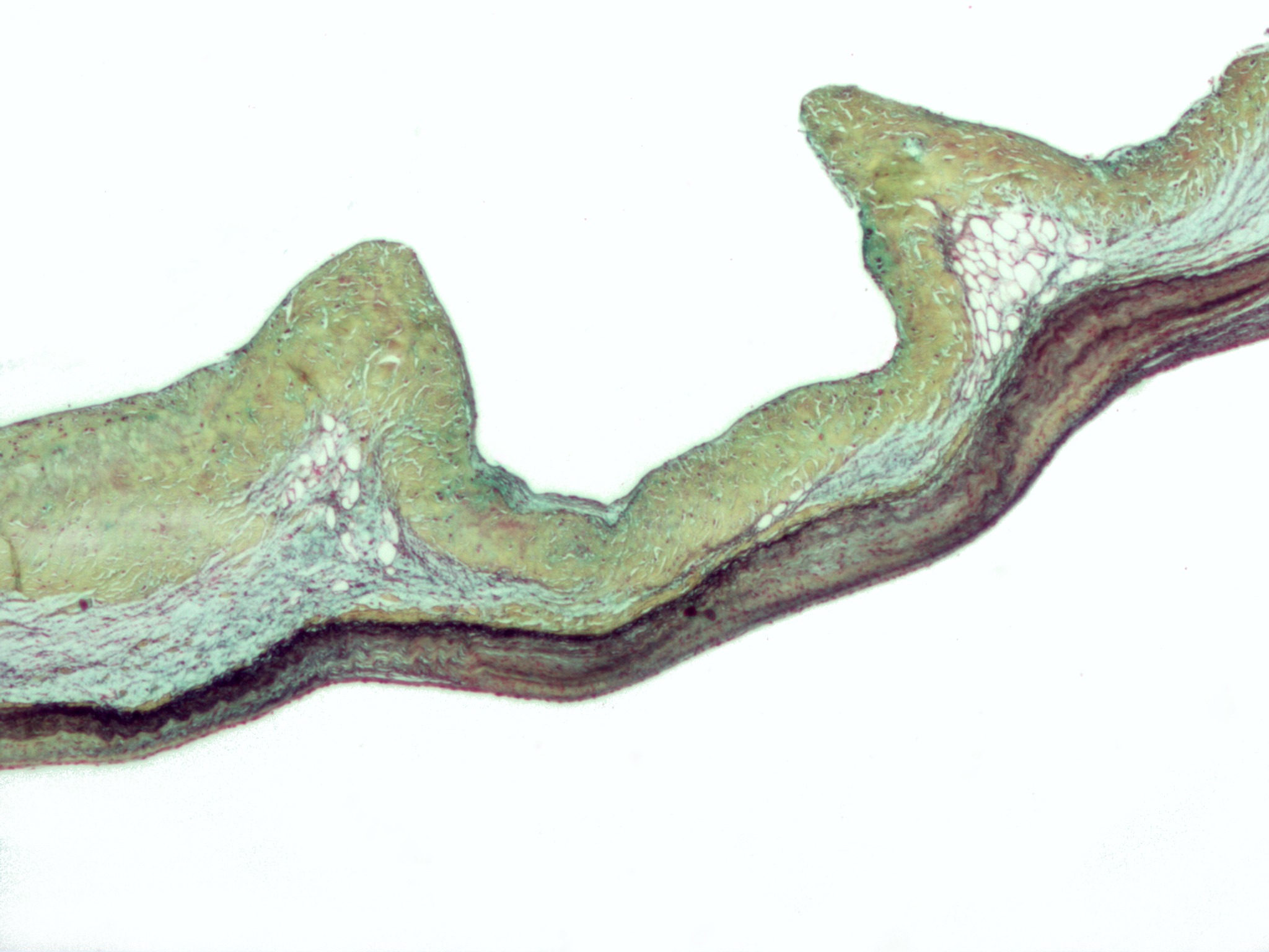
Mönckeberg's arteriosclerosis, or Mönckeberg's sclerosis, is a form of arteriosclerosis or vessel hardening, where calcium deposits are found in the muscular middle layer of the walls of arteries (the tunica media). It is an example of dystrophic calcification. This condition occurs as an age-related degenerative process. However, it can occur in pseudoxanthoma elasticum and idiopathic arterial calcification of infancy as a pathological condition, as well. Its clinical significance and cause are not well understood and its relationship to atherosclerosis and other forms of vascular calcification are the subject of disagreement. Mönckeberg's arteriosclerosis is named after Johann Georg Mönckeberg, who first described it in 1903. Signs and symptoms Typically, Mönckeberg's arteriosclerosis is not associated with symptoms unless complicated by atherosclerosis, calciphylaxis, or accompanied by some other disease. However, the presence of Mönckeberg's arteriosclerosis is associat ... [...More Info...] [...Related Items...] OR: [Wikipedia] [Google] [Baidu] |
Transesophageal Echocardiography
A transesophageal echocardiogram, or TEE (TOE in the United Kingdom and other countries such as Australia and New Zealand, reflecting the British English spelling ''transoesophageal''), is an alternative way to perform an echocardiogram. A specialized probe containing an ultrasound transducer at its tip is passed into the patient's esophagus. This allows image and Doppler evaluation which can be recorded. It is commonly used during cardiac surgery and is an excellent modality for assessing the aorta, although there are some limitations. It has several advantages and some disadvantages compared with a transthoracic echocardiogram (TTE). Details TEE is a semi-invasive procedure in that the probe must enter the body but does not require surgical (i.e., invasive) cutting for this procedure. Before inserting the probe, mild to moderate sedation is induced in the patient to ease the discomfort and to decrease the gag reflex. Usually a local anesthetic spray (e.g., lido ... [...More Info...] [...Related Items...] OR: [Wikipedia] [Google] [Baidu] |
Aortic Insufficiency
Aortic regurgitation (AR), also known as aortic insufficiency (AI), is the leaking of the aortic valve of the heart that causes blood to flow in the reverse direction during ventricular diastole, from the aorta into the left ventricle. As a consequence, the cardiac muscle is forced to work harder than normal. Signs and symptoms Symptoms of aortic regurgitation are similar to those of heart failure and include the following: * Dyspnea on exertion * Orthopnea * Paroxysmal nocturnal dyspnea * Palpitations * Angina pectoris * Cyanosis (in acute cases) Causes In terms of the cause of aortic regurgitation, is often due to the aortic root dilation ('' annuloaortic ectasia''), which is idiopathic in over 80% of cases, but otherwise may result from aging, syphilitic aortitis, osteogenesis imperfecta, aortic dissection, Behçet's disease, reactive arthritis and systemic hypertension.Chapter 1: Diseases of the Cardiovascular system > Section: Valvular Heart Disease in: Aortic root dilation ... [...More Info...] [...Related Items...] OR: [Wikipedia] [Google] [Baidu] |
Hemorrhage
Bleeding, hemorrhage, haemorrhage or blood loss, is blood escaping from the circulatory system from damaged blood vessels. Bleeding can occur internally, or externally either through a natural opening such as the mouth, nose, ear, urethra, vagina or anus, or through a puncture in the skin. Hypovolemia is a massive decrease in blood volume, and death by excessive loss of blood is referred to as exsanguination. Typically, a healthy person can endure a loss of 10–15% of the total blood volume without serious medical difficulties (by comparison, blood donation typically takes 8–10% of the donor's blood volume). The stopping or controlling of bleeding is called hemostasis and is an important part of both first aid and surgery. Types * Upper head ** Intracranial hemorrhage – bleeding in the skull. ** Cerebral hemorrhage – a type of intracranial hemorrhage, bleeding within the brain tissue itself. ** Intracerebral hemorrhage – bleeding in the brain caused by the ruptu ... [...More Info...] [...Related Items...] OR: [Wikipedia] [Google] [Baidu] |
Sensitivity And Specificity
''Sensitivity'' and ''specificity'' mathematically describe the accuracy of a test which reports the presence or absence of a condition. Individuals for which the condition is satisfied are considered "positive" and those for which it is not are considered "negative". *Sensitivity (true positive rate) refers to the probability of a positive test, conditioned on truly being positive. *Specificity (true negative rate) refers to the probability of a negative test, conditioned on truly being negative. If the true condition can not be known, a " gold standard test" is assumed to be correct. In a diagnostic test, sensitivity is a measure of how well a test can identify true positives and specificity is a measure of how well a test can identify true negatives. For all testing, both diagnostic and screening, there is usually a trade-off between sensitivity and specificity, such that higher sensitivities will mean lower specificities and vice versa. If the goal is to return the ratio at w ... [...More Info...] [...Related Items...] OR: [Wikipedia] [Google] [Baidu] |
Femoral Artery
The femoral artery is a large artery in the thigh and the main arterial supply to the thigh and leg. The femoral artery gives off the deep femoral artery or profunda femoris artery and descends along the anteromedial part of the thigh in the femoral triangle. It enters and passes through the adductor canal, and becomes the popliteal artery as it passes through the adductor hiatus in the adductor magnus near the junction of the middle and distal thirds of the thigh. Structure The femoral artery enters the thigh from behind the inguinal ligament as the continuation of the external iliac artery. Here, it lies midway between the anterior superior iliac spine and the symphysis pubis (Mid-inguinal point). Segments In clinical parlance, the femoral artery has the following segments: *The common femoral artery (CFA) is the segment of the femoral artery between the inferior margin of the inguinal ligament and the branching point of the deep femoral artery/profunda femoris artery. Its ... [...More Info...] [...Related Items...] OR: [Wikipedia] [Google] [Baidu] |
Common Iliac Artery
The common iliac artery is a large artery of the abdomen paired on each side. It originates from the aortic bifurcation at the level of the 4th lumbar vertebra. It ends in front of the sacroiliac joint, one on either side, and each bifurcates into the external and internal iliac arteries. Structure The common iliac artery are about 4 cm long in adults and more than a centimeter in diameter. It begins as a branch of the aorta. This is at the level of the 4th lumbar vertebra. It runs inferolaterally, along the medial border of the psoas muscles. It bifurcates into the external iliac artery and the internal iliac artery at the pelvic brim, in front of the sacroiliac joints. The common iliac artery, and all of its branches, exist as paired structures (that is to say, there is one on the left side and one on the right). The distribution of the common iliac artery is basically the pelvis and lower limb (as the femoral artery) on the corresponding side. Relations Both common il ... [...More Info...] [...Related Items...] OR: [Wikipedia] [Google] [Baidu] |
Catheter
In medicine, a catheter (/ˈkæθətər/) is a thin tube made from medical grade materials serving a broad range of functions. Catheters are medical devices that can be inserted in the body to treat diseases or perform a surgical procedure. Catheters are manufactured for specific applications, such as cardiovascular, urological, gastrointestinal, neurovascular and ophthalmic procedures. The process of inserting a catheter is ''catheterization''. In most uses, a catheter is a thin, flexible tube (''soft'' catheter) though catheters are available in varying levels of stiffness depending on the application. A catheter left inside the body, either temporarily or permanently, may be referred to as an "indwelling catheter" (for example, a peripherally inserted central catheter). A permanently inserted catheter may be referred to as a "permcath" (originally a trademark). Catheters can be inserted into a body cavity, duct, or vessel, brain, skin or adipose tissue. Functionally, they all ... [...More Info...] [...Related Items...] OR: [Wikipedia] [Google] [Baidu] |
Lumen (anatomy)
In biology, a lumen (plural lumina) is the inside space of a tubular structure, such as an artery or intestine. It comes . It can refer to: *The interior of a vessel, such as the central space in an artery, vein or capillary through which blood flows. *The interior of the gastrointestinal tract *The pathways of the bronchi in the lungs *The interior of renal tubules and urinary collecting ducts *The pathways of the female genital tract, starting with a single pathway of the vagina, splitting up in two lumina in the uterus, both of which continue through the Fallopian tubes In cell biology, a lumen is a membrane-defined space that is found inside several organelles, cellular components, or structures: *thylakoid, endoplasmic reticulum, Golgi apparatus, lysosome, mitochondrion, or microtubule Transluminal procedures ''Transluminal procedures'' are procedures occurring through lumina, including: *Natural orifice transluminal endoscopic surgery in the lumina of, for example, the ... [...More Info...] [...Related Items...] OR: [Wikipedia] [Google] [Baidu] |


