|
Anterior Ethmoidal Artery
The anterior ethmoidal artery is a branch of the ophthalmic artery in the orbit. It exits the orbit through the anterior ethmoidal foramen. The posterior ethmoidal artery is posterior to it. Structure The anterior ethmoidal artery branches from the ophthalmic artery distal to the posterior ethmoidal artery. It travels with the anterior ethmoidal nerve to exit the medial wall of the orbit at the anterior ethmoidal foramen. It then travels through the anterior ethmoidal canal and gives branches which supply the frontal sinus and anterior and middle ethmoid air cells. Following which, it enters the anterior cranial fossa where it bifurcates into a meningeal branch and nasal branch. The nasal branch travels through cribriform plate to enter the nasal cavity and runs in a groove on the deep surface of the nasal bone. Here it bifurcates into a medial and lateral branch. The lateral branch supplies blood to the lateral wall of the nasal cavity and the medial branch to the nasal septum. ... [...More Info...] [...Related Items...] OR: [Wikipedia] [Google] [Baidu] |
Ophthalmic Artery
The ophthalmic artery (OA) is an artery of the head. It is the first branch of the internal carotid artery distal to the cavernous sinus. Branches of the ophthalmic artery supply all the structures in the orbit around the eye, as well as some structures in the nose, face, and meninges. Occlusion of the ophthalmic artery or its branches can produce sight-threatening conditions. Structure The ophthalmic artery emerges from the internal carotid artery. This is usually just after the internal carotid artery emerges from the cavernous sinus. In some cases, the ophthalmic artery branches just before the internal carotid exits the cavernous sinus. The ophthalmic artery emerges along the medial side of the anterior clinoid process. It runs anteriorly, passing through the optic canal inferolaterally to the optic nerve. It can also pass superiorly to the optic nerve in a minority of cases. In the posterior third of the cone of the orbit, the ophthalmic artery turns sharply and medially t ... [...More Info...] [...Related Items...] OR: [Wikipedia] [Google] [Baidu] |
Ethmoidal Veins
The ethmoidal veins are the venae comitantes Vena comitans is Latin for accompanying vein. It refers to a vein that is usually paired, with both veins lying on the sides of an artery. They are found in close proximity to arteries so that the pulsations of the artery aid venous return. B ... of the ethmoidal arteries. Veins of the head and neck {{circulatory-stub ... [...More Info...] [...Related Items...] OR: [Wikipedia] [Google] [Baidu] |
Ethmoidal Cells
The ethmoid sinuses or ethmoid air cells of the ethmoid bone are one of the four paired paranasal sinuses. The cells are variable in both size and number in the lateral mass of each of the ethmoid bones and cannot be palpated during an extraoral examination. They are divided into anterior and posterior groups. The ethmoid air cells are numerous thin-walled cavities situated in the ethmoidal labyrinth and completed by the frontal, maxilla, lacrimal, sphenoidal, and palatine bones. They lie between the upper parts of the nasal cavities and the orbits, and are separated from these cavities by thin bony lamellae. Groups of sinuses The groups of the ethmoidal air cells drain into the nasal meatuses.Otorhinolaryngology, Head and Neck Surgery, Anniko, Springer, 2010, page 188 * The posterior group the ''posterior ethmoidal sinus'' drains into the superior meatus above the middle nasal concha; sometimes one or more opens into the sphenoidal sinus. * The anterior group the ''anterior ethmo ... [...More Info...] [...Related Items...] OR: [Wikipedia] [Google] [Baidu] |
Frontal Sinus
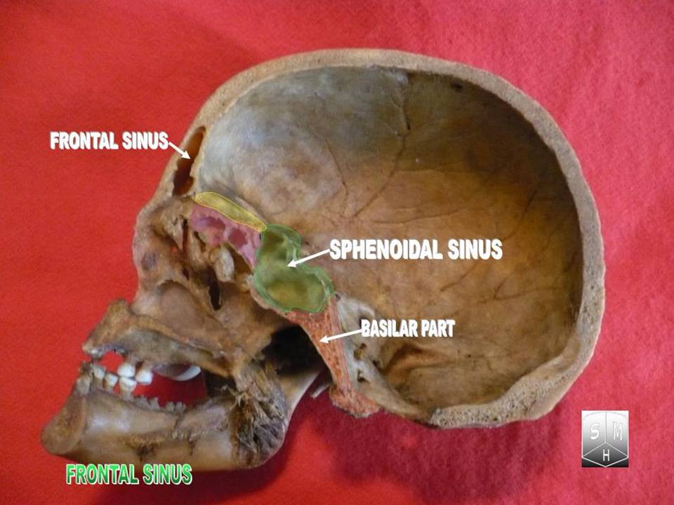
The frontal sinuses are one of the four pairs of paranasal sinuses that are situated behind the brow ridges. Sinuses are mucosa-lined airspaces within the bones of the face and skull. Each opens into the anterior part of the corresponding middle nasal meatus of the nose through the frontonasal duct which traverses the anterior part of the labyrinth of the ethmoid. These structures then open into the semilunar hiatus in the middle meatus. Structure Each frontal sinus is situated between the external and internal plates of the frontal bone.Frontal sinuses are rarely symmetrical. Their average measurements are as follows: height 28 mm, breadth 24 mm, depth 20 mm, creating a space of 6-7 ml. Blood supply The mucous membrane of the frontal sinuses receives arterial supply via the supraorbital artery, and anterior ethmoidal artery. Innervation The mucous membrane in this sinus is innervated by the supraorbital nerve, which contains the postganglionic parasympathetic ... [...More Info...] [...Related Items...] OR: [Wikipedia] [Google] [Baidu] |
Anterior Ethmoidal Foramen
The anterior ethmoidal foramen is a small opening in the ethmoid bone in the skull. Lateral to either olfactory groove are the internal openings of the anterior and posterior ethmoidal foramina (or canals). The anterior ethmoidal foramen, situated about the middle of the lateral margin of the olfactory groove, transmits the anterior ethmoidal artery, vein and nerve. The anterior ethmoidal nerve, a branch of the nasociliary nerve, runs in a groove along the lateral edge of the cribriform plate In mammalian anatomy, the cribriform plate (Latin for lit. ''sieve-shaped''), horizontal lamina or lamina cribrosa is part of the ethmoid bone. It is received into the ethmoidal notch of the frontal bone and roofs in the nasal cavities. It supp ... to the above-mentioned slit-like opening . References External links * () (#5) Foramina of the skull {{musculoskeletal-stub ... [...More Info...] [...Related Items...] OR: [Wikipedia] [Google] [Baidu] |
Posterior Ethmoidal Artery
The posterior ethmoidal artery is an artery of the head which supplies the nasal septum. It is smaller than the anterior ethmoidal artery. Course Once branching from the ophthalmic artery, it passes between the upper border of the medial rectus muscle and superior oblique muscle to enter the posterior ethmoidal canal. It exits into the nasal cavity to supply posterior ethmoidal cells and nasal septum; here it anastomoses with the sphenopalatine artery. There is often a meningeal branch to the dura mater, while it is still contained within the cranium. Supplies This artery supplies the posterior ethmoidal air sinuses, the dura mater of the anterior cranial fossa, and the upper part of the nasal mucosa of the nasal septum The nasal septum () separates the left and right airways of the Human nose, nasal cavity, dividing the two nostrils. It is Depression (kinesiology), depressed by the depressor septi nasi muscle. Structure The fleshy external end of the nasal .... Refer ... [...More Info...] [...Related Items...] OR: [Wikipedia] [Google] [Baidu] |
Anterior Ethmoidal Nerve
The anterior ethmoidal nerve is a nerve of the nose. It is a branch of the nasociliary nerve, itself a branch of the ophthalmic nerve (V1). It provides sensory innervation to some structures around the nasal cavity. Structure The anterior ethmoidal nerve is a terminal branch of the nasociliary nerve, a branch of the ophthalmic nerve (CN V1), itself a branch of the trigeminal nerve (CN V). It branches near the medial wall of the orbit. The anterior ethmoidal nerve arises only after the nasociliary has given off its four branches (the ramus communicans to the ciliary ganglion, the long ciliary nerves, the infratrochlear nerve, and the posterior ethmoidal nerve). It travels through the anterior ethmoidal foramen to reach the anterior cranial fossa. It then moves forward and passes through the cribriform plate to enter the nasal cavity. It gives off branches to the roof of the nasal cavity, and bifurcates into a lateral internal nasal branch and medial internal nasal branch. It send ... [...More Info...] [...Related Items...] OR: [Wikipedia] [Google] [Baidu] |
Ethmoid Sinus
The ethmoid sinuses or ethmoid air cells of the ethmoid bone are one of the four paired paranasal sinuses. The cells are variable in both size and number in the lateral mass of each of the ethmoid bones and cannot be palpated during an extraoral examination. They are divided into anterior and posterior groups. The ethmoid air cells are numerous thin-walled cavities situated in the ethmoidal labyrinth and completed by the frontal, maxilla, lacrimal, sphenoidal, and palatine bones. They lie between the upper parts of the nasal cavities and the orbits, and are separated from these cavities by thin bony lamellae. Groups of sinuses The groups of the ethmoidal air cells drain into the nasal meatuses.Otorhinolaryngology, Head and Neck Surgery, Anniko, Springer, 2010, page 188 * The posterior group the ''posterior ethmoidal sinus'' drains into the superior meatus above the middle nasal concha; sometimes one or more opens into the sphenoidal sinus. * The anterior group the ''anterior ethmo ... [...More Info...] [...Related Items...] OR: [Wikipedia] [Google] [Baidu] |
Anterior Cranial Fossa
The anterior cranial fossa is a depression in the floor of the cranial base which houses the projecting frontal lobes of the brain. It is formed by the orbital part of frontal bone, orbital plates of the Frontal bone, frontal, the cribriform plate of the ethmoid, and the small wings and front part of the body of the Sphenoid bone, sphenoid; it is limited behind by the posterior borders of the small wings of the sphenoid and by the anterior margin of the chiasmatic groove. The lesser wings of the sphenoid separate the anterior and middle cranial fossa, middle fossae. Structure It is traversed by the frontoethmoidal, sphenoethmoidal, and sphenofrontal sutures. Its lateral portions roof in the orbital cavities and support the frontal lobes of the cerebrum; they are convex and marked by depressions for the brain convolutions, and grooves for branches of the meningeal vessels. The central portion corresponds with the roof of the nasal cavity, and is markedly depressed on either side o ... [...More Info...] [...Related Items...] OR: [Wikipedia] [Google] [Baidu] |
Cribriform Plate
In mammalian anatomy, the cribriform plate (Latin for lit. ''sieve-shaped''), horizontal lamina or lamina cribrosa is part of the ethmoid bone. It is received into the ethmoidal notch of the frontal bone and roofs in the nasal cavities. It supports the olfactory bulb, and is perforated by olfactory foramina for the passage of the olfactory nerves to the roof of the nasal cavity to convey smell to the brain. The foramina at the medial part of the groove allow the passage of the nerves to the upper part of the nasal septum while the foramina at the lateral part transmit the nerves to the superior nasal concha. A fractured cribriform plate can result in olfactory dysfunction, septal hematoma, cerebrospinal fluid rhinorrhoea (CSF rhinorrhoea), and possibly infection which can lead to meningitis. CSF rhinorrhoea (clear fluid leaking from the nose) is very serious and considered a medical emergency. Aging can cause the openings in the cribriform plate to close, pinching olfactor ... [...More Info...] [...Related Items...] OR: [Wikipedia] [Google] [Baidu] |
Nasal Cavity
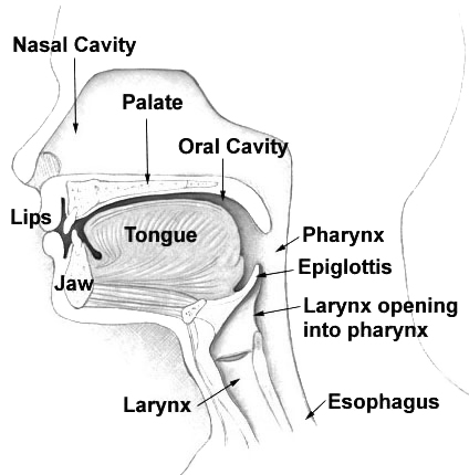
The nasal cavity is a large, air-filled space above and behind the nose in the middle of the face. The nasal septum divides the cavity into two cavities, also known as fossae. Each cavity is the continuation of one of the two nostrils. The nasal cavity is the uppermost part of the respiratory system and provides the nasal passage for inhaled air from the nostrils to the nasopharynx and rest of the respiratory tract. The paranasal sinuses surround and drain into the nasal cavity. Structure The term "nasal cavity" can refer to each of the two cavities of the nose, or to the two sides combined. The lateral wall of each nasal cavity mainly consists of the maxilla. However, there is a deficiency that is compensated for by the perpendicular plate of the palatine bone, the medial pterygoid plate, the labyrinth of ethmoid and the inferior concha. The paranasal sinuses are connected to the nasal cavity through small orifices called ostia. Most of these ostia communicate with the n ... [...More Info...] [...Related Items...] OR: [Wikipedia] [Google] [Baidu] |


