|
Angiodysplasia
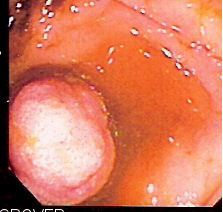
In medicine (gastroenterology), angiodysplasia is a small vascular malformation of the gut. It is a common cause of otherwise unexplained gastrointestinal bleeding and anemia. Lesions are often multiple, and frequently involve the cecum or ascending colon, although they can occur at other places. Treatment may be with colonoscopic interventions, angiography and embolization, medication, or occasionally surgery. Signs and symptoms Although some cases present with black, tarry stool (melena), the blood loss can be subtle, with the anemia symptoms predominating. Pathophysiology Histologically, it resembles telangiectasia and development is related to age and strain on the bowel wall. It is a degenerative lesion, acquired, probably resulting from chronic and intermittent contraction of the colon that is obstructing the venous drainage of the mucosa. As time goes by the veins become more and more tortuous, while the capillaries of the mucosa gradually dilate and precapillary sphinc ... [...More Info...] [...Related Items...] OR: [Wikipedia] [Google] [Baidu] |
Heyde's Syndrome
Heyde's syndrome is a syndrome of gastrointestinal bleeding from angiodysplasia in the presence of aortic stenosis. It is named after Edward C. Heyde, MD, who first noted the association in 1958. It is caused by the induction of Von Willebrand disease type IIA (vWD-2A) by a depletion of Von Willebrand factor (vWF) in blood flowing through the narrowed valvular stenosis. Signs and symptoms Pathophysiology Von Willebrand factor is synthesized in the walls of the blood vessels and circulates freely in the blood in a folded form. When it encounters damage to the wall of a blood vessel, particularly in situations of high velocity blood flow, it binds to the collagen beneath the damaged endothelium and uncoils into its active form. Platelets are attracted to this activated form of von Willebrand factor and they accumulate and block the damaged area, preventing bleeding . In people with aortic valve stenosis, the stenotic aortic valve becomes increasingly narrowed resulting in an ... [...More Info...] [...Related Items...] OR: [Wikipedia] [Google] [Baidu] |
Gut (zoology)
The gastrointestinal tract (GI tract, digestive tract, alimentary canal) is the tract or passageway of the digestive system that leads from the mouth to the anus. The GI tract contains all the major organs of the digestive system, in humans and other animals, including the esophagus, stomach, and intestines. Food taken in through the mouth is digested to extract nutrients and absorb energy, and the waste expelled at the anus as feces. ''Gastrointestinal'' is an adjective meaning of or pertaining to the stomach and intestines. Most animals have a "through-gut" or complete digestive tract. Exceptions are more primitive ones: sponges have small pores ( ostia) throughout their body for digestion and a larger dorsal pore (osculum) for excretion, comb jellies have both a ventral mouth and dorsal anal pores, while cnidarians and acoels have a single pore for both digestion and excretion. The human gastrointestinal tract consists of the esophagus, stomach, and intestines, and is divi ... [...More Info...] [...Related Items...] OR: [Wikipedia] [Google] [Baidu] |
Fecal Occult Blood
Fecal occult blood (FOB) refers to blood in the feces that is not visibly apparent (unlike other types of blood in stool such as melena or hematochezia). A fecal occult blood test (FOBT) checks for hidden (occult) blood in the stool (feces). The American College of Gastroenterology has recommended the abandoning of gFOBT testing as a colorectal cancer screening tool, in favor of the fecal immunochemical test (FIT). The newer and recommended tests look for globin, DNA, or other blood factors including transferrin, while conventional stool guaiac tests look for heme. Medical uses Fecal occult blood testing (FOBT), as its name implies, aims to detect subtle blood loss in the gastrointestinal tract, anywhere from the mouth to the colon. Positive tests ("positive stool") may result from either upper gastrointestinal bleeding or lower gastrointestinal bleeding and warrant further investigation for peptic ulcers or a malignancy (such as colorectal cancer or gastric cancer). The tes ... [...More Info...] [...Related Items...] OR: [Wikipedia] [Google] [Baidu] |
Endoscopy
An endoscopy is a procedure used in medicine to look inside the body. The endoscopy procedure uses an endoscope to examine the interior of a hollow organ or cavity of the body. Unlike many other medical imaging techniques, endoscopes are inserted directly into the organ. There are many types of endoscopies. Depending on the site in the body and type of procedure, an endoscopy may be performed by either a doctor or a surgeon. A patient may be fully conscious or anaesthetised during the procedure. Most often, the term ''endoscopy'' is used to refer to an examination of the upper part of the gastrointestinal tract, known as an esophagogastroduodenoscopy. For nonmedical use, similar instruments are called borescopes. History Adolf Kussmaul was fascinated by sword swallowers who would insert a sword down their throat without gagging. This drew inspiration to insert a camera, the next problem to solve was how to insert a source of light, as they were still relying on candles ... [...More Info...] [...Related Items...] OR: [Wikipedia] [Google] [Baidu] |
Coagulation
Coagulation, also known as clotting, is the process by which blood changes from a liquid to a gel, forming a blood clot. It potentially results in hemostasis, the cessation of blood loss from a damaged vessel, followed by repair. The mechanism of coagulation involves activation, adhesion and aggregation of platelets, as well as deposition and maturation of fibrin. Coagulation begins almost instantly after an injury to the endothelium lining a blood vessel. Exposure of blood to the subendothelial space initiates two processes: changes in platelets, and the exposure of subendothelial tissue factor to plasma factor VII, which ultimately leads to cross-linked fibrin formation. Platelets immediately form a plug at the site of injury; this is called ''primary hemostasis. Secondary hemostasis'' occurs simultaneously: additional coagulation (clotting) factors beyond factor VII ( listed below) respond in a cascade to form fibrin strands, which strengthen the platelet plug. Disorders ... [...More Info...] [...Related Items...] OR: [Wikipedia] [Google] [Baidu] |
Scintigraphy
Scintigraphy (from Latin ''scintilla'', "spark"), also known as a gamma scan, is a diagnostic test in nuclear medicine, where radioisotopes attached to drugs that travel to a specific organ or tissue (radiopharmaceuticals) are taken internally and the emitted gamma radiation is captured by external detectors ( gamma cameras) to form two-dimensional images in a similar process to the capture of x-ray images. In contrast, SPECT and '' positron emission tomography'' (PET) form 3-dimensional images and are therefore classified as separate techniques from scintigraphy, although they also use gamma cameras to detect internal radiation. Scintigraphy is unlike a diagnostic X-ray where external radiation is passed through the body to form an image. Process Scintillography is an imaging method of nuclear events provoked by collisions or charged current interactions among nuclear particles or ionizing radiation and atoms which result in a brief, localised pulse of electromagnetic radi ... [...More Info...] [...Related Items...] OR: [Wikipedia] [Google] [Baidu] |
Mesenteric Artery
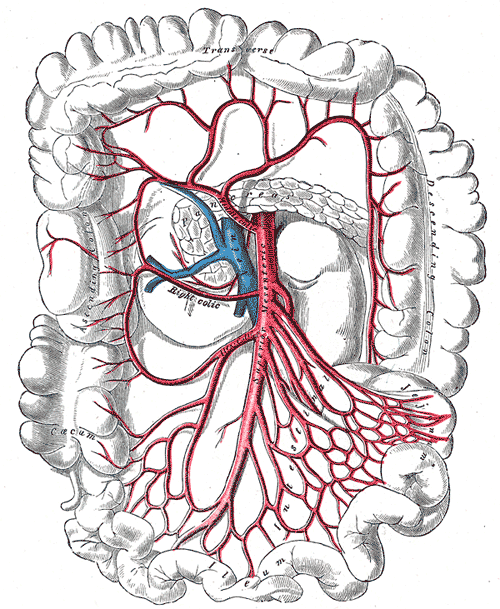
The mesenteric arteries take blood from the aorta and distribute it to a large portion of the gastrointestinal tract. Both the superior and inferior mesenteric arteries arise from the abdominal aorta. Each of these arteries travel through the mesentery, within which they branch several times before reaching the gut. In humans, many of these branches are named, but in smaller animals the branches are more variable. In some animals, including humans, branches of these arteries join with the marginal artery of the colon, which means that occlusion of one of the main arteries does not necessarily lead to the death of the part of the gut that it usually supplies. The term mesenteric artery is also used to describe smaller branches of these vessels which, particularly in smaller animals, provide a significant source of vascular resistance. These branches have a dense innervation by sympathetic nerves, allowing the brain to control their diameter and hence the resistance to blood ... [...More Info...] [...Related Items...] OR: [Wikipedia] [Google] [Baidu] |
Double-balloon Enteroscopy
Double-balloon enteroscopy, also known as push-and-pull enteroscopy, is an endoscopic technique for visualization of the small bowel. It was developed by Hironori Yamamoto in 2001. It is novel in the field of diagnostic gastroenterology as it is the first endoscopic technique that allows for the entire gastrointestinal tract to be visualized in real time. Technique The technique involves the use of a balloon at the end of a special enteroscope camera and an overtube, which is a tube that fits over the endoscope, and which is also fitted with a balloon. The procedure is usually done under general anesthesia, but may be done with the use of conscious sedation. The enteroscope and overtube are inserted through the mouth and passed in conventional fashion (that is, as with gastroscopy) into the small bowel. Following this, the endoscope is advanced a small distance in front of the overtube and the balloon at the end is inflated. Using the assistance of friction at the interfac ... [...More Info...] [...Related Items...] OR: [Wikipedia] [Google] [Baidu] |
Small Bowel
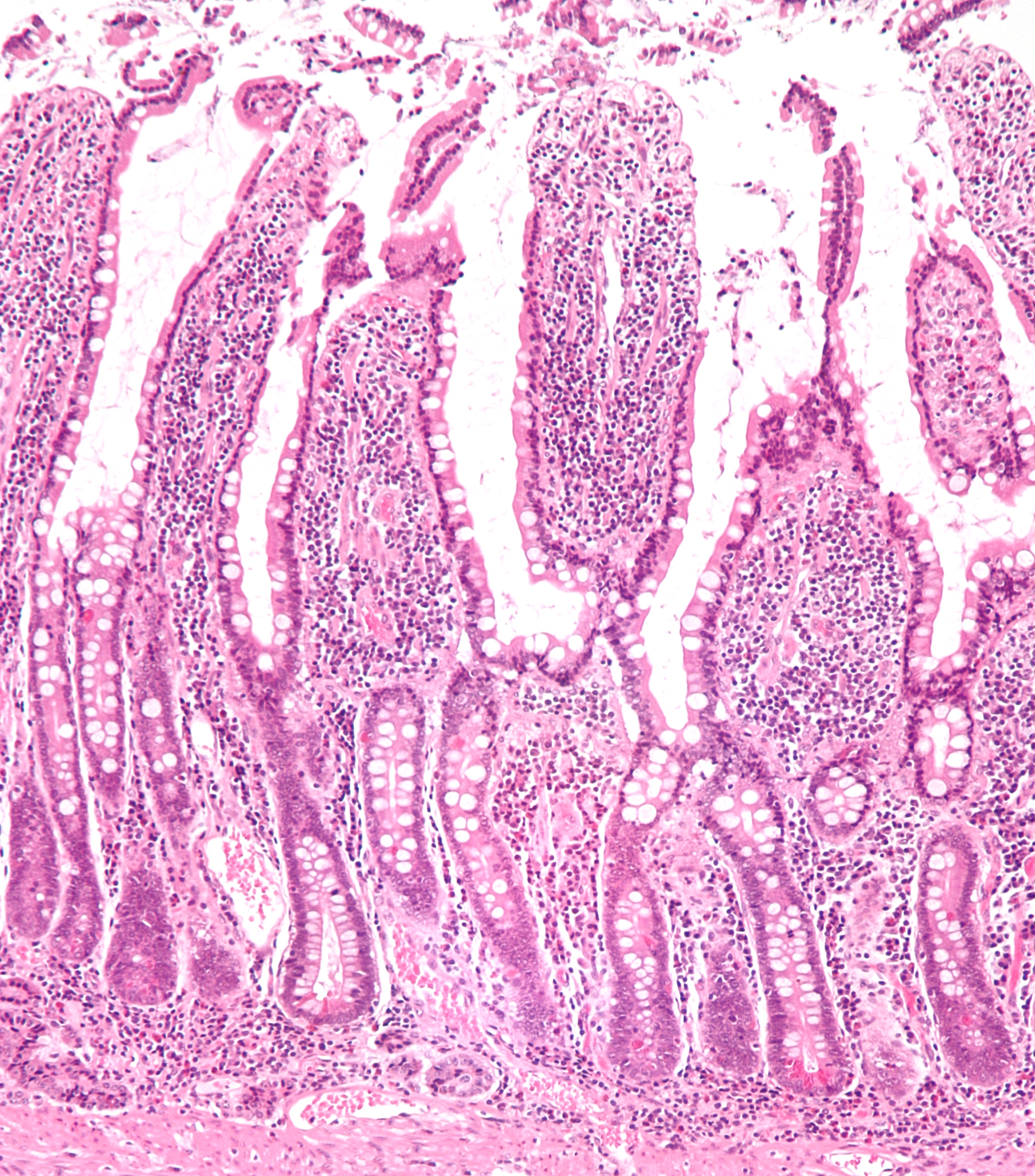
The small intestine or small bowel is an organ in the gastrointestinal tract where most of the absorption of nutrients from food takes place. It lies between the stomach and large intestine, and receives bile and pancreatic juice through the pancreatic duct to aid in digestion. The small intestine is about long and folds many times to fit in the abdomen. Although it is longer than the large intestine, it is called the small intestine because it is narrower in diameter. The small intestine has three distinct regions – the duodenum, jejunum, and ileum. The duodenum, the shortest, is where preparation for absorption through small finger-like protrusions called villi begins. The jejunum is specialized for the absorption through its lining by enterocytes: small nutrient particles which have been previously digested by enzymes in the duodenum. The main function of the ileum is to absorb vitamin B12, bile salts, and whatever products of digestion that were not absorbed by the je ... [...More Info...] [...Related Items...] OR: [Wikipedia] [Google] [Baidu] |
Wireless Capsule Endoscopy
Capsule endoscopy is a medical procedure used to record internal images of the gastrointestinal tract for use in disease diagnosis. Newer developments are also able to take biopsies and release medication at specific locations of the entire gastrointestinal tract. Unlike the more widely used endoscope, capsule endoscopy provides the ability to see the middle portion of the small intestine. It can be applied to the detection of various gastrointestinal cancers, digestive diseases, ulcers, unexplained bleedings, and general abdominal pains. After a patient swallows the capsule, it passes along the gastrointestinal tract, taking a number of images per second which are transmitted wirelessly to an array of receivers connected to a portable recording device carried by the patient. General advantages of capsule endoscopy over standard endoscopy include the minimally invasive procedure setup, ability to visualize more of the gastrointestinal tract, and lower cost of the procedure. ... [...More Info...] [...Related Items...] OR: [Wikipedia] [Google] [Baidu] |
Esophagogastroduodenoscopy
Esophagogastroduodenoscopy (EGD) or oesophagogastroduodenoscopy (OGD), also called by various other names, is a diagnostic endoscopic procedure that visualizes the upper part of the gastrointestinal tract down to the duodenum. It is considered a minimally invasive procedure since it does not require an incision into one of the major body cavities and does not require any significant recovery after the procedure (unless sedation or anesthesia has been used). However, a sore throat is common. Alternative names The words ''esophagogastroduodenoscopy'' (EGD; American English) and ''oesophagogastroduodenoscopy'' (OGD; British English; see spelling differences) are both pronounced . It is also called ''panendoscopy'' (PES) and ''upper GI endoscopy''. It is also often called just ''upper endoscopy'', ''upper GI'', or even just ''endoscopy''; because EGD is the most commonly performed type of endoscopy, the ambiguous term ''endoscopy'' is sometimes informally used to refer to EGD by ... [...More Info...] [...Related Items...] OR: [Wikipedia] [Google] [Baidu] |
Colonoscopy
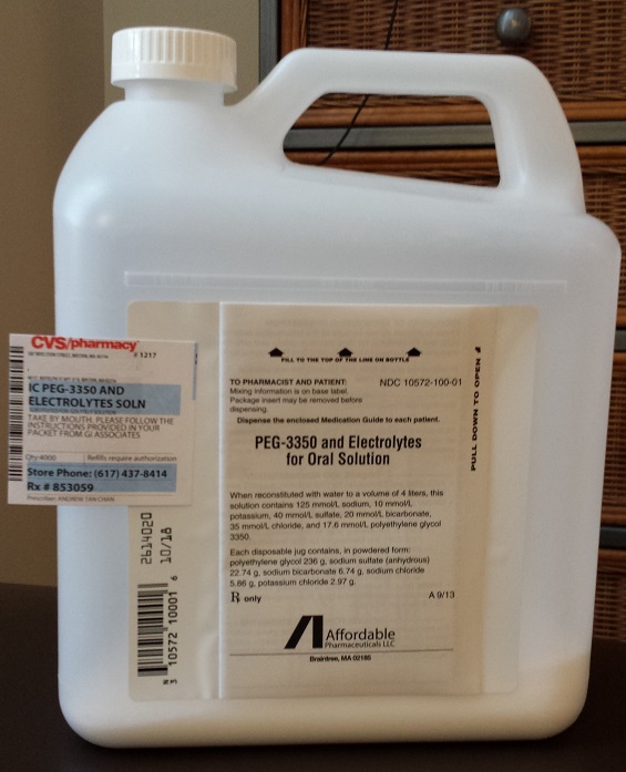
Colonoscopy () or coloscopy () is the endoscopic examination of the large bowel and the distal part of the small bowel with a CCD camera or a fiber optic camera on a flexible tube passed through the anus. It can provide a visual diagnosis (''e.g.,'' ulceration, polyps) and grants the opportunity for biopsy or removal of suspected colorectal cancer lesions. Colonoscopy can remove polyps smaller than one millimeter. Once polyps are removed, they can be studied with the aid of a microscope to determine if they are precancerous or not. Colonoscopy is similar to sigmoidoscopy—the difference being related to which parts of the colon each can examine. A colonoscopy allows an examination of the entire colon (1,200–1,500mm in length). A sigmoidoscopy allows an examination of the distal portion (about 600mm) of the colon, which may be sufficient because benefits to cancer survival of colonoscopy have been limited to the detection of lesions in the distal portion of the colon.< ... [...More Info...] [...Related Items...] OR: [Wikipedia] [Google] [Baidu] |


