|
Anconeus
The anconeus muscle (or anconaeus/anconæus) is a small muscle on the posterior aspect of the elbow joint. Some consider anconeus to be a continuation of the triceps brachii muscle. Some sources consider it to be part of the posterior compartment of the arm, while others consider it part of the posterior compartment of the forearm. The anconeus muscle can easily be palpated just lateral to the olecranon process of the ulna. Structure Anconeus originates on the posterior surface of the lateral epicondyle of the humerus and inserts distally on the superior posterior surface of the ulna and the lateral aspect of the olecranon. Innervation Anconeus is innervated by a branch of the radial nerve (cervical roots 7 and 8) from the posterior cord of the brachial plexus called the nerve to the anconeus. The somatomotor portion of radial nerve innervating anconeus bifurcates from the main branch in the radial groove of the humerus. This innervation pattern follows the rules of innervation ... [...More Info...] [...Related Items...] OR: [Wikipedia] [Google] [Baidu] |
Elbow-joint
The elbow is the region between the arm and the forearm that surrounds the elbow joint. The elbow includes prominent landmarks such as the olecranon, the cubital fossa (also called the chelidon, or the elbow pit), and the lateral and the medial epicondyles of the humerus. The elbow joint is a hinge joint between the arm and the forearm; more specifically between the humerus in the upper arm and the radius and ulna in the forearm which allows the forearm and hand to be moved towards and away from the body. The term ''elbow'' is specifically used for humans and other primates, and in other vertebrates forelimb plus joint is used. The name for the elbow in Latin is ''cubitus'', and so the word cubital is used in some elbow-related terms, as in ''cubital nodes'' for example. Structure Joint The elbow joint has three different portions surrounded by a common joint capsule. These are joints between the three bones of the elbow, the humerus of the upper arm, and the radius and th ... [...More Info...] [...Related Items...] OR: [Wikipedia] [Google] [Baidu] |
Ulna
The ulna (''pl''. ulnae or ulnas) is a long bone found in the forearm that stretches from the elbow to the smallest finger, and when in anatomical position, is found on the medial side of the forearm. That is, the ulna is on the same side of the forearm as the little finger. It runs parallel to the radius, the other long bone in the forearm. The ulna is usually slightly longer than the radius, but the radius is thicker. Therefore, the radius is considered to be the larger of the two. Structure The ulna is a long bone found in the forearm that stretches from the elbow to the smallest finger, and when in anatomical position, is found on the medial side of the forearm. It is broader close to the elbow, and narrows as it approaches the wrist. Close to the elbow, the ulna has a bony process, the olecranon process, a hook-like structure that fits into the olecranon fossa of the humerus. This prevents hyperextension and forms a hinge joint with the trochlea of the humerus. There is ... [...More Info...] [...Related Items...] OR: [Wikipedia] [Google] [Baidu] |
Triceps Brachii Muscle
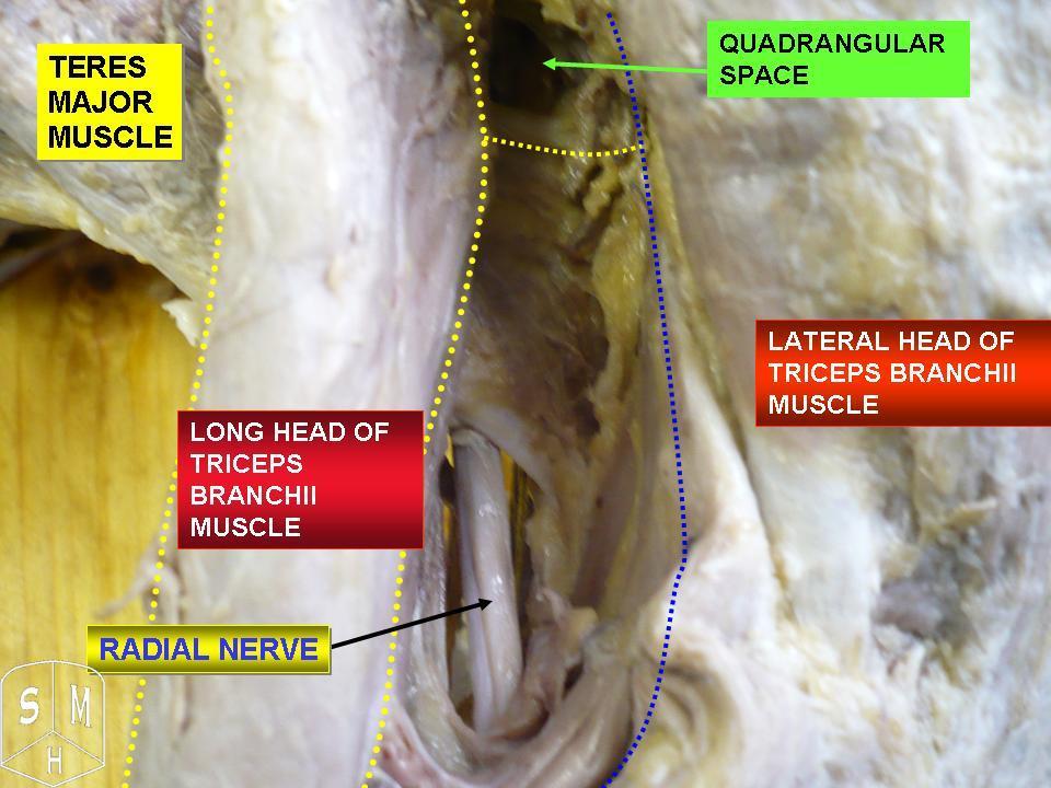
The triceps, or triceps brachii (Latin for "three-headed muscle of the arm"), is a large muscle on the back of the upper limb of many vertebrates. It consists of 3 parts: the medial, lateral, and long head. It is the muscle principally responsible for extension of the elbow joint (straightening of the arm). Structure The long head arises from the infraglenoid tubercle of the scapula. It extends distally anterior to the teres minor and posterior to the teres major. The medial head arises proximally in the humerus, just inferior to the groove of the radial nerve; from the dorsal (back) surface of the humerus; from the medial intermuscular septum; and its distal part also arises from the lateral intermuscular septum. The medial head is mostly covered by the lateral and long heads, and is only visible distally on the humerus. The lateral head arises from the dorsal surface of the humerus, lateral and proximal to the groove of the radial nerve, from the greater tubercle down to ... [...More Info...] [...Related Items...] OR: [Wikipedia] [Google] [Baidu] |
Triceps
The triceps, or triceps brachii (Latin for "three-headed muscle of the arm"), is a large muscle on the back of the upper limb of many vertebrates. It consists of 3 parts: the medial, lateral, and long head. It is the muscle principally responsible for extension of the elbow joint (straightening of the arm). Structure The long head arises from the infraglenoid tubercle of the scapula. It extends distally anterior to the teres minor and posterior to the teres major. The medial head arises proximally in the humerus, just inferior to the groove of the radial nerve; from the dorsal (back) surface of the humerus; from the medial intermuscular septum; and its distal part also arises from the lateral intermuscular septum. The medial head is mostly covered by the lateral and long heads, and is only visible distally on the humerus. The lateral head arises from the dorsal surface of the humerus, lateral and proximal to the groove of the radial nerve, from the greater tubercle down to ... [...More Info...] [...Related Items...] OR: [Wikipedia] [Google] [Baidu] |
Radial Nerve
The radial nerve is a nerve in the human body that supplies the posterior portion of the upper limb. It innervates the medial and lateral heads of the triceps brachii muscle of the arm, as well as all 12 muscles in the posterior osteofascial compartment of the forearm and the associated joints and overlying skin. It originates from the brachial plexus, carrying fibers from the ventral roots of spinal nerves C5, C6, C7, C8 & T1. The radial nerve and its branches provide motor innervation to the dorsal arm muscles (the triceps brachii and the anconeus) and the extrinsic extensors of the wrists and hands; it also provides cutaneous sensory innervation to most of the back of the hand, except for the back of the little finger and adjacent half of the ring finger (which are innervated by the ulnar nerve). The radial nerve divides into a deep branch, which becomes the posterior interosseous nerve, and a superficial branch, which goes on to innervate the dorsum (back) of the hand. Th ... [...More Info...] [...Related Items...] OR: [Wikipedia] [Google] [Baidu] |
Posterior Compartment Of The Forearm
The posterior compartment of the forearm (or extensor compartment) contains twelve muscles which primarily extend the wrist and digits. It is separated from the anterior compartment by the interosseous membrane between the radius and ulna. Structure Muscles There are generally twelve muscles in the posterior compartment of the forearm, which can be further divided into superficial, intermediate, and deep. Most of the muscles in the superficial and the intermediate layers share a common origin which is the outer part of the elbow, the lateral epicondyle of humerus. The deep muscles arise from the distal part of the ulna and the surrounding interosseous membrane. The brachioradialis, flexor of the elbow, is unusual in that it is located in the posterior compartment, but it is actually a muscle of flexor / anterior compartment of the forearm. The anconeus, assisting in extension of the elbow joint, is by some considered part of the posterior compartment of the arm. The majority o ... [...More Info...] [...Related Items...] OR: [Wikipedia] [Google] [Baidu] |
Profunda Brachii Artery
The deep artery of arm (also known as arteria profunda brachii and the deep brachial artery) is a large vessel which arises from the lateral and posterior part of the brachial artery, just below the lower border of the teres major. Structure It follows closely the radial nerve, running at first backward between the long and medial heads of the triceps brachii, then along the groove for the radial nerve (the radial sulcus), where it is covered by the lateral head of the triceps brachii, to the lateral side of the arm; there it pierces the lateral intermuscular septum, and, descending between the brachioradialis and the brachialis to the front of the lateral epicondyle of the humerus, ends by anastomosing with the radial recurrent artery. Branches and anastomoses It gives branches to the deltoid muscle (which, however, primarily is supplied by the posterior circumflex humeral artery) and to the muscles between which it lies; it supplies an occasional nutrient artery which enters ... [...More Info...] [...Related Items...] OR: [Wikipedia] [Google] [Baidu] |
Brachial Plexus
The brachial plexus is a network () of nerves formed by the anterior rami of the lower four cervical nerves and first thoracic nerve ( C5, C6, C7, C8, and T1). This plexus extends from the spinal cord, through the cervicoaxillary canal in the neck, over the first rib, and into the armpit, it supplies afferent and efferent nerve fibers the to chest, shoulder, arm, forearm, and hand. Structure The brachial plexus is divided into five ''roots'', three ''trunks'', six ''divisions'' (three anterior and three posterior), three ''cords'', and five ''branches''. There are five "terminal" branches and numerous other "pre-terminal" or "collateral" branches, such as the subscapular nerve, the thoracodorsal nerve, and the long thoracic nerve, that leave the plexus at various points along its length. A common structure used to identify part of the brachial plexus in cadaver dissections is the M or W shape made by the musculocutaneous nerve, lateral cord, median nerve, medial cord, and ... [...More Info...] [...Related Items...] OR: [Wikipedia] [Google] [Baidu] |
Olecranon Fossa
The olecranon fossa is a deep triangular depression on the posterior side of the humerus, superior to the trochlea. It provides space for the olecranon of the ulna during extension of the forearm. Structure The olecranon fossa is located on the posterior side of the distal humerus. The joint capsule of the elbow attaches to the humerus just proximal to the olecranon fossa. Function The olecranon fossa provides space for the olecranon of the ulna during extension of the forearm, from which it gets its name. Other animals The olecranon fossa is present in various mammals, including dogs The dog (''Canis familiaris'' or ''Canis lupus familiaris'') is a domesticated descendant of the wolf. Also called the domestic dog, it is derived from the extinct Pleistocene wolf, and the modern wolf is the dog's nearest living relative. Do .... Additional images File:Slide1bgbg.JPG, Elbow joint. Deep dissection. Posterior view. File:Slide2bgbg.JPG, Elbow joint. Deep dissection. ... [...More Info...] [...Related Items...] OR: [Wikipedia] [Google] [Baidu] |
Middle Collateral Artery
The medial collateral artery (also known as the middle collateral artery) is a branch of profunda brachii artery that descends in the middle head of the triceps brachii and assists in forming the anastomosis with the interosseous recurrent artery above the olecranon of the ulna near the elbow. See also * radial collateral artery * superior ulnar collateral artery * inferior ulnar collateral artery The inferior ulnar collateral artery (anastomotica magna artery) is an artery in the arm. It arises about 5 cm. above the elbow from the brachial artery. Course It passes medialward upon the Brachialis, and piercing the medial intermuscular ... External links Arteries of the upper limb {{circulatory-stub ... [...More Info...] [...Related Items...] OR: [Wikipedia] [Google] [Baidu] |
Nomina Anatomica
''Nomina Anatomica'' (''NA'') was the international standard on human anatomic terminology from 1895 until it was replaced by ''Terminologia Anatomica'' in 1998. In the late nineteenth century some 30,000 terms for various body parts were in use. The same structures were described by different names, depending (among other things) on the anatomist's school and national tradition. Vernacular translations of Latin and Greek, as well as various eponymous terms, were barriers to effective international communication. There was disagreement and confusion among anatomists regarding anatomical terminology. Editions The first and last entries in the following table are not NA editions, but they are included for the sake of continuity. Although these early editions were authorized by different bodies, they are sometimes considered part of the same series. Before these codes of terminology, approved at anatomists congresses, the usage of anatomical terms was based on authoritative work ... [...More Info...] [...Related Items...] OR: [Wikipedia] [Google] [Baidu] |
Heterotopic Ossification
Heterotopic ossification (HO) is the process by which bone tissue forms outside of the skeleton in muscles and soft tissue. Symptoms In traumatic heterotopic ossification (traumatic myositis ossificans), the patient may complain of a warm, tender, firm swelling in a muscle and decreased range of motion in the joint served by the muscle involved. There is often a history of a blow or other trauma to the area a few weeks to a few months earlier. Patients with traumatic neurological injuries, severe neurologic disorders or severe burns who develop heterotopic ossification experience limitation of motion in the areas affected. Causes Heterotopic ossification of varying severity can be caused by surgery or trauma to the hips and legs. About every third patient who has total hip arthroplasty (joint replacement) or a severe fracture of the long bones of the lower leg will develop heterotopic ossification, but is uncommonly symptomatic. Between 50% and 90% of patients who developed ... [...More Info...] [...Related Items...] OR: [Wikipedia] [Google] [Baidu] |


