|
Adaptation (eye)
In visual physiology, adaptation is the ability of the retina of the eye to adjust to various levels of light. Natural night vision, or scotopic vision, is the ability to see under low-light conditions. In humans, rod cells are exclusively responsible for night vision as cone cells are only able to function at higher illumination levels. Night vision is of lower quality than day vision because it is limited in resolution and colors cannot be discerned; only shades of gray are seen. In order for humans to transition from day to night vision they must undergo a dark adaptation period of up to two hours in which each eye adjusts from a high to a low luminescence "setting", increasing sensitivity hugely, by many orders of magnitude. This adaptation period is different between rod and cone cells and results from the regeneration of photopigments to increase retinal sensitivity. Light adaptation, in contrast, works very quickly, within seconds. Efficiency The human eye can functi ... [...More Info...] [...Related Items...] OR: [Wikipedia] [Google] [Baidu] |
Visual Perception
Visual perception is the ability to interpret the surrounding Biophysical environment, environment through photopic vision (daytime vision), color vision, scotopic vision (night vision), and mesopic vision (twilight vision), using light in the visible spectrum reflected by objects in the environment. This is different from visual acuity, which refers to how clearly a person sees (for example "20/20 vision"). A person can have problems with visual perceptual processing even if they have 20/20 vision. The resulting perception is also known as vision, sight, or eyesight (adjectives ''visual'', ''optical'', and ''ocular'', respectively). The various physiological components involved in vision are referred to collectively as the visual system, and are the focus of much research in linguistics, psychology, cognitive science, neuroscience, and molecular biology, collectively referred to as vision science. Visual system In humans and a number of other mammals, light enters the eye t ... [...More Info...] [...Related Items...] OR: [Wikipedia] [Google] [Baidu] |
Fovea Centralis
The fovea centralis is a small, central pit composed of closely packed cones in the eye. It is located in the center of the macula lutea of the retina. The fovea is responsible for sharp central vision (also called foveal vision), which is necessary in humans for activities for which visual detail is of primary importance, such as reading and driving. The fovea is surrounded by the ''parafovea'' belt and the ''perifovea'' outer region. The parafovea is the intermediate belt, where the ganglion cell layer is composed of more than five layers of cells, as well as the highest density of cones; the perifovea is the outermost region where the ganglion cell layer contains two to four layers of cells, and is where visual acuity is below the optimum. The perifovea contains an even more diminished density of cones, having 12 per 100 micrometres versus 50 per 100 micrometres in the most central fovea. That, in turn, is surrounded by a larger peripheral area, which delivers highly comp ... [...More Info...] [...Related Items...] OR: [Wikipedia] [Google] [Baidu] |
Visual Phototransduction
Visual phototransduction is the sensory transduction process of the visual system by which light is detected to yield nerve impulses in the rod cells and cone cells in the retina of the eye in humans and other vertebrates. It relies on the visual cycle, a sequence of biochemical reactions in which a molecule of retinal bound to opsin undergoes photoisomerization, initiates a cascade that signals detection of the photon, and is indirectly restored to its photosensitive isomer for reuse. Phototransduction in some invertebrates such as fruit flies relies on similar processes. Photoreceptors The photoreceptor cells involved in vertebrate vision are the rods, the cones, and the photosensitive ganglion cells (ipRGCs). These cells contain a chromophore ( 11-''cis''-retinal, the aldehyde of vitamin A1 and light-absorbing portion) that is bound to a cell membrane protein, opsin. Rods deal with low light level and do not mediate color vision. Cones, on the other hand, can code the c ... [...More Info...] [...Related Items...] OR: [Wikipedia] [Google] [Baidu] |
Photobleaching
In optics, photobleaching (sometimes termed fading) is the photochemical alteration of a dye or a fluorophore molecule such that it is permanently unable to fluoresce. This is caused by cleaving of covalent bonds or non-specific reactions between the fluorophore and surrounding molecules. Such irreversible modifications in covalent bonds are caused by transition from a singlet state to the triplet state of the fluorophores. The number of excitation cycles to achieve full bleaching varies. In microscopy, photobleaching may complicate the observation of fluorescent molecules, since they will eventually be destroyed by the light exposure necessary to stimulate them into fluorescing. This is especially problematic in time-lapse microscopy. However, photobleaching may also be used prior to applying the (primarily antibody-linked) fluorescent molecules, in an attempt to quench autofluorescence. This can help improve the signal-to-noise ratio. Photobleaching may also be exploited to ... [...More Info...] [...Related Items...] OR: [Wikipedia] [Google] [Baidu] |
Rhodopsin
Rhodopsin, also known as visual purple, is a protein encoded by the RHO gene and a G-protein-coupled receptor (GPCR). It is the opsin of the rod cells in the retina and a light-sensitive receptor protein that triggers visual phototransduction in rods. Rhodopsin mediates dim light vision and thus is extremely sensitive to light. When rhodopsin is exposed to light, it immediately photobleaches. In humans, it is regenerated fully in about 30 minutes, after which the rods are more sensitive. Defects in the rhodopsin gene cause eye diseases such as retinitis pigmentosa and congenital stationary night blindness. Names Rhodopsin was discovered by Franz Christian Boll in 1876. The name rhodospsin derives from Ancient Greek () for "rose", due to its pinkish color, and () for "sight". It was coined in 1878 by the German physiologist Wilhelm Friedrich Kühne (1837-1900). When George Wald discovered that rhodopsin is a holoprotein, consisting of retinal and an apoprotein, ... [...More Info...] [...Related Items...] OR: [Wikipedia] [Google] [Baidu] |
Photoreceptor Cell
A photoreceptor cell is a specialized type of neuroepithelial cell found in the retina that is capable of visual phototransduction. The great biological importance of photoreceptors is that they convert light (visible electromagnetic radiation) into signals that can stimulate biological processes. To be more specific, photoreceptor proteins in the cell absorb photons, triggering a change in the cell's membrane potential. There are currently three known types of photoreceptor cells in mammalian eyes: rods, cones, and intrinsically photosensitive retinal ganglion cells. The two classic photoreceptor cells are rods and cones, each contributing information used by the visual system to form an image of the environment, sight. Rods primarily mediate scotopic vision (dim conditions) whereas cones primarily mediate to photopic vision (bright conditions), but the processes in each that supports phototransduction is similar. A third class of mammalian photoreceptor cell was di ... [...More Info...] [...Related Items...] OR: [Wikipedia] [Google] [Baidu] |
Tapetum Lucidum
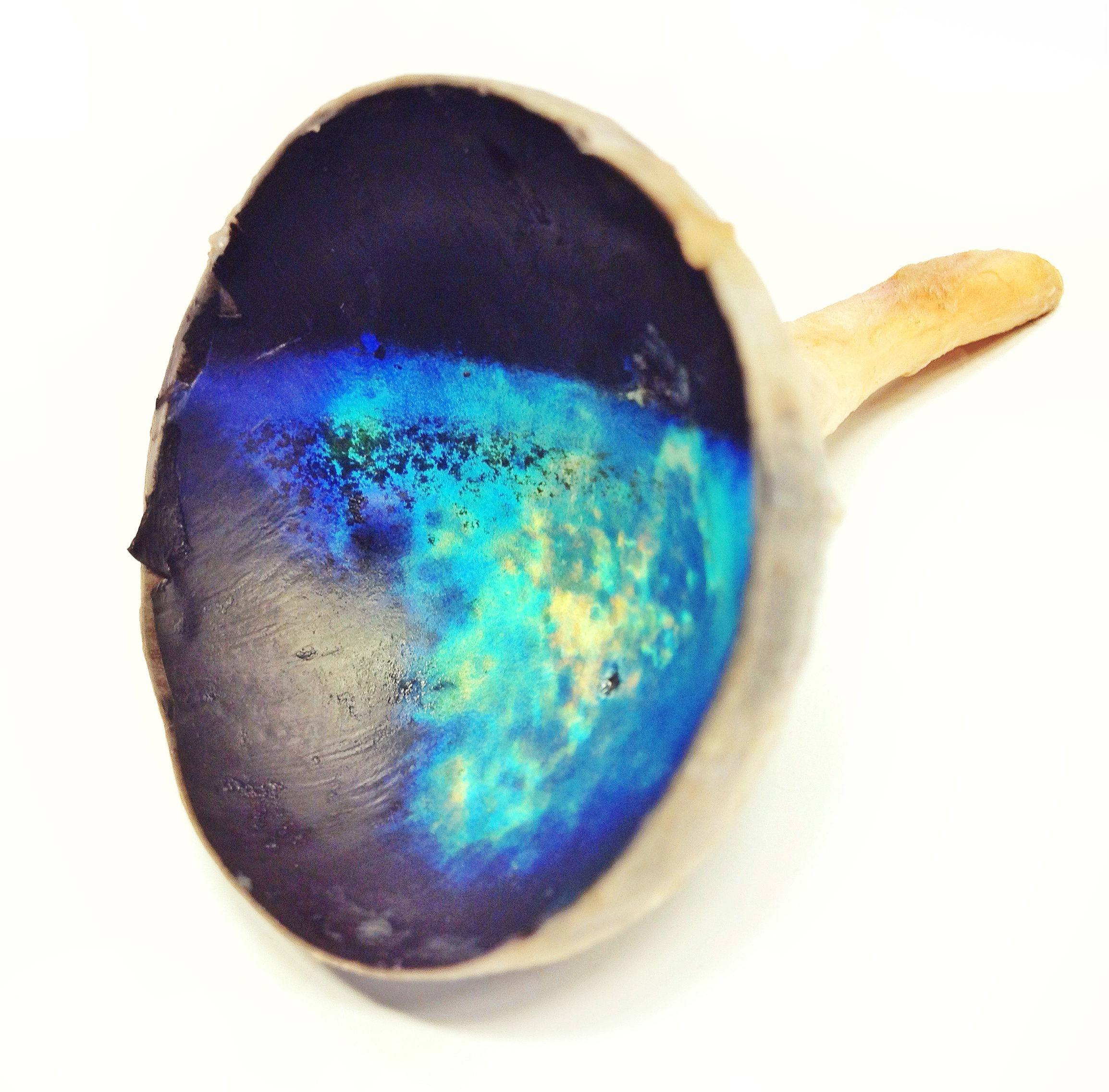
The ''tapetum lucidum'' ( ; ; ) is a layer of tissue in the eye of many vertebrates and some other animals. Lying immediately behind the retina, it is a retroreflector. It reflects visible light back through the retina, increasing the light available to the photoreceptors (although slightly blurring the image). The tapetum lucidum contributes to the superior night vision of some animals. Many of these animals are nocturnal, especially carnivores, while others are deep sea animals. Similar adaptations occur in some species of spiders. Haplorhine primates, including humans, are diurnal and lack a ''tapetum lucidum''. Function and mechanism Presence of a ''tapetum lucidum'' enables animals to see in dimmer light than would otherwise be possible. The ''tapetum lucidum'', which is iridescent, reflects light roughly on the interference principles of thin-film optics, as seen in other iridescent tissues. However, the ''tapetum lucidum'' cells are leucophores, not iridophores ... [...More Info...] [...Related Items...] OR: [Wikipedia] [Google] [Baidu] |
Mesopic Vision
Mesopic vision, sometimes also called twilight vision, is a combination of photopic and scotopic vision under low-light (but not necessarily dark) conditions. Mesopic levels range approximately from 0.01 to 3.0 cd/m2 in luminance. Most nighttime outdoor and street lighting conditions are in the mesopic range. Human eyes respond to certain light levels differently. This is because under high light levels typical during daytime (photopic vision), the eye uses cones to process light. Under very low light levels, corresponding to moonless nights without artificial lighting (scotopic vision), the eye uses rods to process light. At many nighttime levels, a combination of both cones and rods supports vision. Photopic vision facilitates excellent color perception, whereas colors are barely perceptible under scotopic vision. Mesopic vision falls between these two extremes. In most nighttime environments, enough ambient light prevents true scotopic vision. In the words of Duco ... [...More Info...] [...Related Items...] OR: [Wikipedia] [Google] [Baidu] |
Photopic Vision
Photopic vision is the vision of the eye under well-lit conditions (luminance levels from 10 to 108 cd/m2). In humans and many other animals, photopic vision allows color perception, mediated by cone cells, and a significantly higher visual acuity and temporal resolution than available with scotopic vision. The human eye uses three types of cones to sense light in three bands of color. The biological pigments of the cones have maximum absorption values at wavelengths of about 420 nm (blue), 534 nm (bluish-green), and 564 nm (yellowish-green). Their sensitivity ranges overlap to provide vision throughout the visible spectrum. The maximum efficacy is 683 lm/W at a wavelength of 555 nm (green). By definition, light at a frequency of hertz has a luminous efficacy of 683 lm/W. The wavelengths for when a person is in photopic vary with the intensity of light. For the blue-green region (500 nm), 50% of the light reaches the image point of the retina ... [...More Info...] [...Related Items...] OR: [Wikipedia] [Google] [Baidu] |
Luminance
Luminance is a photometric measure of the luminous intensity per unit area of light travelling in a given direction. It describes the amount of light that passes through, is emitted from, or is reflected from a particular area, and falls within a given solid angle. Brightness is the term for the ''subjective'' impression of the ''objective'' luminance measurement standard (see for the importance of this contrast). The SI unit for luminance is candela per square metre (cd/m2). A non-SI term for the same unit is the nit. The unit in the Centimetre–gram–second system of units (CGS) (which predated the SI system) is the stilb, which is equal to one candela per square centimetre or 10 kcd/m2. Description Luminance is often used to characterize emission or reflection from flat, diffuse surfaces. Luminance levels indicate how much luminous power could be detected by the human eye looking at a particular surface from a particular angle of view. Luminance is thus ... [...More Info...] [...Related Items...] OR: [Wikipedia] [Google] [Baidu] |


