|
Acute Posterior Multifocal Placoid Pigment Epitheliopathy
Acute posterior multifocal placoid pigment epitheliopathy (APMPPE) is an acquired inflammatory uveitis that belongs to the heterogenous group of white dot syndromes in which light-coloured (yellowish-white) lesions begin to form in the macular area of the retina. Early in the course of the disease, the lesions cause acute and marked vision loss (if it interferes with the optic nerve) that ranges from mild to severe but is usually transient in nature. APMPPE is classified as an inflammatory disorder that is usually bilateral and acute in onset but self-limiting. The lesions leave behind some pigmentation, but visual acuity eventually improves even without any treatment (providing scarring doesn't interfere with the optic nerve). It occurs equally between men and women with a male to female ratio of 1.2:1. Mean onset age is 27, but has been seen in people aged 16 to 40. It is known to occur after or concurrently with a systemic infection (but not always), showing that it is related ... [...More Info...] [...Related Items...] OR: [Wikipedia] [Google] [Baidu] |
Uveitis
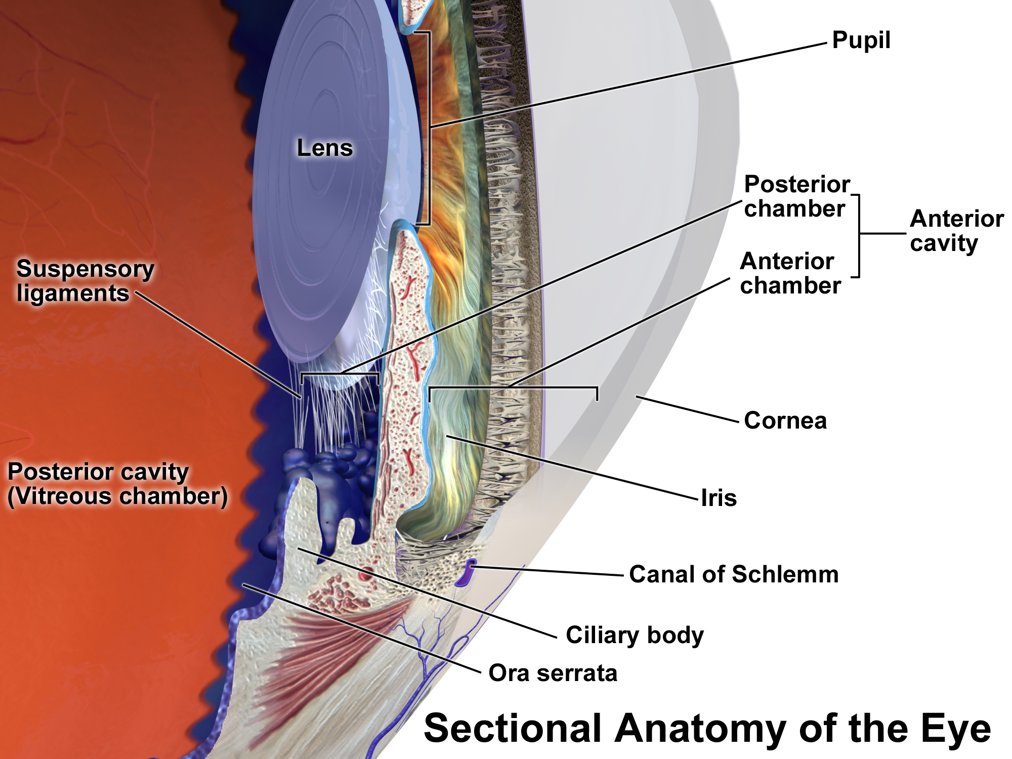
Uveitis () is inflammation of the uvea, the pigmented layer of the eye between the inner retina and the outer fibrous layer composed of the sclera and cornea. The uvea consists of the middle layer of pigmented vascular structures of the eye and includes the iris, ciliary body, and choroid. Uveitis is described anatomically, by the part of the eye affected, as anterior, intermediate or posterior, or panuveitic if all parts are involved. Anterior uveitis ( iridocyclytis) is the most common, with the incidence of uveitis overall affecting approximately 1:4500, most commonly those between the ages of 20-60. Symptoms include eye pain, eye redness, floaters and blurred vision, and ophthalmic examination may show dilated ciliary blood vessels and the presence of cells in the anterior chamber. Uveitis may arise spontaneously, have a genetic component, or be associated with an autoimmune disease or infection. While the eye is a relatively protected environment, its immune mechanisms ... [...More Info...] [...Related Items...] OR: [Wikipedia] [Google] [Baidu] |
Ophthalmoscopy
Ophthalmoscopy, also called funduscopy, is a test that allows a health professional to see inside the fundus of the eye and other structures using an ophthalmoscope (or funduscope). It is done as part of an eye examination and may be done as part of a routine physical examination. It is crucial in determining the health of the retina, optic disc, and vitreous humor. The pupil is a hole through which the eye's interior will be viewed. Opening the pupil wider (dilating it) is a simple and effective way to better see the structures behind it. Therefore, dilation of the pupil ( mydriasis) is often accomplished with medicated eye drops before funduscopy. However, although dilated fundus examination is ideal, undilated examination is more convenient and is also helpful (albeit not as comprehensive), and it is the most common type in primary care. An alternative or complement to ophthalmoscopy is to perform a fundus photography, where the image can be analysed later by a professional. ... [...More Info...] [...Related Items...] OR: [Wikipedia] [Google] [Baidu] |
White Dot Syndromes
White dot syndromes are inflammatory diseases characterized by the presence of white dots on the fundus (eye), fundus, the interior surface of the eye. Tewari A, Elliot D. White Dot Syndromes. 2007. Emedicine from WebMD. The majority of individuals affected with white dot syndromes are younger than fifty years of age. Some symptoms include blurred vision and visual field loss.Quillen DA, Davis JB, Gottlieb JL, Blodi BA, Callanan DG, Chang TS, et al. The white dot syndromes. American Journal of Ophthalmology. 2004;137(3):538-50. There are many theories for the etiology of white dot syndromes including infectious, viral, genetics and autoimmune. Classically recognized white dot syndromes include:Forrester JV, IOIS, Okada AA, BenEzra D. Posterior segment intraocular inflammation: guidelines. 1998:184. [...More Info...] [...Related Items...] OR: [Wikipedia] [Google] [Baidu] |
Fovea Centralis
The fovea centralis is a small, central pit composed of closely packed cones in the eye. It is located in the center of the macula lutea of the retina. The fovea is responsible for sharp central vision (also called foveal vision), which is necessary in humans for activities for which visual detail is of primary importance, such as reading and driving. The fovea is surrounded by the ''parafovea'' belt and the ''perifovea'' outer region. The parafovea is the intermediate belt, where the ganglion cell layer is composed of more than five layers of cells, as well as the highest density of cones; the perifovea is the outermost region where the ganglion cell layer contains two to four layers of cells, and is where visual acuity is below the optimum. The perifovea contains an even more diminished density of cones, having 12 per 100 micrometres versus 50 per 100 micrometres in the most central fovea. That, in turn, is surrounded by a larger peripheral area, which delivers highly compres ... [...More Info...] [...Related Items...] OR: [Wikipedia] [Google] [Baidu] |
Dysesthesia
Dysesthesia is an unpleasant, abnormal sense of touch. Its etymology comes from the Greek word "dys," meaning "bad," and "aesthesis," which means "sensation" (abnormal sensation). It often presents as painIASP Pain Terminology . but may also present as an inappropriate, but not discomforting, sensation. It is caused by lesions of the nervous system, peripheral or central, and it involves sensations, whether spontaneous or evoked, such as burning, wetness, itching, electric shock, and pins and needles. Dysesthesia can include sensations in any bodily tissue, including most often the mouth, scalp, skin, or legs. It is sometimes described as feeling like acid under the skin. Bu ... [...More Info...] [...Related Items...] OR: [Wikipedia] [Google] [Baidu] |
Vasculitis
Vasculitis is a group of disorders that destroy blood vessels by inflammation. Both arteries and veins are affected. Lymphangitis (inflammation of lymphatic vessels) is sometimes considered a type of vasculitis. Vasculitis is primarily caused by leukocyte migration and resultant damage. Although both occur in vasculitis, inflammation of veins (phlebitis) or arteries (arteritis) on their own are separate entities. Signs and symptoms Possible signs and symptoms include: * General symptoms: Fever, unintentional weight loss * Skin: Palpable purpura, livedo reticularis * Muscles and joints: Muscle pain or inflammation, joint pain or joint swelling * Nervous system: Mononeuritis multiplex, headache, stroke, tinnitus, reduced visual acuity, acute visual loss * Heart and arteries: Heart attack, high blood pressure, gangrene * Respiratory tract: Nosebleeds, bloody cough, lung infiltrates * GI tract: Abdominal pain, bloody stool, perforations (hole in the GI tract) * Kidneys: Inflamma ... [...More Info...] [...Related Items...] OR: [Wikipedia] [Google] [Baidu] |
Antimetabolite
An antimetabolite is a chemical that inhibits the use of a metabolite, which is another chemical that is part of normal metabolism. Such substances are often similar in structure to the metabolite that they interfere with, such as the antifolates that interfere with the use of folic acid; thus, competitive inhibition can occur, and the presence of antimetabolites can have toxic effects on cells, such as halting cell growth and cell division, so these compounds are used as chemotherapy for cancer. Function Cancer treatment Antimetabolites can be used in cancer treatment, as they interfere with DNA production and therefore cell division and tumor growth. Because cancer cells spend more time dividing than other cells, inhibiting cell division harms tumor cells more than other cells. Antimetabolite drugs are commonly used to treat leukemia, cancers of the breast, ovary, and the gastrointestinal tract, as well as other types of cancers. In the Anatomical Therapeutic Chemical Class ... [...More Info...] [...Related Items...] OR: [Wikipedia] [Google] [Baidu] |
Prednisone
Prednisone is a glucocorticoid medication mostly used to immunosuppressive drug, suppress the immune system and decrease inflammation in conditions such as asthma, COPD, and rheumatologic diseases. It is also used to treat high blood calcium due to cancer and adrenal insufficiency along with other steroids. It is taken Oral administration, by mouth. Common side effects with long-term use include cataracts, Osteoporosis, bone loss, easy bruising, muscle weakness, and oral candidiasis, thrush. Other side effects include weight gain, swelling, high blood sugar, increased risk of infection, and psychosis. It is generally considered safe in pregnancy and low doses appear to be safe when breastfeeding. After prolonged use, prednisone needs to be stopped gradually. Prednisone is a prodrug and must be converted to prednisolone by the liver before it becomes active. Prednisolone then binds to glucocorticoid receptors, activating them and triggering changes in gene expression. Pred ... [...More Info...] [...Related Items...] OR: [Wikipedia] [Google] [Baidu] |
Fundus Photography
Fundus photography involves photographing the rear of an eye, also known as the fundus. Specialized fundus cameras consisting of an intricate microscope attached to a flash enabled camera are used in fundus photography. The main structures that can be visualized on a fundus photo are the central and peripheral retina, optic disc and macula. Fundus photography can be performed with colored filters, or with specialized dyes including fluorescein and indocyanine green. The models and technology of fundus photography have advanced and evolved rapidly over the last century. Since the equipment is sophisticated and challenging to manufacture to clinical standards, only a few manufacturers/brands are available in the market: Welch Allyn, Digisight, Volk, Topcon, Zeiss, Canon, Nidek, Kowa, CSO, CenterVue, Ezer and Optos are some example of fundus camera manufacturers. History The concept of fundus photography was first introduced in the mid 19th century, after the introduction of ... [...More Info...] [...Related Items...] OR: [Wikipedia] [Google] [Baidu] |
HLA-DR2
HLA-DR2 (DR2) of the HLA- DR serotype system, is a broad antigen serotype that is now preferentially covered by HLA-DR15 and HLA-DR16 serotype group. This serotype primarily recognizes gene products of the HLA-DRB1*15 and HLA-DRB1*16 allele groups. Serology Disease associations DR2 serotypes are associated with Goodpasture syndrome, systemic lupus erythematosus, multiple sclerosis, and narcolepsy, tuberculoid leprosy (multi-drug-resistant tuberculosis or leprosy), ulcerative colitis(Japanese), primary biliary cirrhosis and autoimmune hepatitis. DR2 is also found in all patients that test positive for anti-anti-Asn-RNA-synthetase and chronic interstitial lung disease. Genetic linkage DR2 is linked to the HLA-DR51 HLA-DR51 is a HLA-DR serotype that recognizes the antigens encoded by the minor DR locus HLA-DRB5. DRB3, DRB4, and DRB5 are minor DR beta encoding loci, they have been recognized as having distinct evolution, having diverged from DRB1 approximate .... Reference ... [...More Info...] [...Related Items...] OR: [Wikipedia] [Google] [Baidu] |
White Dot Syndromes
White dot syndromes are inflammatory diseases characterized by the presence of white dots on the fundus (eye), fundus, the interior surface of the eye. Tewari A, Elliot D. White Dot Syndromes. 2007. Emedicine from WebMD. The majority of individuals affected with white dot syndromes are younger than fifty years of age. Some symptoms include blurred vision and visual field loss.Quillen DA, Davis JB, Gottlieb JL, Blodi BA, Callanan DG, Chang TS, et al. The white dot syndromes. American Journal of Ophthalmology. 2004;137(3):538-50. There are many theories for the etiology of white dot syndromes including infectious, viral, genetics and autoimmune. Classically recognized white dot syndromes include:Forrester JV, IOIS, Okada AA, BenEzra D. Posterior segment intraocular inflammation: guidelines. 1998:184. [...More Info...] [...Related Items...] OR: [Wikipedia] [Google] [Baidu] |
HLA-B7
HLA-B7 (B7) is an HLA- B serotype. The serotype identifies the more common HLA-B*07 gene products. (For terminology help see: HLA-serotype tutorial) B7, previously HL-A7, was one of the first 'HL-A' antigens recognized, largely because of the frequency of B*0702 in Northern and Western Europe and the United States. B7 is found in two major haplotypes in Europe, where it reaches peak frequency in Ireland. One haplotype A3-B7-DR15-DQ1 can be found over a vast region and is in apparent selective disequilibrium. B7 is a risk factor for cervical cancer, sarcoidosis, and early-onset spondylarthropathies. Serology Alleles In disease Cervical cancer HLA-B7 along with HLA-DQ8 increased risk for cervical cancer in at risk Costa Rican women and Asian Indians Sarcoidosis A weak relationship between HLA-B7 and sarcoidosis has been known for 30+ years, yet has not consistently been reproducible in all studies however. A common serologically defined haplotype in Europeans is HLA A3- ... [...More Info...] [...Related Items...] OR: [Wikipedia] [Google] [Baidu] |


