|
Acetabular Angle
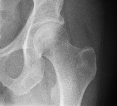
In vertebrate anatomy, hip (or "coxa"Latin ''coxa'' was used by Celsus in the sense "hip", but by Pliny the Elder in the sense "hip bone" (Diab, p 77) in medical terminology) refers to either an anatomical region or a joint. The hip region is located lateral and anterior to the gluteal region, inferior to the iliac crest, and overlying the greater trochanter of the femur, or "thigh bone". In adults, three of the bones of the pelvis have fused into the hip bone or acetabulum which forms part of the hip region. The hip joint, scientifically referred to as the acetabulofemoral joint (''art. coxae''), is the joint between the head of the femur and acetabulum of the pelvis and its primary function is to support the weight of the body in both static (e.g., standing) and dynamic (e.g., walking or running) postures. The hip joints have very important roles in retaining balance, and for maintaining the pelvic inclination angle. Pain of the hip may be the result of numerous causes, i ... [...More Info...] [...Related Items...] OR: [Wikipedia] [Google] [Baidu] |
Vertebrate
Vertebrates () comprise all animal taxa within the subphylum Vertebrata () ( chordates with backbones), including all mammals, birds, reptiles, amphibians, and fish. Vertebrates represent the overwhelming majority of the phylum Chordata, with currently about 69,963 species described. Vertebrates comprise such groups as the following: * jawless fish, which include hagfish and lampreys * jawed vertebrates, which include: ** cartilaginous fish (sharks, rays, and ratfish) ** bony vertebrates, which include: *** ray-fins (the majority of living bony fish) *** lobe-fins, which include: **** coelacanths and lungfish **** tetrapods (limbed vertebrates) Extant vertebrates range in size from the frog species ''Paedophryne amauensis'', at as little as , to the blue whale, at up to . Vertebrates make up less than five percent of all described animal species; the rest are invertebrates, which lack vertebral columns. The vertebrates traditionally include the hagfish, which do no ... [...More Info...] [...Related Items...] OR: [Wikipedia] [Google] [Baidu] |
Triradiate Cartilages
The triradiate cartilage (in Latin cartilago ypsiloformis) is the 'Y'-shaped epiphyseal plate between the ilium, ischium and pubis to form the acetabulum of the os coxae. Human development In children, the triradiate cartilage closes at an approximate bone age of 12 years for girls and 14 years for boys. Clinical use Evaluating the position of the triradiate cartilage on an AP radiograph of the pelvis with both Perkin's line and Hilgenreiner's line Hilgenreiner's line is a horizontal line drawn on an AP radiograph of the pelvis running between the inferior aspects of both triradiate cartilages of the acetabulums. It is named for Heinrich Hilgenreiner. Clinical Use Used in conjunction wi ... can help establish a diagnosis of developmental dysplasia of the hip. References See also {{Pelvis Pelvis ... [...More Info...] [...Related Items...] OR: [Wikipedia] [Google] [Baidu] |
Osteoporosis
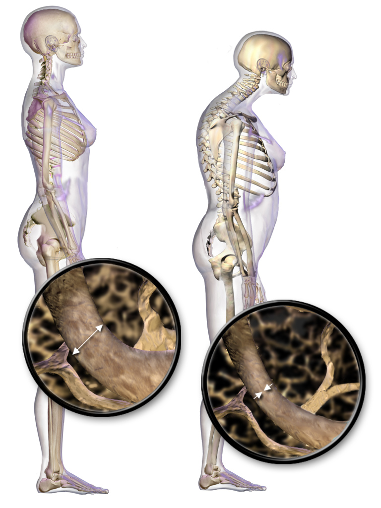
Osteoporosis is a systemic skeletal disorder characterized by low bone mass, micro-architectural deterioration of bone tissue leading to bone fragility, and consequent increase in fracture risk. It is the most common reason for a broken bone among the elderly. Bones that commonly break include the vertebrae in the spine, the bones of the forearm, and the hip. Until a broken bone occurs there are typically no symptoms. Bones may weaken to such a degree that a break may occur with minor stress or spontaneously. After the broken bone heals, the person may have chronic pain and a decreased ability to carry out normal activities. Osteoporosis may be due to lower-than-normal maximum bone mass and greater-than-normal bone loss. Bone loss increases after the menopause due to lower levels of estrogen, and after ' andropause' due to lower levels of testosterone. Osteoporosis may also occur due to a number of diseases or treatments, including alcoholism, anorexia, hyperthyroidism, ... [...More Info...] [...Related Items...] OR: [Wikipedia] [Google] [Baidu] |
Ligament Of Head Of Femur
In human anatomy, the ligament of the head of the femur (round ligament of the femur, ligamentum teres femoris, the foveal ligament, or Fillmore’s ligament) is a ligament located in the hip. It is triangular in shape and somewhat flattened. The ligament is implanted by its apex into the antero- superior part of the fovea capitis femoris and its base is attached by two bands, one into either side of the acetabular notch, and between these bony attachments it blends with the transverse ligament.''Gray's Anatomy'' (1918), see infobox It is ensheathed by the synovial membrane, and varies greatly in strength in different subjects; occasionally only the synovial fold exists, and in rare cases even this is absent. The ligament of the head of the femur contains within it the acetabular branch of the obturator artery. Function The ligament is made tense when the thigh is semiflexed and the limb then adducted or rotated outward; it is, on the other hand, relaxed when the limb is abducte ... [...More Info...] [...Related Items...] OR: [Wikipedia] [Google] [Baidu] |
Acetabular Labrum
The acetabular labrum (glenoidal labrum of the hip joint or cotyloid ligament in older texts) is a ring of cartilage that surrounds the acetabulum of the hip. The anterior portion is most vulnerable when the labrum tears. It provides an articulating surface for the acetabulum, allowing the head of the femur to articulate with the pelvis. Acetabular labrum tear Mechanisms of Injury It is estimated that 75% of acetabular labrum tears have an unknown cause. Tears of the labrum have been credited to a variety of causes such as excessive force, hip dislocation, capsular hip hypermobility, hip dysplasia, and hip degeneration. A tight iliopsoas tendon has also been attributed to labrum tears by causing compression or traction injuries that eventually lead to a labrum tear.Smith, M., Panchal, H., Ruberte, R., & Sekiya, J. (2011). Effect of acetabular labrum tears on hip stability and labral strain in a joint compression model. The American Journal of Sports Medicine, 39, 103S-110S. Most ... [...More Info...] [...Related Items...] OR: [Wikipedia] [Google] [Baidu] |
Ball And Socket Joint
The ball-and-socket joint (or spheroid joint) is a type of synovial joint in which the ball-shaped surface of one rounded bone fits into the cup-like depression of another bone. The distal bone is capable of motion around an indefinite number of axes, which have one common center. This enables the joint to move in many directions. An enarthrosis is a special kind of spheroidal joint in which the socket covers the sphere beyond its equator.Platzer, Werner (2008) ''Color Atlas of Human Anatomy'', Volume 1p.28/ref> Examples Examples of this form of articulation are found in the hip, where the round head of the femur (ball) rests in the cup-like acetabulum (socket) of the pelvis; and in the shoulder joint, where the rounded upper extremity of the humerus (ball) rests in the cup-like glenoid fossa (socket) of the shoulder blade.And the phalanges (toes, fingers)Introduction to Joints: Synovial Joints - Ball and Socket Joints (The shoulder also includes a sternoclavicular joint ... [...More Info...] [...Related Items...] OR: [Wikipedia] [Google] [Baidu] |
Triradiate Cartilage
The triradiate cartilage (in Latin cartilago ypsiloformis) is the 'Y'-shaped epiphyseal plate between the ilium, ischium and pubis to form the acetabulum of the os coxae. Human development In children, the triradiate cartilage closes at an approximate bone age of 12 years for girls and 14 years for boys. Clinical use Evaluating the position of the triradiate cartilage on an AP radiograph of the pelvis with both Perkin's line and Hilgenreiner's line Hilgenreiner's line is a horizontal line drawn on an AP radiograph of the pelvis running between the inferior aspects of both triradiate cartilages of the acetabulums. It is named for Heinrich Hilgenreiner. Clinical Use Used in conjunction wi ... can help establish a diagnosis of developmental dysplasia of the hip. References See also {{Pelvis Pelvis ... [...More Info...] [...Related Items...] OR: [Wikipedia] [Google] [Baidu] |
Ischium
The ischium () forms the lower and back region of the (''os coxae''). Situated below the ilium and behind the pubis, it is one of three regions whose fusion creates the . The superior portion of this region forms approximately one-third of the acetabulum. |
Pubis (bone)
In vertebrates, the pubic region ( la, pubis) is the most forward-facing (ventral and anterior) of the three main regions making up the coxal bone. The left and right pubic regions are each made up of three sections, a superior ramus, inferior ramus, and a body. Structure The pubic region is made up of a ''body'', ''superior ramus'', and ''inferior ramus'' (). The left and right coxal bones join at the pubic symphysis. It is covered by a layer of fat, which is covered by the mons pubis. The pubis is the lower limit of the suprapubic region. In the female, the pubic region is anterior to the urethral sponge. Body The body forms the wide, strong, middle and flat part of the pubic region. The bodies of the left and right pubic regions join at the pubic symphysis. The rough upper edge is the pubic crest, ending laterally in the pubic tubercle. This tubercle, found roughly 3 cm from the pubic symphysis, is a distinctive feature on the lower part of the abdominal wall; important ... [...More Info...] [...Related Items...] OR: [Wikipedia] [Google] [Baidu] |
Ilium (bone)
The ilium () (plural ilia) is the uppermost and largest part of the hip bone, and appears in most vertebrates including mammals and birds, but not bony fish. All reptiles have an ilium except snakes, although some snake species have a tiny bone which is considered to be an ilium. The ilium of the human is divisible into two parts, the body and the wing; the separation is indicated on the top surface by a curved line, the arcuate line, and on the external surface by the margin of the acetabulum. The name comes from the Latin (''ile'', ''ilis''), meaning "groin" or "flank". Structure The ilium consists of the body and wing. Together with the ischium and pubis, to which the ilium is connected, these form the pelvic bone, with only a faint line indicating the place of union. The body ( la, corpus) forms less than two-fifths of the acetabulum; and also forms part of the acetabular fossa. The internal surface of the body is part of the wall of the lesser pelvis and gives ... [...More Info...] [...Related Items...] OR: [Wikipedia] [Google] [Baidu] |
Cartilage
Cartilage is a resilient and smooth type of connective tissue. In tetrapods, it covers and protects the ends of long bones at the joints as articular cartilage, and is a structural component of many body parts including the rib cage, the neck and the bronchial tubes, and the intervertebral discs. In other taxa, such as chondrichthyans, but also in cyclostomes, it may constitute a much greater proportion of the skeleton. It is not as hard and rigid as bone, but it is much stiffer and much less flexible than muscle. The matrix of cartilage is made up of glycosaminoglycans, proteoglycans, collagen fibers and, sometimes, elastin. Because of its rigidity, cartilage often serves the purpose of holding tubes open in the body. Examples include the rings of the trachea, such as the cricoid cartilage and carina. Cartilage is composed of specialized cells called chondrocytes that produce a large amount of collagenous extracellular matrix, abundant ground substance that is rich in pro ... [...More Info...] [...Related Items...] OR: [Wikipedia] [Google] [Baidu] |


