|
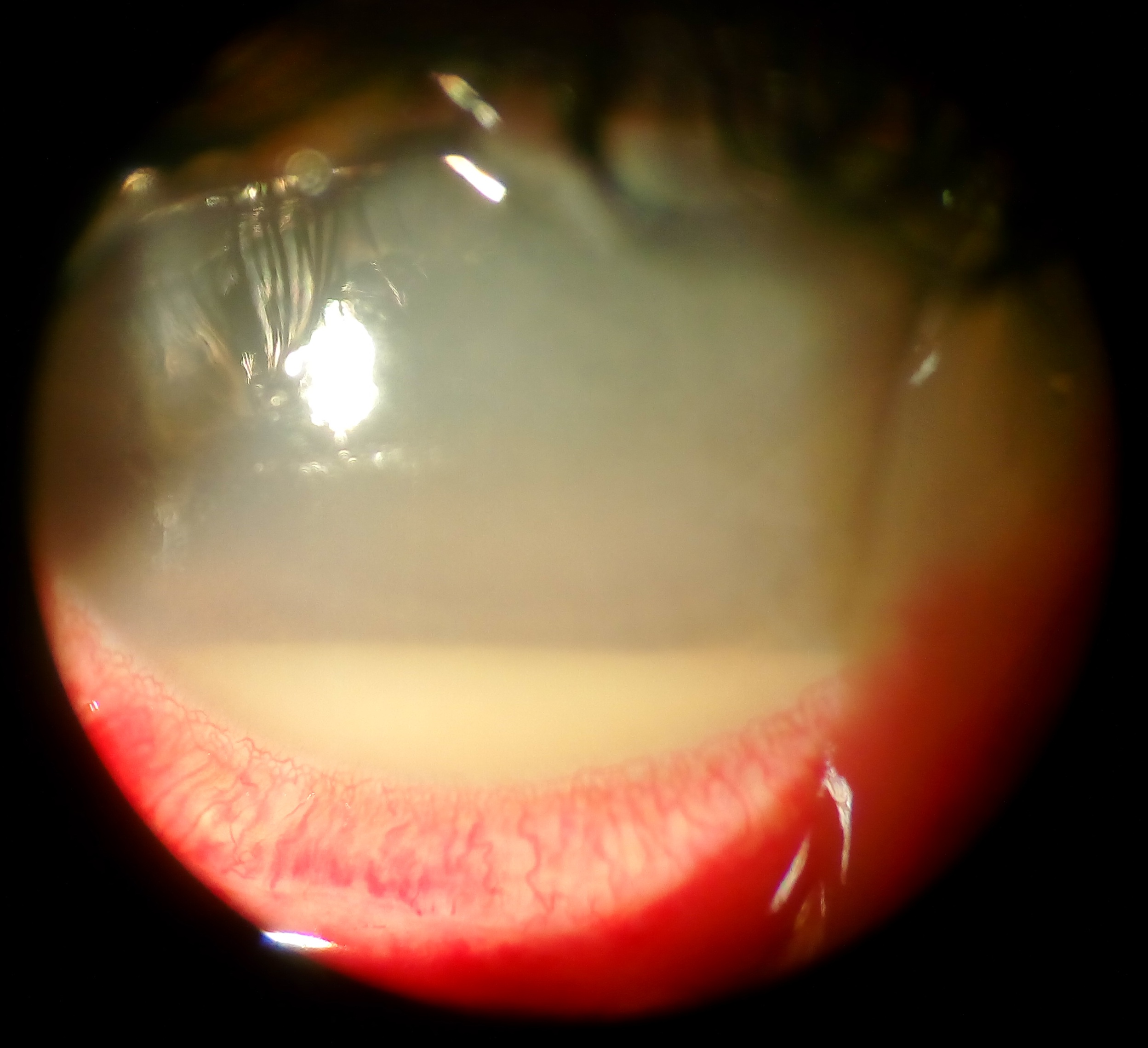
Acanthamoeba Keratitis
''Acanthamoeba'' keratitis (AK) is a rare disease in which amoebae of the genus ''Acanthamoeba'' invade the clear portion of the front (cornea) of the eye. It affects roughly 100 people in the United States each year. ''Acanthamoeba'' are protozoa found nearly ubiquitously in soil and water and can cause infections of the skin, eyes, and central nervous system. Infection of the cornea by ''Acanthamoeba'' is difficult to treat with conventional medications, and AK may cause permanent visual impairment or blindness, due to damage to the cornea or through damage to other structures important to vision. Recently, AK has been recognized as an orphan disease and a funded project, orphan diseases ''Acanthamoeba'' keratitis (ODAK), has tested the effects of a diverse range drugs and biocides on AK. Pathogenesis In the United States, ''Acanthamoeba'' keratitis is nearly always associated with soft contact lens use. ''Acanthamoeba'' spp. is most commonly introduced to the eye by contact l ... [...More Info...] [...Related Items...] OR: [Wikipedia] [Google] [Baidu] |
Fluorescein
Fluorescein is an organic compound and dye based on the xanthene tricyclic structural motif, formally belonging to triarylmethine dyes family. It is available as a dark orange/red powder slightly soluble in water and alcohol. It is widely used as a fluorescent tracer for many applications. The color of its aqueous solutions is green by reflection and orange by transmission (its spectral properties are dependent on pH of the solution), as can be noticed in bubble levels, for example, in which fluorescein is added as a colorant to the alcohol filling the tube in order to increase the visibility of the air bubble contained within (thus enhancing the precision of the instrument). More concentrated solutions of fluorescein can even appear red (because under these conditions nearly all incident emission is re-absorbed by the solution). It is on the World Health Organization's List of Essential Medicines. Uses Fluorescein sodium, the sodium salt of fluorescein, is used extensi ... [...More Info...] [...Related Items...] OR: [Wikipedia] [Google] [Baidu] |
Anterior Chamber Of Eyeball
The anterior chamber (Optometric Abbreviations#AC, AC) is the aqueous humor-filled space inside the human eye, eye between the iris (anatomy), iris and the cornea's innermost surface, the Corneal endothelium, endothelium. Hyphema, Uveitis, anterior uveitis and glaucoma are three main pathologies in this area. In hyphema, blood fills the anterior chamber as a result of a hemorrhage, most commonly after a blunt eye injury. Anterior uveitis is an inflammatory process affecting the iris (anatomy), iris and ciliary body, with resulting inflammatory signs in the anterior chamber. In glaucoma, blockage of the trabecular meshwork prevents the normal outflow of aqueous humour, resulting in increased intraocular pressure, progressive damage to the optic nerve head, and eventually blindness. The depth of the anterior chamber of the eye varies between 1.5 and 4.0 mm, averaging 3.0 mm. It tends to become shallower at older age and in eyes with Far-sightedness, hypermetropia (far sight ... [...More Info...] [...Related Items...] OR: [Wikipedia] [Google] [Baidu] |
Polymerase Chain Reaction
The polymerase chain reaction (PCR) is a method widely used to rapidly make millions to billions of copies (complete or partial) of a specific DNA sample, allowing scientists to take a very small sample of DNA and amplify it (or a part of it) to a large enough amount to study in detail. PCR was invented in 1983 by the American biochemist Kary Mullis at Cetus Corporation; Mullis and biochemist Michael Smith (chemist), Michael Smith, who had developed other essential ways of manipulating DNA, were jointly awarded the Nobel Prize in Chemistry in 1993. PCR is fundamental to many of the procedures used in genetic testing and research, including analysis of Ancient DNA, ancient samples of DNA and identification of infectious agents. Using PCR, copies of very small amounts of DNA sequences are exponentially amplified in a series of cycles of temperature changes. PCR is now a common and often indispensable technique used in medical laboratory research for a broad variety of applications ... [...More Info...] [...Related Items...] OR: [Wikipedia] [Google] [Baidu] |
Giemsa Stain
Giemsa stain (), named after German chemist and bacteriologist Gustav Giemsa, is a nucleic acid stain used in cytogenetics and for the histopathological diagnosis of malaria and other parasites. Uses It is specific for the phosphate groups of DNA and attaches itself to regions of DNA where there are high amounts of adenine-thymine bonding. Giemsa stain is used in Giemsa banding, commonly called G-banding, to stain chromosomes and often used to create a Karyotype, karyogram (chromosome map). It can identify chromosomal aberrations such as chromosomal translocation, translocations and chromosomal inversion, rearrangements. It stains the trophozoite ''Trichomonas vaginalis'', which presents with greenish discharge and motile cells on wet prep. Giemsa stain is also a Differential staining, differential stain, such as when it is combined with Wright's stain, Wright stain to form Wright-Giemsa stain. It can be used to study the adherence of pathogenic bacteria to human cells. It dif ... [...More Info...] [...Related Items...] OR: [Wikipedia] [Google] [Baidu] |
Gram Stain
In microbiology and bacteriology, Gram stain (Gram staining or Gram's method), is a method of staining used to classify bacterial species into two large groups: gram-positive bacteria and gram-negative bacteria. The name comes from the Danish bacteriologist Hans Christian Gram, who developed the technique in 1884. Gram staining differentiates bacteria by the chemical and physical properties of their cell walls. Gram-positive cells have a thick layer of peptidoglycan in the cell wall that retains the primary stain, crystal violet. Gram-negative cells have a thinner peptidoglycan layer that allows the crystal violet to wash out on addition of ethanol. They are stained pink or red by the counterstain, commonly safranin or fuchsine. Lugol's iodine solution is always added after addition of crystal violet to strengthen the bonds of the stain with the cell membrane. Gram staining is almost always the first step in the preliminary identification of a bacterial organism. While Gram s ... [...More Info...] [...Related Items...] OR: [Wikipedia] [Google] [Baidu] |
Hypopyon
Hypopyon is a medical condition involving inflammatory cells in the anterior chamber of the eye. It is an exudate rich in white blood cells, seen in the anterior chamber, usually accompanied by redness of the conjunctiva and the underlying episclera. It is a sign of inflammation of the anterior uvea and iris, i.e. iritis, which is a form of anterior uveitis. The exudate settles at the dependent aspect of the eye due to gravity. It can be sterile (in bacterial corneal ulcer) or not sterile (fungal corneal ulcer). Differential diagnosis Hypopyon can be present in a corneal ulcer. It can occur as a result of Behçet's disease, endophthalmitis, panuveitis/panophthalmitis, or adverse reactions to some drugs (such as rifabutin). Hypopyon is also known as ''sterile pus'' because it occurs due to the release of toxins and not by the actual invasion of pathogens. The toxins secreted by the pathogens mediate the outpouring of leukocytes that settle in the anterior chamber of the eye. A ... [...More Info...] [...Related Items...] OR: [Wikipedia] [Google] [Baidu] |
Corneal Perforation
Corneal perforation is an anomaly in the cornea resulting from damage to the corneal surface. A corneal perforation means that the cornea has been penetrated, thus leaving the cornea damaged. The cornea is a clear part of the eye which controls and focuses the entry of light into the eye. Damage to the cornea due to corneal perforation can cause decreased visual acuity. Signs and symptoms Corneal perforation may cause difficulty in seeing and persistent eye pain. Physical examination may reveal discoloration of the cornea. Causes Perforation of the cornea may occur due to diseases of the cornea, injury during eye surgery, or infection of the eye, which may occur after surgery or procedures. Pellucid marginal degeneration may cause corneal thinning, leading to perforation. Diagnosis Corneal perforation can be diagnosed by using the Seidel test. Any aqueous leakage is revealed during the Seidel test confirms corneal perforation. A fluorescence strip is wiped over the wound. ... [...More Info...] [...Related Items...] OR: [Wikipedia] [Google] [Baidu] |
Corneal Ulceration
Corneal ulcer is an inflammatory or, more seriously, infective condition of the cornea involving disruption of its epithelial layer with involvement of the corneal stroma. It is a common condition in humans particularly in the tropics and the agrarian societies. In developing countries, children afflicted by Vitamin A deficiency are at high risk for corneal ulcer and may become blind in both eyes, which may persist lifelong. In ophthalmology, a corneal ulcer usually refers to having an infectious cause while the term corneal abrasion refers more to physical abrasions. Types Superficial and deep corneal ulcers Corneal ulcers are a common human eye disease. They are caused by trauma, particularly with vegetable matter, as well as chemical injury, contact lenses and infections. Other eye conditions can cause corneal ulcers, such as entropion, distichiasis, corneal dystrophy, and keratoconjunctivitis sicca (dry eye). Many micro-organisms cause infective corneal ulcer. Among them ar ... [...More Info...] [...Related Items...] OR: [Wikipedia] [Google] [Baidu] |
Herpes Simplex Virus
Herpes simplex virus 1 and 2 (HSV-1 and HSV-2), also known by their taxonomical names ''Human alphaherpesvirus 1'' and '' Human alphaherpesvirus 2'', are two members of the human ''Herpesviridae'' family, a set of viruses that produce viral infections in the majority of humans. Both HSV-1 and HSV-2 are very common and contagious. They can be spread when an infected person begins shedding the virus. As of 2016, about 67% of the world population under the age of 50 had HSV-1. In the United States, about 47.8% and 11.9% are estimated to have HSV-1 and HSV-2, respectively, though actual prevalence may be much higher. Because it can be transmitted through any intimate contact, it is one of the most common sexually transmitted infections. Symptoms Many of those who are infected ''never'' develop symptoms. Symptoms, when they occur, may include watery blisters in the skin or mucous membranes of the mouth, lips, nose, genitals, or eyes (herpes simplex keratitis). Lesions heal with a ... [...More Info...] [...Related Items...] OR: [Wikipedia] [Google] [Baidu] |
Varicella Zoster Virus
Varicella-zoster virus (VZV), also known as human herpesvirus 3 (HHV-3, HHV3) or ''Human alphaherpesvirus 3'' (taxonomically), is one of nine known herpes viruses that can infect humans. It causes chickenpox (varicella) commonly affecting children and young adults, and shingles (herpes zoster) in adults but rarely in children. VZV infections are species-specific to humans. The virus can survive in external environments for a few hours. VZV multiplies in the tonsils, and causes a wide variety of symptoms. Similar to the herpes simplex viruses, after primary infection with VZV (chickenpox), the virus lies dormant in neurons, including the cranial nerve ganglia, dorsal root ganglia, and autonomic ganglia. Many years after the person has recovered from initial chickenpox infection, VZV can ''reactivate'' to cause shingles. Epidemiology Chickenpox Primary varicella zoster virus infection results in chickenpox (varicella), which may result in complications including encephaliti ... [...More Info...] [...Related Items...] OR: [Wikipedia] [Google] [Baidu] |
Conjunctival Hyperemia
Conjunctivitis, also known as pink eye, is inflammation of the outermost layer of the white part of the eye and the inner surface of the eyelid. It makes the eye appear pink or reddish. Pain, burning, scratchiness, or itchiness may occur. The affected eye may have increased tears or be "stuck shut" in the morning. Swelling of the white part of the eye may also occur. Itching is more common in cases due to allergies. Conjunctivitis can affect one or both eyes. The most common infectious causes are viral followed by bacterial. The viral infection may occur along with other symptoms of a common cold. Both viral and bacterial cases are easily spread between people. Allergies to pollen or animal hair are also a common cause. Diagnosis is often based on signs and symptoms. Occasionally, a sample of the discharge is sent for culture. Prevention is partly by handwashing. Treatment depends on the underlying cause. In the majority of viral cases, there is no specific treatment. Most ... [...More Info...] [...Related Items...] OR: [Wikipedia] [Google] [Baidu] |
Photophobia
Photophobia is a medical symptom of abnormal intolerance to visual perception of light. As a medical symptom photophobia is not a morbid fear or phobia, but an experience of discomfort or pain to the eyes due to light exposure or by presence of actual physical sensitivity of the eyes, though the term is sometimes additionally applied to abnormal or irrational fear of light such as heliophobia. The term ''photophobia'' comes from the Greek language, Greek φῶς (''phōs''), meaning "light", and φόβος (''phóbos''), meaning "fear". Causes Patients may develop photophobia as a result of several different medical conditions, related to the human eye, eye, the nervous system, genetic, or other causes. Photophobia may manifest itself in an increased response to light starting at any step in the visual system, such as: *Too much light entering the eye. Too much light can enter the eye if it is damaged, such as with corneal abrasion and retinal damage, or if its pupil(s) is unabl ... [...More Info...] [...Related Items...] OR: [Wikipedia] [Google] [Baidu] |


.jpg)


