|
Abductor Hallucis
The abductor hallucis muscle is an intrinsic muscle of the foot. It participates in the abduction and flexion of the great toe. Structure The abductor hallucis muscle is located in the medial border of the foot and contributes to form the prominence that is observed on the region. It is inserted behind on the tuberosity of the calcaneus, the flexor retinaculum, and the plantar aponeurosis. Its muscle body, relatively thick behind, flattens as it goes forward. It ends in a common tendon with the medial head of the flexor hallucis brevis that inserts on the medial surface of the base of the first proximal phalanx and its related sesamoid bone. Its medial surface is superficial and covered with the muscle's fascia and the skin. Nerve supply Abductor hallucis is supplied by the medial plantar nerve. The nerves that supply it enter the muscle from its upper border. Additional images Image:Sobo 1909 309.png, Superficial dissection of the sole of the foot, showing the medial ... [...More Info...] [...Related Items...] OR: [Wikipedia] [Google] [Baidu] |
Plantar Aponeurosis
The plantar fascia is the thick connective tissue (aponeurosis) which supports the arch on the bottom (plantar side) of the foot. It runs from the tuberosity of the calcaneus (heel bone) forward to the heads of the metatarsal bones (the bone between each toe and the bones of the mid-foot). Structure The plantar fascia is a broad structure that spans between the medial calcaneal tubercle and the proximal phalanges of the toes. Recent studies suggest that the plantar fascia is actually an aponeurosis rather than true fascia. The Dorland’s Medical Dictionary defines an aponeurosis as: (i) a white, flattened or ribbon-like tendinous expansion, serving mainly to connect a muscle with the parts that it moves, (ii) a term formerly applied to certain fasciae. Further, it defines the plantar aponeurosis as bands of fibrous connective tissue radiating toward the bases of the toes from the medial process of the tuber calcanei (posterior half of the calcaneus). The plantar fascia is ma ... [...More Info...] [...Related Items...] OR: [Wikipedia] [Google] [Baidu] |
Foot Muscles
The foot ( : feet) is an anatomical structure found in many vertebrates. It is the terminal portion of a limb which bears weight and allows locomotion. In many animals with feet, the foot is a separate organ at the terminal part of the leg made up of one or more segments or bones, generally including claws or nails. Etymology The word "foot", in the sense of meaning the "terminal part of the leg of a vertebrate animal" comes from "Old English fot "foot," from Proto-Germanic *fot (source also of Old Frisian fot, Old Saxon fot, Old Norse fotr, Danish fod, Swedish fot, Dutch voet, Old High German fuoz, German Fuß, Gothic fotus "foot"), from PIE root *ped- "foot". The "plural form feet is an instance of i-mutation." Structure The human foot is a strong and complex mechanical structure containing 26 bones, 33 joints (20 of which are actively articulated), and more than a hundred muscles, tendons, and ligaments.Podiatry Channel, ''Anatomy of the foot and ankle'' The joints of the ... [...More Info...] [...Related Items...] OR: [Wikipedia] [Google] [Baidu] |
Sole (foot)
The sole is the bottom of the foot. In humans the sole of the foot is anatomically referred to as the plantar aspect. Structure The glabrous skin on the sole of the foot lacks the hair and pigmentation found elsewhere on the body, and it has a high concentration of sweat pores. The sole contains the thickest layers of skin on the body due to the weight that is continually placed on it. It is crossed by a set of creases that form during the early stages of embryonic development. Like those of the palm, the sweat pores of the sole lack sebaceous glands. The sole is a sensory organ by which we can perceive the ground while standing and walking. The subcutaneous tissue in the sole has adapted to deal with the high local compressive forces on the heel and the ball (between the toes and the arch) by developing a system of "pressure chambers." Each chamber is composed of internal fibrofatty tissue covered by external collagen connective tissue. The septa (internal walls) ... [...More Info...] [...Related Items...] OR: [Wikipedia] [Google] [Baidu] |
Foot
The foot ( : feet) is an anatomical structure found in many vertebrates. It is the terminal portion of a limb which bears weight and allows locomotion. In many animals with feet, the foot is a separate organ at the terminal part of the leg made up of one or more segments or bones, generally including claws or nails. Etymology The word "foot", in the sense of meaning the "terminal part of the leg of a vertebrate animal" comes from "Old English fot "foot," from Proto-Germanic *fot (source also of Old Frisian fot, Old Saxon fot, Old Norse fotr, Danish fod, Swedish fot, Dutch voet, Old High German fuoz, German Fuß, Gothic fotus "foot"), from PIE root *ped- "foot". The "plural form feet is an instance of i-mutation." Structure The human foot is a strong and complex mechanical structure containing 26 bones, 33 joints (20 of which are actively articulated), and more than a hundred muscles, tendons, and ligaments.Podiatry Channel, ''Anatomy of the foot and ankle'' The joints of the ... [...More Info...] [...Related Items...] OR: [Wikipedia] [Google] [Baidu] |
Medial Plantar Nerve
The medial plantar nerve (internal plantar nerve) is the larger of the two terminal divisions of the tibial nerve (medial and lateral plantar nerve), which accompanies the medial plantar artery. From its origin under the laciniate ligament it passes under cover of the abductor hallucis muscle, and, appearing between this muscle and the flexor digitorum brevis, gives off a proper digital plantar nerve and finally divides opposite the bases of the metatarsal bones into three common digital plantar nerves. Branches The branches of the medial plantar nerve are: (1) cutaneous, (2) muscular, (3) articular, (4) a proper digital nerve to the medial side of the great toe, and (5) three common digital nerves. Cutaneous branches The cutaneous branches pierce the plantar aponeurosis between the abductor hallucis and the flexor digitorum brevis and are distributed to the skin of the sole of the foot. Muscular branches The muscular branches supply muscles on the medial side of the sole, incl ... [...More Info...] [...Related Items...] OR: [Wikipedia] [Google] [Baidu] |
Fascia
A fascia (; plural fasciae or fascias; adjective fascial; from Latin: "band") is a band or sheet of connective tissue, primarily collagen, beneath the skin that attaches to, stabilizes, encloses, and separates muscles and other internal organs. Fascia is classified by layer, as superficial fascia, deep fascia, and ''visceral'' or ''parietal'' fascia, or by its function and anatomical location. Like ligaments, aponeuroses, and tendons, fascia is made up of fibrous connective tissue containing closely packed bundles of collagen fibers oriented in a wavy pattern parallel to the direction of pull. Fascia is consequently flexible and able to resist great unidirectional tension forces until the wavy pattern of fibers has been straightened out by the pulling force. These collagen fibers are produced by fibroblasts located within the fascia. Fasciae are similar to ligaments and tendons as they have collagen as their major component. They differ in their location and function: ligament ... [...More Info...] [...Related Items...] OR: [Wikipedia] [Google] [Baidu] |
Sesamoid Bone
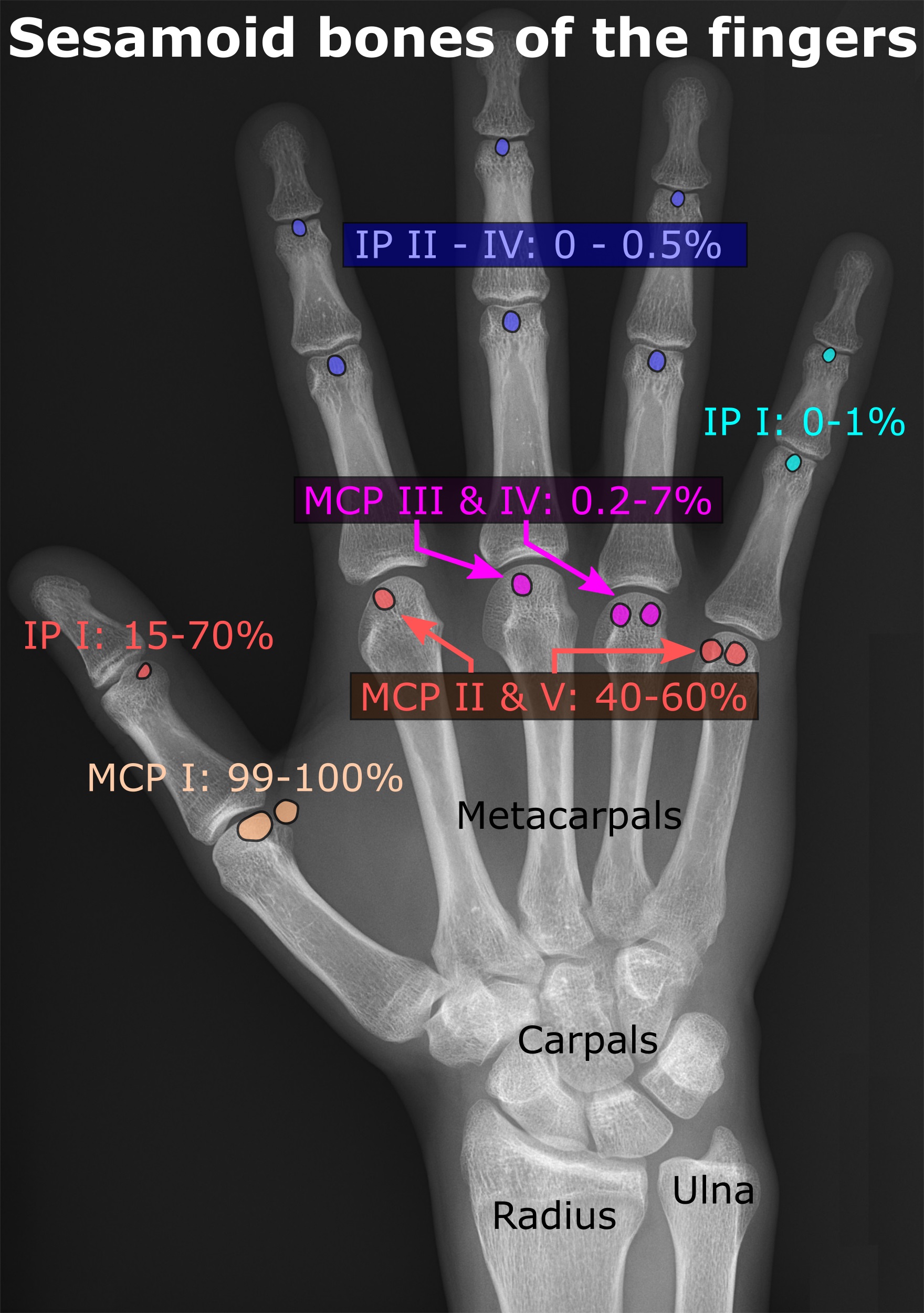
In anatomy, a sesamoid bone () is a bone embedded within a tendon or a muscle. Its name is derived from the Arabic word for ' sesame seed', indicating the small size of most sesamoids. Often, these bones form in response to strain, or can be present as a normal variant. The patella is the largest sesamoid bone in the body. Sesamoids act like pulleys, providing a smooth surface for tendons to slide over, increasing the tendon's ability to transmit muscular forces. Structure Sesamoid bones can be found on joints throughout the body, including: * In the knee—the patella (within the quadriceps tendon). This is the largest sesamoid bone. * In the hand—two sesamoid bones are commonly found in the distal portions of the first metacarpal bone (within the tendons of adductor pollicis and flexor pollicis brevis). There is also commonly a sesamoid bone in distal portions of the second metacarpal bone. * In the wrist—The pisiform of the wrist is a sesamoid bone (within the tend ... [...More Info...] [...Related Items...] OR: [Wikipedia] [Google] [Baidu] |
Phalanx Bone
The phalanges (singular: ''phalanx'' ) are digital bones in the hands and feet of most vertebrates. In primates, the thumbs and big toes have two phalanges while the other digits have three phalanges. The phalanges are classed as long bones. Structure The phalanges are the bones that make up the fingers of the hand and the toes of the foot. There are 56 phalanges in the human body, with fourteen on each hand and foot. Three phalanges are present on each finger and toe, with the exception of the thumb and large toe, which possess only two. The middle and far phalanges of the fifth toes are often fused together (symphalangism). The phalanges of the hand are commonly known as the finger bones. The phalanges of the foot differ from the hand in that they are often shorter and more compressed, especially in the proximal phalanges, those closest to the torso. A phalanx is named according to whether it is proximal, middle, or distal and its associated finger or toe. The proximal ... [...More Info...] [...Related Items...] OR: [Wikipedia] [Google] [Baidu] |
Flexor Hallucis Brevis
A flexor is a muscle that flexes a joint. In anatomy, flexion (from the Latin verb ''flectere'', to bend) is a joint movement that decreases the angle between the bones that converge at the joint. For example, one’s elbow joint flexes when one brings their hand closer to the shoulder. Flexion is typically instigated by muscle contraction of a flexor. Flexors Upper limb *of the humerus bone (the bone in the upper arm) at the shoulder **Pectoralis major **Anterior deltoid **Coracobrachialis ** Biceps brachii * of the forearm at the elbow ** Brachialis **Brachioradialis ** Biceps brachii *of carpus (the carpal bones) at the wrist **flexor carpi radialis **flexor carpi ulnaris **palmaris longus *of the hand **flexor pollicis longus muscle **flexor pollicis brevis muscle **flexor digitorum profundus muscle **flexor digitorum superficialis muscle Lower limb Hip The hip flexors are (in descending order of importance to the action of flexing the hip joint):Platzer (2004), p 246 *Co ... [...More Info...] [...Related Items...] OR: [Wikipedia] [Google] [Baidu] |
Calcaneus
In humans and many other primates, the calcaneus (; from the Latin ''calcaneus'' or ''calcaneum'', meaning heel) or heel bone is a bone of the tarsus of the foot which constitutes the heel. In some other animals, it is the point of the hock. Structure In humans, the calcaneus is the largest of the tarsal bones and the largest bone of the foot. Its long axis is pointed forwards and laterally. The talus bone, calcaneus, and navicular bone are considered the proximal row of tarsal bones. In the calcaneus, several important structures can be distinguished:Platzer (2004), p 216 There is a large calcaneal tuberosity located posteriorly on plantar surface with medial and lateral tubercles on its surface. Besides, there is another peroneal tubecle on its lateral surface. On its lower edge on either side are its lateral and medial processes (serving as the origins of the abductor hallucis and abductor digiti minimi). The Achilles tendon is inserted into a roughened area on its superio ... [...More Info...] [...Related Items...] OR: [Wikipedia] [Google] [Baidu] |
Flexor Retinaculum Of Foot
The flexor retinaculum of foot (laciniate ligament, internal annular ligament) is a strong fibrous band in the foot. Structure The flexor retinaculum of the foot extends from the medial malleolus above, to the calcaneus below. This converts a series of bony grooves into canals for the passage of the tendons of the flexor muscles and the posterior tibial vessels and tibial nerve into the sole of the foot, known as the tarsal tunnel. It is continuous by its upper border with the deep fascia of the leg, and by its lower border with the plantar aponeurosis and the fibers of origin of the abductor hallucis muscle. Enumerated from the medial side, the four canals which it forms transmit the tendons of the tibialis posterior and flexor digitorum longus muscles; the posterior tibial artery and tibial nerve, which run through a broad space beneath the ligament; and lastly, in a canal formed partly by the talus, the tendon of the flexor hallucis longus. Clinical significance Tarsal ... [...More Info...] [...Related Items...] OR: [Wikipedia] [Google] [Baidu] |


