|
Abductor Digiti Minimi Muscle (foot)
The abductor digiti minimi (abductor minimi digiti, abductor digiti quinti) is a muscle which lies along the lateral (outer) border of the foot, and is in relation by its medial margin with the lateral plantar artery, vein and nerves. Its homolog in the arm is the abductor digiti minimi muscle in the hand. Origin and insertion It arises, by a broad origin, from the lateral process of the tuberosity of the calcaneus, from the under surface of the calcaneus between the two processes of the tuberosity, from the forepart of the medial process, from the plantar aponeurosis, and from the intermuscular septum between it and the flexor digitorum brevis. Its tendon, after gliding over a smooth facet on the under surface of the base of the fifth metatarsal bone, is inserted, with the flexor digiti quinti brevis, into the fibular side of the base of the first phalanx of the fifth toe. Innervation The abductor digiti minimi is innervated by the lateral plantar nerve, a branch of th ... [...More Info...] [...Related Items...] OR: [Wikipedia] [Google] [Baidu] |
Sole (foot)
The sole is the bottom of the foot. In humans the sole of the foot is anatomically referred to as the plantar aspect. Structure The glabrous skin on the sole of the foot lacks the hair and pigmentation found elsewhere on the body, and it has a high concentration of sweat pores. The sole contains the thickest layers of skin on the body due to the weight that is continually placed on it. It is crossed by a set of creases that form during the early stages of embryonic development. Like those of the palm, the sweat pores of the sole lack sebaceous glands. The sole is a sensory organ by which we can perceive the ground while standing and walking. The subcutaneous tissue in the sole has adapted to deal with the high local compressive forces on the heel and the ball (between the toes and the arch) by developing a system of "pressure chambers." Each chamber is composed of internal fibrofatty tissue covered by external collagen connective tissue. The septa (internal walls) ... [...More Info...] [...Related Items...] OR: [Wikipedia] [Google] [Baidu] |
Medial Intermuscular Septum Of Thigh
The medial intermuscular septum of thigh is a fold of deep fascia in the thigh. It is between the vastus medialis, and the adductors and pectineus. It separates the anterior compartment of the thigh from the medial compartment of the thigh. See also * Lateral intermuscular septum of thigh *Anterior compartment of thigh *Medial compartment of thigh The medial compartment of thigh is one of the fascial compartments of the thigh and contains the hip adductor muscles and the gracilis muscle. The obturator nerve is the primary nerve supplying this compartment. The obturator artery is the bl ... References External links Topographical Anatomy of the Lower Limb - Listed Alphabeticallyfrom UAMS Department of Neurobiology and Developmental Sciences from anatomy.med.umich.edu Lower limb anatomy {{musculoskeletal-stub ... [...More Info...] [...Related Items...] OR: [Wikipedia] [Google] [Baidu] |
Coronal Plane
The coronal plane (also known as the frontal plane) is an anatomical plane that divides the body into dorsal and ventral sections. It is perpendicular to the sagittal and transverse planes. Details The coronal plane is an example of a longitudinal plane. For a human, the mid-coronal plane would transect a standing body into two halves (front and back, or anterior and posterior) in an imaginary line that cuts through both shoulders. The description of the coronal plane applies to most animals as well as humans even though humans walk upright and the various planes are usually shown in the vertical orientation. The sternal plane (''planum sternale'') is a coronal plane which transects the front of the sternum. Etymology The term is derived from Latin ''corona'' ('garland, crown'), from Ancient Greek κορώνη (''korōnē'', 'garland, wreath'). The coronal plane is so-called because it lies in the direction of Coronal suture. Additional images File:Coronal plane CT scan o ... [...More Info...] [...Related Items...] OR: [Wikipedia] [Google] [Baidu] |
Foot
The foot ( : feet) is an anatomical structure found in many vertebrates. It is the terminal portion of a limb which bears weight and allows locomotion. In many animals with feet, the foot is a separate organ at the terminal part of the leg made up of one or more segments or bones, generally including claws or nails. Etymology The word "foot", in the sense of meaning the "terminal part of the leg of a vertebrate animal" comes from "Old English fot "foot," from Proto-Germanic *fot (source also of Old Frisian fot, Old Saxon fot, Old Norse fotr, Danish fod, Swedish fot, Dutch voet, Old High German fuoz, German Fuß, Gothic fotus "foot"), from PIE root *ped- "foot". The "plural form feet is an instance of i-mutation." Structure The human foot is a strong and complex mechanical structure containing 26 bones, 33 joints (20 of which are actively articulated), and more than a hundred muscles, tendons, and ligaments.Podiatry Channel, ''Anatomy of the foot and ankle'' The joints of t ... [...More Info...] [...Related Items...] OR: [Wikipedia] [Google] [Baidu] |
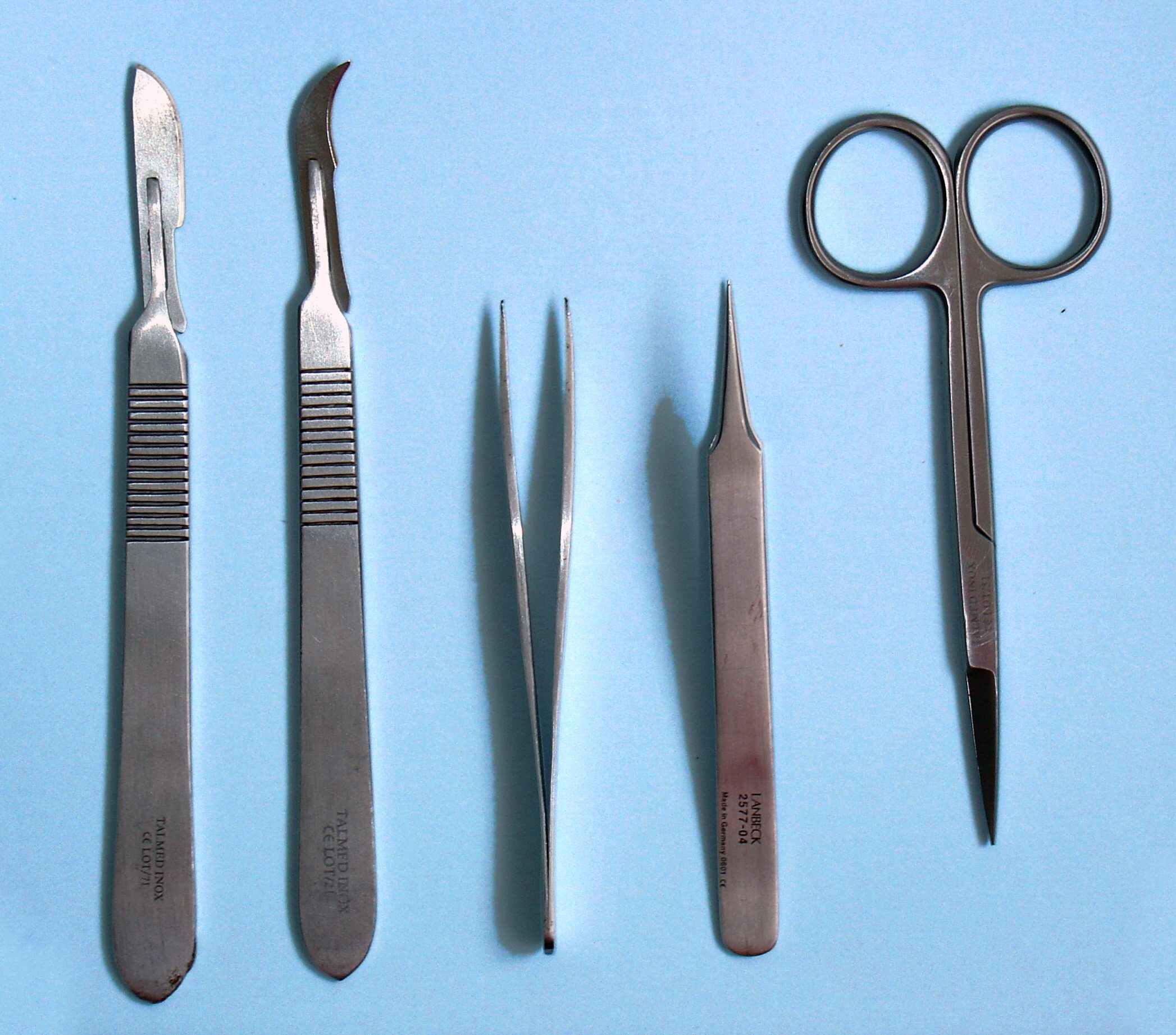
Dissection
Dissection (from Latin ' "to cut to pieces"; also called anatomization) is the dismembering of the body of a deceased animal or plant to study its anatomical structure. Autopsy is used in pathology and forensic medicine to determine the cause of death in humans. Less extensive dissection of plants and smaller animals preserved in a formaldehyde solution is typically carried out or demonstrated in biology and natural science classes in middle school and high school, while extensive dissections of cadavers of adults and children, both fresh and preserved are carried out by medical students in medical schools as a part of the teaching in subjects such as anatomy, pathology and forensic medicine. Consequently, dissection is typically conducted in a morgue or in an anatomy lab. Dissection has been used for centuries to explore anatomy. Objections to the use of cadavers have led to the use of alternatives including virtual dissection of computer models. Overview Plant and animal ... [...More Info...] [...Related Items...] OR: [Wikipedia] [Google] [Baidu] |
Cadaver
A cadaver or corpse is a dead human body that is used by medical students, physicians and other scientists to study anatomy, identify disease sites, determine causes of death, and provide tissue to repair a defect in a living human being. Students in medical school study and dissect cadavers as a part of their education. Others who study cadavers include archaeologists and arts students. The term ''cadaver'' is used in courts of law (and, to a lesser extent, also by media outlets such as newspapers) to refer to a dead body, as well as by recovery teams searching for bodies in natural disasters. The word comes from the Latin word ''cadere'' ("to fall"). Related terms include ''cadaverous'' (resembling a cadaver) and ''cadaveric spasm'' (a muscle spasm causing a dead body to twitch or jerk). A cadaver graft (also called “postmortem graft”) is the grafting of tissue from a dead body onto a living human to repair a defect or disfigurement. Cadavers can be observed for their sta ... [...More Info...] [...Related Items...] OR: [Wikipedia] [Google] [Baidu] |
Polydactyly
Polydactyly or polydactylism (), also known as hyperdactyly, is an anomaly in humans and animals resulting in supernumerary fingers and/or toes. Polydactyly is the opposite of oligodactyly (fewer fingers or toes). Signs and symptoms In humans/animals this condition can present itself on one or both hands or feet. The extra digit is usually a small piece of soft tissue that can be removed. Occasionally it contains bone without joints; rarely it may be a complete functioning digit. The extra digit is most common on the ulnar (little finger) side of the hand, less common on the radial ( thumb) side, and very rarely within the middle three digits. These are respectively known as postaxial (little finger), preaxial (thumb), and central (ring, middle, index fingers) polydactyly. The extra digit is most commonly an abnormal fork in an existing digit, or it may rarely originate at the wrist as a normal digit does. The incidence of congenital deformities in newborns is approximately 2 ... [...More Info...] [...Related Items...] OR: [Wikipedia] [Google] [Baidu] |
Metatarsophalangeal Articulations
The metatarsophalangeal joints (MTP joints), also informally known as toe knuckles, are the joints between the metatarsal bones of the foot and the proximal bones (proximal phalanges) of the toes. They are condyloid joints, meaning that an elliptical or rounded surface (of the metatarsal bones) comes close to a shallow cavity (of the proximal phalanges). The ligaments are the plantar and two collateral. Movements The movements permitted in the metatarsophalangeal joints are flexion, extension, abduction, adduction and circumduction. File:The feet of C. H. Unthan, the armless fiddler Wellcome L0034227.jpg, Left: toes adducted (pulled towards the center) and spread (abducted); right, both feet clenched (plantar flexed) File:Footgym rings1.jpg, The upper foot is clenching (plantarflexing) at the MTP joints and at the joints of the toes; the central foot is lifting the toes (dorsiflexing) at the MTP joints; and the foot flat on the ground off to the side is in a neutral position. ... [...More Info...] [...Related Items...] OR: [Wikipedia] [Google] [Baidu] |
Flexion
Motion, the process of movement, is described using specific anatomical terms. Motion includes movement of organs, joints, limbs, and specific sections of the body. The terminology used describes this motion according to its direction relative to the anatomical position of the body parts involved. Anatomists and others use a unified set of terms to describe most of the movements, although other, more specialized terms are necessary for describing unique movements such as those of the hands, feet, and eyes. In general, motion is classified according to the anatomical plane it occurs in. ''Flexion'' and ''extension'' are examples of ''angular'' motions, in which two axes of a joint are brought closer together or moved further apart. ''Rotational'' motion may occur at other joints, for example the shoulder, and are described as ''internal'' or ''external''. Other terms, such as ''elevation'' and ''depression'', describe movement above or below the horizontal plane. Many anatomic ... [...More Info...] [...Related Items...] OR: [Wikipedia] [Google] [Baidu] |
Tibial Nerve
The tibial nerve is a branch of the sciatic nerve. The tibial nerve passes through the popliteal fossa to pass below the arch of soleus. Structure Popliteal fossa The tibial nerve is the larger terminal branch of the sciatic nerve with root values of L4, L5, S1, S2, and S3. It lies superficial (or posterior) to the popliteal vessels, extending from the superior angle to the inferior angle of the popliteal fossa, crossing the popliteal vessels from lateral to medial side. It gives off branches as shown below: * Muscular branches - Muscular branches arise from the distal part of the popliteal fossa. It supplies the medial and lateral heads of gastrocnemius, soleus, plantaris and popliteus muscles. Nerve to popliteus crosses the popliteus muscle, runs downwards and laterally, winds around the lower border of the popliteus to supply the deep (or anterior) surface of the popliteus. This nerve also supplies the tibialis posterior muscle, superior tibiofibular joint, tibia bone, ... [...More Info...] [...Related Items...] OR: [Wikipedia] [Google] [Baidu] |
Lateral Plantar Nerve
The lateral plantar nerve (external plantar nerve) is a branch of the tibial nerve, in turn a branch of the sciatic nerve and supplies the skin of the fifth toe and lateral half of the fourth, as well as most of the deep muscles, its distribution being similar to that of the ulnar nerve in the hand. It passes obliquely forward with the lateral plantar artery to the lateral side of the foot, lying between the flexor digitorum brevis and quadratus plantae and, in the interval between the flexor muscle and the abductor digiti minimi, divides into a superficial and a deep branch. Before its division, it supplies the quadratus plantae and abductor digiti minimi. It divides into deep and superficial branches. Additional images File:Gray357.png, Coronal section through right talocrural and talocalcaneal joint In human anatomy, the subtalar joint, also known as the talocalcaneal joint, is a joint of the foot. It occurs at the meeting point of the talus and the calcaneus. Th ... [...More Info...] [...Related Items...] OR: [Wikipedia] [Google] [Baidu] |
Phalanx Bones
The phalanges (singular: ''phalanx'' ) are digital bones in the hands and feet of most vertebrates. In primates, the thumbs and big toes have two phalanges while the other digits have three phalanges. The phalanges are classed as long bones. Structure The phalanges are the bones that make up the fingers of the hand and the toes of the foot. There are 56 phalanges in the human body, with fourteen on each hand and foot. Three phalanges are present on each finger and toe, with the exception of the thumb and large toe, which possess only two. The middle and far phalanges of the fifth toes are often fused together (symphalangism). The phalanges of the hand are commonly known as the finger bones. The phalanges of the foot differ from the hand in that they are often shorter and more compressed, especially in the proximal phalanges, those closest to the torso. A phalanx is named according to whether it is proximal, middle, or distal and its associated finger or toe. The proximal ... [...More Info...] [...Related Items...] OR: [Wikipedia] [Google] [Baidu] |


