|
Axonogenesis
Axon guidance (also called axon pathfinding) is a subfield of neural development concerning the process by which neurons send out axons to reach their correct targets. Axons often follow very precise paths in the nervous system, and how they manage to find their way so accurately is an area of ongoing research. Axon growth takes place from a region called the growth cone and reaching the axon target is accomplished with relatively few guidance molecules. Growth cone receptors respond to the guidance cues. Mechanisms Growing axons have a highly motile structure at the growing tip called the growth cone, which responds to signals in the extracellular environment that instruct the axon in which direction to grow. These signals, called guidance cues, can be fixed in place or diffusible; they can attract or repel axons. Growth cones contain receptors that recognize these guidance cues and interpret the signal into a chemotropic response. The general theoretical framework is that whe ... [...More Info...] [...Related Items...] OR: [Wikipedia] [Google] [Baidu] |
Neural Development
The development of the nervous system, or neural development (neurodevelopment), refers to the processes that generate, shape, and reshape the nervous system of animals, from the earliest stages of embryonic development to adulthood. The field of neural development draws on both neuroscience and developmental biology to describe and provide insight into the cellular and molecular mechanisms by which complex nervous systems develop, from nematodes and fruit flies to mammals. Defects in neural development can lead to malformations such as holoprosencephaly, and a wide variety of neurological disorders including limb paresis and paralysis, balance and vision disorders, and seizures, and in humans other disorders such as Rett syndrome, Down syndrome and intellectual disability. Overview of vertebrate brain development The vertebrate central nervous system (CNS) is derived from the ectoderm—the outermost germ layer of the embryo. A part of the dorsal ectoderm becomes speci ... [...More Info...] [...Related Items...] OR: [Wikipedia] [Google] [Baidu] |
Plexins
A plexin is a protein which acts as a receptor for semaphorin family signaling proteins. It is classically known for its expression on the surface of axon growth cones and involvement in signal transduction to steer axon growth away from the source of semaphorin. Plexin also has implications in development of other body systems by activating GTPase enzymes to induce a number of intracellular biochemical changes leading to a variety of downstream effects. Structure Extracellular All plexins have an extracellular SEMA domain at their N-terminus. This is a structural motif common among all semaphorins and plexins and is responsible for this binding of semaphorin dimers, which are the native conformation for these ligands in vivo. This is followed by alternating plexin, semaphorin, and integrin (PSI) domains and immunoglobulin-like, plexin, and transcription factors (IPT) domains. Each of these is named for the proteins in which their structure is conserved. Collectively, the ex ... [...More Info...] [...Related Items...] OR: [Wikipedia] [Google] [Baidu] |
Xenopus
''Xenopus'' () (Gk., ξενος, ''xenos''=strange, πους, ''pous''=foot, commonly known as the clawed frog) is a genus of highly aquatic frogs native to sub-Saharan Africa. Twenty species are currently described within it. The two best-known species of this genus are ''Xenopus laevis'' and ''Xenopus tropicalis'', which are commonly studied as model organisms for developmental biology, cell biology, toxicology, neuroscience and for modelling human disease and birth defects. The genus is also known for its polyploidy, with some species having up to 12 sets of chromosomes. Characteristics ''Xenopus laevis'' is a rather inactive creature. It is incredibly hardy and can live up to 15 years. At times the ponds that ''Xenopus laevis'' is found in dry up, compelling it, in the dry season, to burrow into the mud, leaving a tunnel for air. It may lie dormant for up to a year. If the pond dries up in the rainy season, ''Xenopus laevis'' may migrate long distances to another pond, main ... [...More Info...] [...Related Items...] OR: [Wikipedia] [Google] [Baidu] |
Limb Development
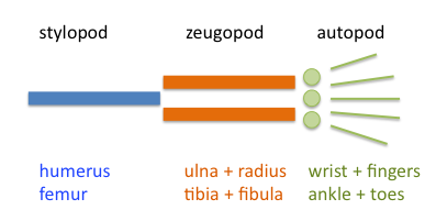
Limb development in vertebrates is an area of active research in both developmental and evolutionary biology, with much of the latter work focused on the transition from fin to limb. Limb formation begins in the morphogenetic limb field, as mesenchymal cells from the lateral plate mesoderm proliferate to the point that they cause the ectoderm above to bulge out, forming a limb bud. Fibroblast growth factor (FGF) induces the formation of an organizer at the end of the limb bud, called the apical ectodermal ridge (AER), which guides further development and controls cell death. Programmed cell death is necessary to eliminate webbing between digits. The limb field is a region specified by expression of certain Hox genes, a subset of homeotic genes, and T-box transcription factors – Tbx5 for forelimb or wing development, and Tbx4 for leg or hindlimb development. Establishment of the forelimb field (but not hindlimb field) requires retinoic acid signaling in the developing trunk of ... [...More Info...] [...Related Items...] OR: [Wikipedia] [Google] [Baidu] |
Pioneer Neuron
A pioneer neuron is a cell that is a derivative of the preplate in the early stages of corticogenesis of the brain. Pioneer neurons settle in the marginal zone of the cortex and project to sub-cortical levels. In the rat, pioneer neurons are only present in prenatal brains. Unlike Cajal-Retzius cells, these neurons are reelin-negative. Pioneer neurons are born in the ventricular neuroepithelium all over the cortical primordium. In the rat cortex, they appear at embryonic day (E) 11.5 in the lateral aspect of the telencephalic vesicle and cover its whole surface on E12. These cells, which show intense immunoreactivity for calbindin and calretinin, are characterized by their large size and axonal projection. They remain in the marginal zone after the formation of the cortical plate; they project first into the ventricular zone, and then into the subplate and the internal capsule. Therefore, these cells are the origin of the earliest efferent pathway of the developing cortex. Funct ... [...More Info...] [...Related Items...] OR: [Wikipedia] [Google] [Baidu] |
Pioneer Axon
Pioneer axon is the classification given to axons that are the first to grow in a particular region. They originate from pioneer neurons, and have the main function of laying down the initial growing path that subsequent growing axons, dubbed follower axons, from other neurons will eventually follow. Several theories relating to the structure and function of pioneer axons are currently being explored. The first theory is that pioneer axons are specialized structures, and that they play a crucial role in guiding follower axons. The second is that pioneer axons are no different from follower axons, and that they play no role in guiding follower axons. Anatomically, there are no differences between pioneer and follower axons, although there are morphological differences. The mechanisms of pioneer axons and their role in axon guidance is currently being explored. In addition, many studies are being conducted in model organisms, such grasshoppers, zebrafish, and fruit flies to study t ... [...More Info...] [...Related Items...] OR: [Wikipedia] [Google] [Baidu] |
Commissure
A commissure () is the location at which two objects abut or are joined. The term is used especially in the fields of anatomy and biology. * The most common usage of the term refers to the brain's commissures, of which there are five. Such a commissure is a bundle of commissural fibers as a tract that crosses the midline at its level of origin or entry (as opposed to a decussation of fibers that cross obliquely). The five are the anterior commissure, posterior commissure, corpus callosum, commissure of fornix (hippocampal commissure), and habenular commissure. They consist of fibre tracts that connect the two cerebral hemispheres and span the longitudinal fissure. In the spinal cord there are the anterior white commissure, and the gray commissure. ''Commissural neurons'' refer to neuronal cells that grow their axons across the midline of the nervous system within the brain and the spinal cord. * ''Commissure'' also often refers to cardiac anatomy of heart valves. In the heart, a co ... [...More Info...] [...Related Items...] OR: [Wikipedia] [Google] [Baidu] |
Neurotrophins
Neurotrophins are a family of proteins that induce the survival, development, and function of neurons. They belong to a class of growth factors, secreted proteins that can signal particular cells to survive, differentiate, or grow. Growth factors such as neurotrophins that promote the survival of neurons are known as neurotrophic factors. Neurotrophic factors are secreted by target tissue and act by preventing the associated neuron from initiating programmed cell death – allowing the neurons to survive. Neurotrophins also induce differentiation of progenitor cells, to ''form'' neurons. Although the vast majority of neurons in the mammalian brain are formed prenatally, parts of the adult brain (for example, the hippocampus) retain the ability to grow new neurons from neural stem cells, a process known as neurogenesis. Neurotrophins are chemicals that help to stimulate and control neurogenesis. Terminology According to the United States National Library of Medicine's medical sub ... [...More Info...] [...Related Items...] OR: [Wikipedia] [Google] [Baidu] |
Olfactory Bulb
The olfactory bulb (Latin: ''bulbus olfactorius'') is a grey matter, neural structure of the vertebrate forebrain involved in olfaction, the sense of odor, smell. It sends olfactory information to be further processed in the amygdala, the orbitofrontal cortex (OFC) and the hippocampus where it plays a role in emotion, memory and learning. The bulb is divided into two distinct structures: the main olfactory bulb and the accessory olfactory bulb. The main olfactory bulb connects to the amygdala via the piriform cortex of the primary olfactory cortex and directly projects from the main olfactory bulb to specific amygdala areas. The accessory olfactory bulb resides on the dorsal-posterior region of the main olfactory bulb and forms a parallel pathway. Destruction of the olfactory bulb results in ipsilateral anosmia, while irritative lesions of the uncus can result in olfactory and gustatory hallucinations. Structure In most vertebrates, the olfactory bulb is the most Anatomical term ... [...More Info...] [...Related Items...] OR: [Wikipedia] [Google] [Baidu] |
Cell-adhesion Molecule
Cell adhesion molecules (CAMs) are a subset of cell surface proteins that are involved in the binding of cells with other cells or with the extracellular matrix (ECM), in a process called cell adhesion. In essence, CAMs help cells stick to each other and to their surroundings. CAMs are crucial components in maintaining tissue structure and function. In fully developed animals, these molecules play an integral role in generating force and movement and consequently ensuring that organs are able to execute their functions normally. In addition to serving as "molecular glue", CAMs play important roles in the cellular mechanisms of growth, contact inhibition, and apoptosis. Aberrant expression of CAMs may result in a wide range of pathologies, ranging from frostbite to cancer. Structure CAMs are typically single-pass transmembrane receptors and are composed of three conserved domains: an intracellular domain that interacts with the cytoskeleton, a transmembrane domain, and an extrac ... [...More Info...] [...Related Items...] OR: [Wikipedia] [Google] [Baidu] |
Cadherin
Cadherins (named for "calcium-dependent adhesion") are a type of cell adhesion molecule (CAM) that is important in the formation of adherens junctions to allow cells to adhere to each other . Cadherins are a class of type-1 transmembrane proteins, and they are dependent on calcium (Ca2+) ions to function, hence their name. Cell-cell adhesion is mediated by extracellular cadherin domains, whereas the intracellular cytoplasmic tail associates with numerous adaptors and signaling proteins, collectively referred to as the cadherin adhesome. The cadherin family is essential in maintaining the cell-cell contact and regulating cytoskeletal complexes. The cadherin superfamily includes cadherins, protocadherins, desmogleins, desmocollins, and more. In structure, they share ''cadherin repeats'', which are the extracellular Ca2+-binding domains. There are multiple classes of cadherin molecules, each designated with a prefix (in general, noting the types of tissue with which it is associated). ... [...More Info...] [...Related Items...] OR: [Wikipedia] [Google] [Baidu] |
Extracellular Matrix
In biology, the extracellular matrix (ECM), also called intercellular matrix, is a three-dimensional network consisting of extracellular macromolecules and minerals, such as collagen, enzymes, glycoproteins and hydroxyapatite that provide structural and biochemical support to surrounding cells. Because multicellularity evolved independently in different multicellular lineages, the composition of ECM varies between multicellular structures; however, cell adhesion, cell-to-cell communication and differentiation are common functions of the ECM. The animal extracellular matrix includes the interstitial matrix and the basement membrane. Interstitial matrix is present between various animal cells (i.e., in the intercellular spaces). Gels of polysaccharides and fibrous proteins fill the Interstitial fluid, interstitial space and act as a compression buffer against the stress placed on the ECM. Basement membranes are sheet-like depositions of ECM on which various epithelial cells rest ... [...More Info...] [...Related Items...] OR: [Wikipedia] [Google] [Baidu] |


