|
Axial Skeleton
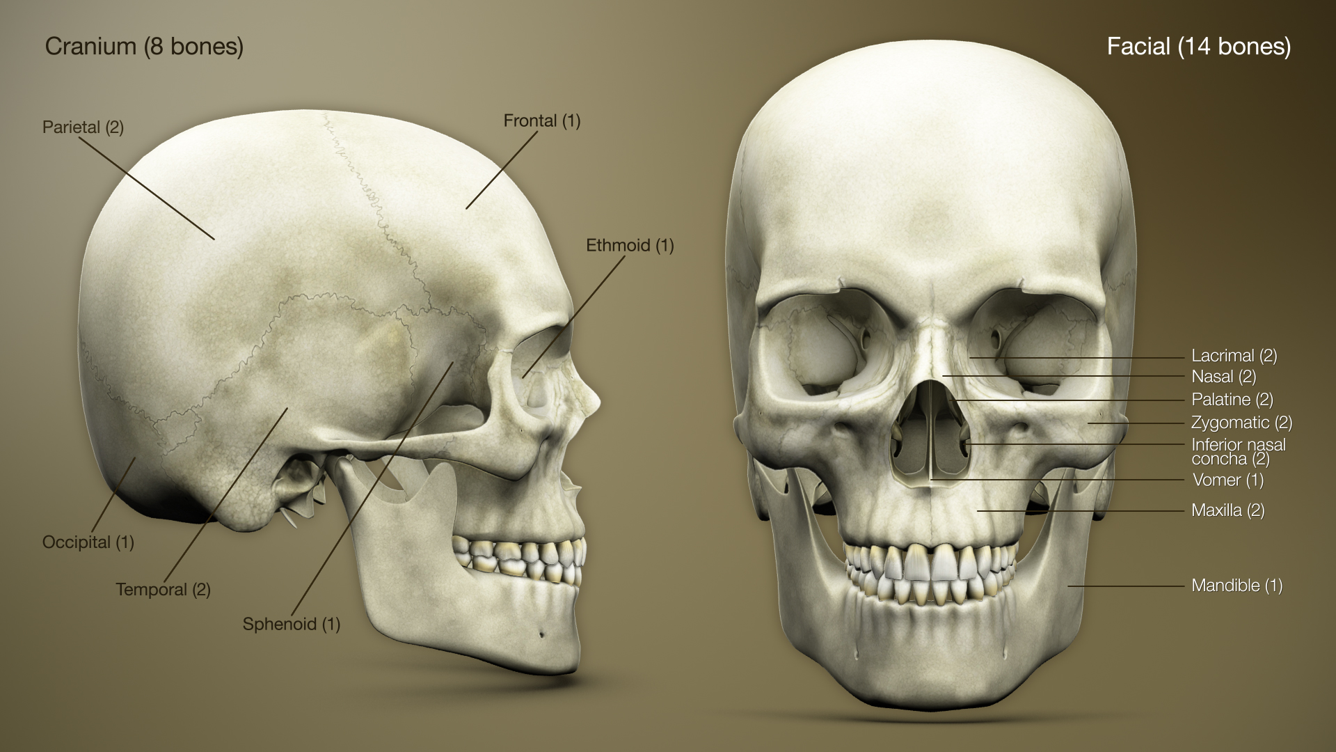
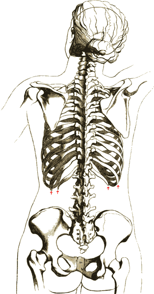
The axial skeleton is the core part of the endoskeleton made of the bones of the head and trunk of vertebrates. In the human skeleton, it consists of 80 bones and is composed of the skull (28 bones, including the cranium, mandible and the middle ear ossicles), the vertebral column (26 bones, including vertebrae, sacrum and coccyx), the rib cage (25 bones, including ribs and sternum), and the hyoid bone. The axial skeleton is joined to the appendicular skeleton (which support the limbs) via the shoulder girdles and the pelvis. Structure Flat bones house the brain and other vital organs. This article mainly deals with the axial skeletons of humans; however, it is important to understand its evolutionary lineage. The human axial skeleton consists of 81 different bones forming the medial core of the body and connects the pelvis to the body, where the appendicular skeleton attaches. As the skeleton grows older the bones get weaker with the exception of the skull. The skull ... [...More Info...] [...Related Items...] OR: [Wikipedia] [Google] [Baidu] |
3D Medical Animation Axial Skeleton
3D, 3-D, 3d, or Three D may refer to: Science, technology, and mathematics * A three-dimensional space in mathematics Relating to three-dimensionality * 3D computer graphics, computer graphics that use a three-dimensional representation of geometric data * 3D display, a type of information display that conveys depth to the viewer * 3D film, a motion picture that gives the illusion of three-dimensional perception * 3D modeling, developing a representation of any three-dimensional surface or object * 3D printing, making a three-dimensional solid object of a shape from a digital model * 3D television, television that conveys depth perception to the viewer * 3D projection * 3D rendering * 3D scanning, making a digital representation of three-dimensional objects * Video game graphics#3D, 3D video game * Stereoscopy, any technique capable of recording three-dimensional visual information or creating the illusion of depth in an image * Three-dimensional space Other uses in science and ... [...More Info...] [...Related Items...] OR: [Wikipedia] [Google] [Baidu] |
Rib Cage
The rib cage or thoracic cage is an endoskeletal enclosure in the thorax of most vertebrates that comprises the ribs, vertebral column and sternum, which protect the vital organs of the thoracic cavity, such as the heart, lungs and great vessels and support the shoulder girdle to form the core part of the axial skeleton. A typical human thoracic cage consists of 12 pairs of ribs and the adjoining costal cartilages, the sternum (along with the manubrium and xiphoid process), and the 12 thoracic vertebrae articulating with the ribs. The thoracic cage also provides attachments for extrinsic skeletal muscles of the neck, upper limbs, upper abdomen and back, and together with the overlying skin and associated fascia and muscles, makes up the thoracic wall. In tetrapods, the rib cage intrinsically holds the muscles of respiration ( diaphragm, intercostal muscles, etc.) that are crucial for active inhalation and forced exhalation, and therefore has a major ventilatory fu ... [...More Info...] [...Related Items...] OR: [Wikipedia] [Google] [Baidu] |
Ribs
The rib cage or thoracic cage is an endoskeletal enclosure in the thorax of most vertebrates that comprises the ribs, vertebral column and sternum, which protect the vital organs of the thoracic cavity, such as the heart, lungs and great vessels and support the shoulder girdle to form the core (anatomy), core part of the axial skeleton. A typical human skeleton, human thoracic cage consists of 12 pairs of ribs and the adjoining costal cartilages, the sternum (along with the manubrium and xiphoid process), and the 12 thoracic vertebrae articulating with the ribs. The thoracic cage also provides attachments for extrinsic skeletal muscles of the neck, upper limbs, upper abdomen and back, and together with the overlying skin and associated fascia and muscles, makes up the thoracic wall. In tetrapods, the rib cage intrinsically holds the muscles of respiration (thoracic diaphragm, diaphragm, intercostal muscles, etc.) that are crucial for active inhalation and forced exhalation, and ... [...More Info...] [...Related Items...] OR: [Wikipedia] [Google] [Baidu] |
Neck
The neck is the part of the body in many vertebrates that connects the head to the torso. It supports the weight of the head and protects the nerves that transmit sensory and motor information between the brain and the rest of the body. Additionally, the neck is highly flexible, allowing the head to turn and move in all directions. Anatomically, the human neck is divided into four compartments: vertebral, visceral, and two vascular compartments. Within these compartments, the neck houses the cervical vertebrae, the cervical portion of the spinal cord, upper parts of the respiratory and digestive tracts, endocrine glands, nerves, arteries and veins. The muscles of the neck, which are separate from the compartments, form the boundaries of the neck triangles. In anatomy, the neck is also referred to as the or . However, when the term ''cervix'' is used alone, it often refers to the uterine cervix, the neck of the uterus. Therefore, the adjective ''cervical'' ... [...More Info...] [...Related Items...] OR: [Wikipedia] [Google] [Baidu] |
Middle Ear Ossicles
The ossicles (also called auditory ossicles) are three irregular bones in the middle ear of humans and other mammals, and are among the smallest bones in the human body. Although the term "ossicle" literally means "tiny bone" (from Latin ''ossiculum'') and may refer to any small bone throughout the body, it typically refers specifically to the malleus, incus and stapes ("hammer, anvil, and stirrup") of the middle ear. The auditory ossicles serve as a kinematic chain to transmit and amplify (intensify) sound vibrations collected from the air by the ear drum to the fluid-filled labyrinth (cochlea). The absence or pathology of the auditory ossicles would constitute a moderate-to-severe conductive hearing loss. Structure The ossicles are, in order from the eardrum to the inner ear (from superficial to deep): the malleus, incus, and stapes, terms that in Latin are translated as "the hammer, anvil, and stirrup". * The malleus () articulates with the incus through the incudomalleola ... [...More Info...] [...Related Items...] OR: [Wikipedia] [Google] [Baidu] |
Skull
The skull, or cranium, is typically a bony enclosure around the brain of a vertebrate. In some fish, and amphibians, the skull is of cartilage. The skull is at the head end of the vertebrate. In the human, the skull comprises two prominent parts: the neurocranium and the facial skeleton, which evolved from the first pharyngeal arch. The skull forms the frontmost portion of the axial skeleton and is a product of cephalization and vesicular enlargement of the brain, with several special senses structures such as the eyes, ears, nose, tongue and, in fish, specialized tactile organs such as barbels near the mouth. The skull is composed of three types of bone: cranial bones, facial bones and ossicles, which is made up of a number of fused flat and irregular bones. The cranial bones are joined at firm fibrous junctions called sutures and contains many foramina, fossae, processes, and sinuses. In zoology, the openings in the skull are called fenestrae, the most ... [...More Info...] [...Related Items...] OR: [Wikipedia] [Google] [Baidu] |
Cervical Vertebrae
In tetrapods, cervical vertebrae (: vertebra) are the vertebrae of the neck, immediately below the skull. Truncal vertebrae (divided into thoracic and lumbar vertebrae in mammals) lie caudal (toward the tail) of cervical vertebrae. In sauropsid species, the cervical vertebrae bear cervical ribs. In lizards and saurischian dinosaurs, the cervical ribs are large; in birds, they are small and completely fused to the vertebrae. The vertebral transverse processes of mammals are homologous to the cervical ribs of other amniotes. Most mammals have seven cervical vertebrae, with the only three known exceptions being the manatee with six, the two-toed sloth with five or six, and the three-toed sloth with nine. In humans, cervical vertebrae are the smallest of the true vertebrae and can be readily distinguished from those of the thoracic or lumbar regions by the presence of a transverse foramen, an opening in each transverse process, through which the vertebral artery, verteb ... [...More Info...] [...Related Items...] OR: [Wikipedia] [Google] [Baidu] |
Sobotta's Atlas Of Human Anatomy
Robert Heinrich Johannes Sobotta (31 January 1869 in Berlin – 20 April 1945 in Bonn) was a German anatomist. He studied medicine in Berlin, where he subsequently worked as a second assistant at the institute of anatomy. From 1895 he served as prosector at the institute for comparative anatomy, embryology and histology at Würzburg. In 1903 he became an associate professor and in 1912 a full professor of topographical anatomy. In 1916 he relocated to the University of Königsberg as director of the anatomical institute, afterwards performing similar duties at the University of Bonn (from 1919).Uni Giessen.de (biography in German) He is remembered today for the ''Sobotta atlas of human anatomy'', a masterpiece of macroscopic anatomy acclaimed for its high quality and detail. First issued in 1904 with the ti ... [...More Info...] [...Related Items...] OR: [Wikipedia] [Google] [Baidu] |
Pelvis
The pelvis (: pelves or pelvises) is the lower part of an Anatomy, anatomical Trunk (anatomy), trunk, between the human abdomen, abdomen and the thighs (sometimes also called pelvic region), together with its embedded skeleton (sometimes also called bony pelvis or pelvic skeleton). The pelvic region of the trunk includes the bony pelvis, the pelvic cavity (the space enclosed by the bony pelvis), the pelvic floor, below the pelvic cavity, and the perineum, below the pelvic floor. The pelvic skeleton is formed in the area of the back, by the sacrum and the coccyx and anteriorly and to the left and right sides, by a pair of hip bones. The two hip bones connect the spine with the lower limbs. They are attached to the sacrum posteriorly, connected to each other anteriorly, and joined with the two femurs at the hip joints. The gap enclosed by the bony pelvis, called the pelvic cavity, is the section of the body underneath the abdomen and mainly consists of the reproductive organs and ... [...More Info...] [...Related Items...] OR: [Wikipedia] [Google] [Baidu] |
Shoulder Girdle
The shoulder girdle or pectoral girdle is the set of bones in the appendicular skeleton which connects to the arm on each side. In humans, it consists of the clavicle and scapula; in those species with three bones in the shoulder, it consists of the clavicle, scapula, and coracoid. Some mammalian species (such as the dog and the horse) have only the scapula. The pectoral girdles are to the upper limbs as the pelvic girdle is to the lower limbs; the girdles are the part of the appendicular skeleton that anchor the appendages to the axial skeleton. In humans, the only true anatomical joints between the shoulder girdle and the axial skeleton are the sternoclavicular joints on each side. No anatomical joint exists between each scapula and the rib cage; instead the muscular connection or physiological joint between the two permits great mobility of the shoulder girdle compared to the compact pelvic girdle; because the upper limb is not usually involved in weight bearing, its stabil ... [...More Info...] [...Related Items...] OR: [Wikipedia] [Google] [Baidu] |
Limb (anatomy)
A limb (from Old English ''lim'', meaning "body part") is a jointed, muscled appendage of a tetrapod vertebrate animal used for weight-bearing, terrestrial locomotion and physical interaction with other objects. The distalmost portion of a limb is known as its extremity. The limbs' bony endoskeleton, known as the appendicular skeleton, is homologous among all tetrapods, who use their limbs for walking, running and jumping, swimming, climbing, grasping, touching and striking. All tetrapods have four limbs that are organized into two bilaterally symmetrical pairs, with one pair at each end of the torso, which phylogenetically correspond to the four paired fins ( pectoral and pelvic fins) of their fish ( sarcopterygian) ancestors. The cranial pair (i.e. closer to the head) of limbs are known as the forelimbs or ''front legs'', and the caudal pair (i.e. closer to the tail or coccyx) are the hindlimbs or ''back legs''. In animals with a more erect bipedal ... [...More Info...] [...Related Items...] OR: [Wikipedia] [Google] [Baidu] |
Appendicular Skeleton
The appendicular skeleton is the portion of the vertebrate endoskeleton consisting of the bones, cartilages and ligaments that support the paired appendages ( fins, flippers or limbs). In most terrestrial vertebrates (except snakes, legless lizards and caecillians), the appendicular skeleton and the associated skeletal muscles are the predominant locomotive structures. There are 126 bones in the human appendicular skeleton, includes the skeletal elements within the shoulder and pelvic girdles, upper and lower limbs, and hands and feet. These bones have shared ancestry (are homologous) to those in the forelimbs and hindlimbs of all other tetrapods, which are in turn homologous to the pectoral and pelvic fins in fish. Etymology The adjective "appendicular" comes from Latin ''appendicula'', meaning "small addition". It is the diminutive of ''appendix'', which comes from the prefix ''ad-'' (meaning "to") + and the word root ''pendere'' (meaning "to hang", from PIE root ''* ... [...More Info...] [...Related Items...] OR: [Wikipedia] [Google] [Baidu] |


