|
Astrocytomas
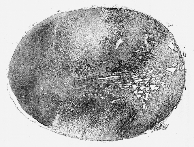
Astrocytoma is a type of brain tumor. Astrocytomas (also astrocytomata) originate from a specific kind of star-shaped glial cell in the cerebrum called an astrocyte. This type of tumor does not usually spread outside the brain and spinal cord, and it does not usually affect other organs. After glioblastomas, astrocytomas are the second most common glioma and can occur in most parts of the brain and occasionally in the spinal cord. Within the astrocytomas, two broad classes are recognized in literature, those with: * Narrow zones of infiltration (mostly noninvasive tumors; e.g., pilocytic astrocytoma, subependymal giant cell astrocytoma, pleomorphic xanthoastrocytoma), that often are clearly outlined on diagnostic images * Diffuse zones of infiltration (e.g., high-grade astrocytoma), that share various features, including the ability to arise at any location in the central nervous system, but with a preference for the cerebral hemispheres; they occur usually in adults, and have an ... [...More Info...] [...Related Items...] OR: [Wikipedia] [Google] [Baidu] |
Anaplastic Astrocytoma
Anaplastic astrocytoma is a rare WHO grade III type of astrocytoma, which is a type of cancer of the brain. In the United States, the annual incidence rate for anaplastic astrocytoma is 0.44 per 100,000 people. Signs and symptoms Initial presenting symptoms most commonly are headache, depressed mental status, focal neurological deficits, and/or seizures. The growth rate and mean interval between onset of symptoms and diagnosis is approximately 1.5–2 years but is highly variable, being intermediate between that of low-grade astrocytomas and glioblastomas. Seizures are less common among patients with anaplastic astrocytomas compared to low-grade lesions. Causes The best-known risk factor is exposure to ionizing radiation, and CT scan radiation is an important cause.Smoll NR, Brady Z, Scurrah KJ, Lee C, Berrington de González A, Mathews JD. Computed tomography scan radiation and brain cancer incidence. Neuro-Oncology. 2023 Jan 14;https://doi.org/10.1093/neuonc/noad012Smoll NR, Bra ... [...More Info...] [...Related Items...] OR: [Wikipedia] [Google] [Baidu] |
Pilocytic Astrocytoma
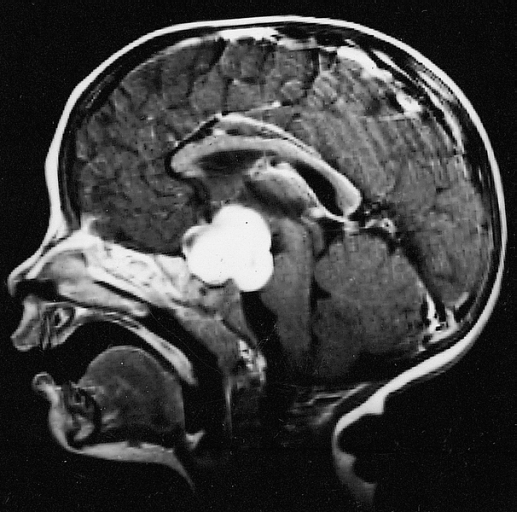
Pilocytic astrocytoma (and its variant pilomyxoid astrocytoma) is a brain tumor that occurs most commonly in children and young adults (in the first 20 years of life). They usually arise in the cerebellum, near the brainstem, in the hypothalamic region, or the optic chiasm, but they may occur in any area where astrocytes are present, including the cerebral hemispheres and the spinal cord. These tumors are usually slow growing and benign, corresponding to WHO malignancy grade 1. Signs and symptoms Children affected by pilocytic astrocytoma can present with different symptoms that might include failure to thrive (lack of appropriate weight gain/ weight loss), headache, nausea, vomiting, irritability, torticollis (tilt neck or wry neck), difficulty to coordinate movements, and visual complaints (including nystagmus). The complaints may vary depending on the location and size of the neoplasm. The most common symptoms are associated with increased intracranial pressure due to the siz ... [...More Info...] [...Related Items...] OR: [Wikipedia] [Google] [Baidu] |
Grading Of The Tumors Of The Central Nervous System
The concept of grading of the tumors of the central nervous system, agreeing for such the regulation of the "progressiveness" of these neoplasias (from benign and localized tumors to malignant and infiltrating tumors), dates back to 1926 and was introduced by P. Bailey and H. Cushing, in the elaboration of what turned out the first systematic classification of gliomas. In the following, the grading systems present in the current literature are introduced. Then, through a table, the more relevant are compared. ICD-O scale The first edition of the International Classification of Diseases (ICD) dates back to 1893. The current review (ICD-10) dates back to 1994, came into use in the U.S. in 2015, and is revised yearly, being very comprehensive. In 1976 the World Health Organization (WHO) published the first edition of the International Classification of Diseases for Oncology (ICD-O), which is now at the third edition (ICD-O-3, 2000). In this last edition, the Arabic numeral after t ... [...More Info...] [...Related Items...] OR: [Wikipedia] [Google] [Baidu] |
Pleomorphic Xanthoastrocytoma
Pleomorphic xanthoastrocytoma (PXA) is a brain tumor that occurs most frequently in children and teenagers. At Boston Children's Hospital, the average age at diagnosis is 12 years. Pleomorphic xanthoastrocytoma usually develops within the supratentorial region (the area of the brain located above the tentorium cerebelli). It is generally located superficially (in the uppermost sections) in the cerebral hemispheres and involves the leptomeninges. It rarely arises from the spinal cord. These tumors are formed through the mitosis of astrocytes. They are found in the area of the temples, in the brain's frontal lobe or on top of the parietal lobe. In about 20% of cases, tumors exist in more than one lobe. Symptoms and signs Children with PXA can present with a variety of symptoms. Complaints may vary, and patients may report symptoms that have been occurring for many months and are often linked with more common diseases. (For example, headaches are a common complaint.) Some children ... [...More Info...] [...Related Items...] OR: [Wikipedia] [Google] [Baidu] |
Fibrillary Astrocytoma
Fibrillary astrocytomas are a group of primary slow-growing brain tumors that typically occur in adults between the ages of 20 and 50. Symptoms Seizures, frequent mood changes, and headaches are among the earliest symptoms of the tumor. Hemiparesis (physical weakness on one side of the body) is also common. Pathology Fibrillary astrocytomas arise from neoplastic astrocytes, a type of glial cell found in the central nervous system. They may occur anywhere in the brain, or even in the spinal cord, but are most commonly found in the cerebral hemispheres. As the alternative name "diffuse astrocytoma" implies, the outline of the tumour is not clearly visible in scans, because the borders of the neoplasm tend to send out tiny microscopic fibrillary tentacles that spread into the surrounding brain tissue. These tentacles intermingle with healthy brain cells, making complete surgical removal difficult. However, they are low-grade tumors, with a slow rate of growth, so patients commonly ... [...More Info...] [...Related Items...] OR: [Wikipedia] [Google] [Baidu] |
Astrocyte
Astrocytes (from Ancient Greek , , "star" and , , "cavity", "cell"), also known collectively as astroglia, are characteristic star-shaped glial cells in the brain and spinal cord. They perform many functions, including biochemical control of endothelial cells that form the blood–brain barrier, provision of nutrients to the nervous tissue, maintenance of extracellular ion balance, regulation of cerebral blood flow, and a role in the repair and scarring process of the brain and spinal cord following infection and traumatic injuries. The proportion of astrocytes in the brain is not well defined; depending on the counting technique used, studies have found that the astrocyte proportion varies by region and ranges from 20% to around 40% of all glia. Another study reports that astrocytes are the most numerous cell type in the brain. Astrocytes are the major source of cholesterol in the central nervous system. Apolipoprotein E transports cholesterol from astrocytes to neurons and ot ... [...More Info...] [...Related Items...] OR: [Wikipedia] [Google] [Baidu] |
Oligodendroglioma
Oligodendrogliomas are a type of glioma that are believed to originate from the oligodendrocytes of the brain or from a oligodendrocyte progenitor cell, glial precursor cell. They occur primarily in adults (9.4% of all primary brain and central nervous system tumors) but are also found in children (4% of all primary brain tumors). With a 0.2 incidence (epidemiology), incidence rate out of 100,000 adults, oligodendrogliomas comprise approximately 5% of all central nervous system tumors. Signs and symptoms Oligodendroglioma arise mainly in the frontal lobe and in 50–80% of cases, the first symptom is the onset of seizure activity without any prior symptoms . Headaches combined with increased intracranial pressure are also a common symptom of oligodendroglioma. Depending on the location of the tumor, many different neurological and neuropsychological deficits can be induced, including, but not limited to, visual loss, muscle weakness, motor weakness, cognitive decline, and anxiety. ... [...More Info...] [...Related Items...] OR: [Wikipedia] [Google] [Baidu] |
Brain Tumor
A brain tumor (sometimes referred to as brain cancer) occurs when a group of cells within the Human brain, brain turn cancerous and grow out of control, creating a mass. There are two main types of tumors: malignant (cancerous) tumors and benign tumor, benign (non-cancerous) tumors. These can be further classified as primary tumors, which start within the brain, and metastasis, secondary tumors, which most commonly have spread from tumors located outside the brain, known as brain metastasis tumors. All types of brain tumors may produce symptoms that vary depending on the size of the tumor and the part of the brain that is involved. Where symptoms exist, they may include headaches, seizures, problems with visual perception, vision, vomiting and cognition, mental changes. Other symptoms may include difficulty walking, speaking, with sensations, or unconsciousness. The cause of most brain tumors is unknown, though up to 4% of brain cancers may be caused by CT scan radiation. Uncommo ... [...More Info...] [...Related Items...] OR: [Wikipedia] [Google] [Baidu] |
Glioblastoma
Glioblastoma, previously known as glioblastoma multiforme (GBM), is the most aggressive and most common type of cancer that originates in the brain, and has a very poor prognosis for survival. Initial signs and symptoms of glioblastoma are nonspecific. They may include headaches, personality changes, nausea, and symptoms similar to those of a stroke. Symptoms often worsen rapidly and may progress to unconsciousness. The cause of most cases of glioblastoma is not known. Uncommon risk factors include genetic disorders, such as neurofibromatosis and Li–Fraumeni syndrome, and previous radiation therapy. Glioblastomas represent 15% of all brain tumors. They are thought to arise from astrocytes. The diagnosis typically is made by a combination of a CT scan, MRI scan, and tissue biopsy. There is no known method of preventing the cancer. Treatment usually involves surgery, after which chemotherapy and radiation therapy are used. The medication temozolomide is frequently used a ... [...More Info...] [...Related Items...] OR: [Wikipedia] [Google] [Baidu] |
Glioma
A glioma is a type of primary tumor that starts in the glial cells of the brain or spinal cord. They are malignant but some are extremely slow to develop. Gliomas comprise about 30% of all brain and central nervous system tumors and 80% of all malignant brain tumors. They are a few common types that include astrocytoma (cancer of astrocytes), glioblastoma (an aggressive form of astrocytoma), oligodendroglioma (cancer of oligodendrocytes), and ependymoma (cancer of ependymal cells). Signs and symptoms Symptoms of gliomas depend on the part of the central nervous system (CNS) that is affected. A brain glioma can cause headaches, vomiting, memory loss, seizures, vision problems, speech difficulties, and cranial nerve disorders as a result of increased intracranial pressure. Cognitive impairments such as vision loss arise in glioma patients when a tumor arises in or around their optic nerve. Spinal cord gliomas can cause pain, weakness, or numbness in the extremities ... [...More Info...] [...Related Items...] OR: [Wikipedia] [Google] [Baidu] |
Subependymoma
A subependymoma is a type of brain tumor; specifically, it is a rare form of ependymal tumor. They are usually in middle aged people. Earlier, they were called subependymal astrocytomas. The prognosis for a subependymoma is better than for most ependymal tumors, and it is considered a grade I tumor in the World Health Organization (WHO) classification. They are classically found within the fourth ventricle, typically have a well demarcated interface to normal tissue and do not usually extend into the brain parenchyma, like ependymomas often do. Symptoms and signs Patients are often asymptomatic, and are incidentally diagnosed. Larger tumours are often with increased intracranial pressure. Pathology These tumours are small, no more than two centimeters across, coming from the ependyma. The best way to distinguish it from a subependymal giant cell astrocytoma is the size. Diagnosis The diagnosis is based on tissue, e.g. a biopsy. Histologically subependymomas consistent ... [...More Info...] [...Related Items...] OR: [Wikipedia] [Google] [Baidu] |
Mitosis
Mitosis () is a part of the cell cycle in eukaryote, eukaryotic cells in which replicated chromosomes are separated into two new Cell nucleus, nuclei. Cell division by mitosis is an equational division which gives rise to genetically identical cells in which the total number of chromosomes is maintained. Mitosis is preceded by the S phase of interphase (during which DNA replication occurs) and is followed by telophase and cytokinesis, which divide the cytoplasm, organelles, and cell membrane of one cell into two new cell (biology), cells containing roughly equal shares of these cellular components. The different stages of mitosis altogether define the mitotic phase (M phase) of a cell cycle—the cell division, division of the mother cell into two daughter cells genetically identical to each other. The process of mitosis is divided into stages corresponding to the completion of one set of activities and the start of the next. These stages are preprophase (specific to plant ce ... [...More Info...] [...Related Items...] OR: [Wikipedia] [Google] [Baidu] |


