|
Aponeurosis Of The Abdominal External Oblique Muscle
The aponeurosis of the abdominal external oblique muscle is a thin but strong membranous structure, the fibers of which are directed downward and medially. It is joined with that of the opposite muscle along the middle line, and covers the whole of the front of the abdomen; above, it is covered by and gives origin to the lower fibers of the pectoralis major; below, its fibers are closely aggregated together, and extend obliquely across from the anterior superior iliac spine to the pubic tubercle and the pectineal line to form the inguinal ligament. In the middle line, it interlaces with the aponeurosis of the opposite muscle, forming the linea alba, which extends from the xiphoid process to the pubic symphysis. That portion of the aponeurosis which extends between the anterior superior iliac spine and the pubic tubercle is a thick band, folded inward, and continuous below with the fascia lata; it is called the inguinal ligament. The portion which is reflected from the inguinal ... [...More Info...] [...Related Items...] OR: [Wikipedia] [Google] [Baidu] |
Superficial Inguinal Ring
The inguinal canals are the two passages in the anterior abdominal wall of humans and animals which in males convey the spermatic cords and in females the round ligament of the uterus. The inguinal canals are larger and more prominent in males. There is one inguinal canal on each side of the midline. Structure The inguinal canals are situated just above the medial half of the inguinal ligament. In both sexes the canals transmit the ilioinguinal nerves. The canals are approximately 3.75 to 4 cm long. , angled anteroinferiorly and medially. In males, its diameter is normally 2 cm (±1 cm in standard deviation) at the deep inguinal ring.The diameter has been estimated to be ±2.2cm ±1.08cm in Africans, and 2.1 cm ±0.41cm in Europeans. A first-order approximation is to visualize each canal as a cylinder. Walls To help define the boundaries, these canals are often further approximated as boxes with six sides. Not including the two rings, the remaining four sides are usually cal ... [...More Info...] [...Related Items...] OR: [Wikipedia] [Google] [Baidu] |
Linea Alba (abdomen)
The linea alba ( la, white line) is a fibrous structure that runs down the midline of the abdomen in humans and other vertebrates. Structure In humans, the linea alba runs from the xiphoid process to the pubic symphysis down the midline of the abdomen. The name means ''white line'' as it is composed mostly of collagen connective tissue, which has a white appearance. It is formed by the fusion of the aponeuroses of the muscles of the anterior abdominal wall. It separates the left and right rectus abdominis muscles. In muscular individuals, its presence can be seen on the skin, forming the depression between the left and right halves of a " six pack". Function The Linea alba stabilizes the anterior abdominal wall, as it balances contractile forces from the muscles attached to it. Clinical significance A median incision through the linea alba is a common surgical approach for abdominal surgery. This is because it consists of mostly connective tissue, and does not contain an ... [...More Info...] [...Related Items...] OR: [Wikipedia] [Google] [Baidu] |
Pubic Crest
Medial to the pubic tubercle is the pubic crest, which extends from this process to the medial end of the pubic bone. It gives attachment to the conjoint tendon, the rectus abdominis, the abdominal external oblique muscle, and the pyramidalis muscle. The point of junction of the crest with the medial border of the bone is called the ''angle'' to it, as well as to the symphysis, the superior crus of the subcutaneous inguinal ring The inguinal canals are the two passages in the anterior abdominal wall of humans and animals which in males convey the spermatic cords and in females the round ligament of the uterus. The inguinal canals are larger and more prominent in males. T ... is attached. References External links * Bones of the pelvis Pubis (bone) {{musculoskeletal-stub ... [...More Info...] [...Related Items...] OR: [Wikipedia] [Google] [Baidu] |
Reflected Inguinal Ligament
The reflected inguinal ligament (triangular fascia) is a layer of tendinous fibers of a triangular shape, formed by an expansion from the lacunar ligament and the inferior crus of the subcutaneous inguinal ring. It passes medialward behind the spermatic cord, and expands into a somewhat fan-shaped band, lying behind the superior crus of the subcutaneous inguinal ring, and in front of the inguinal aponeurotic falx, and interlaces with the ligament of the other side of the linea alba. See also * inguinal ligament The inguinal ligament (), also known as Poupart's ligament or groin ligament, is a band running from the pubic tubercle to the anterior superior iliac spine. It forms the base of the inguinal canal through which an indirect inguinal hernia may ... Additional Images File:Slide4Nemo.JPG, Anterior abdominal wall.Intermediate dissection.Anterior view References External links Ligaments {{ligament-stub ... [...More Info...] [...Related Items...] OR: [Wikipedia] [Google] [Baidu] |
Lacunar Ligament
The lacunar ligament, also named Gimbernat’s ligament, is a ligament in the inguinal region. It connects the inguinal ligament to the pectineal ligament, near the point where they both insert on the pubic tubercle. Structure The lacunar ligament is the part of the aponeurosis of the external oblique muscle that is reflected backward and laterally and is attached to the pectineal line of the pubis. It is about 1.25 cm. long, larger in the male than in the female, almost horizontal in direction in the erect posture, and of a triangular form with the base directed laterally. Its ''base'' is concave, thin, and sharp, and forms the medial boundary of the femoral ring. Its ''apex'' corresponds to the pubic tubercle. Its ''posterior margin'' is attached to the pectineal line, and is continuous with the pectineal ligament. Its ''anterior margin'' is attached to the inguinal ligament. Its surfaces are directed upward and downward. Clinical significance The lacunar ligament ... [...More Info...] [...Related Items...] OR: [Wikipedia] [Google] [Baidu] |
Inguinal Ligament
The inguinal ligament (), also known as Poupart's ligament or groin ligament, is a band running from the pubic tubercle to the anterior superior iliac spine. It forms the base of the inguinal canal through which an indirect inguinal hernia may develop. Structure The inguinal ligament runs from the anterior superior iliac crest of the ilium to the pubic tubercle of the pubic bone. It is formed by the external abdominal oblique aponeurosis and is continuous with the fascia lata of the thigh. There is some dispute over the attachments. Structures that pass deep to the inguinal ligament include: *Psoas major, iliacus, pectineus *Femoral nerve, artery, and vein *Lateral cutaneous nerve of thigh *Lymphatics Function The ligament serves to contain soft tissues as they course anteriorly from the trunk to the lower extremity. This structure demarcates the superior border of the femoral triangle. It demarcates the inferior border of the inguinal triangle. The midpoint of the ingui ... [...More Info...] [...Related Items...] OR: [Wikipedia] [Google] [Baidu] |
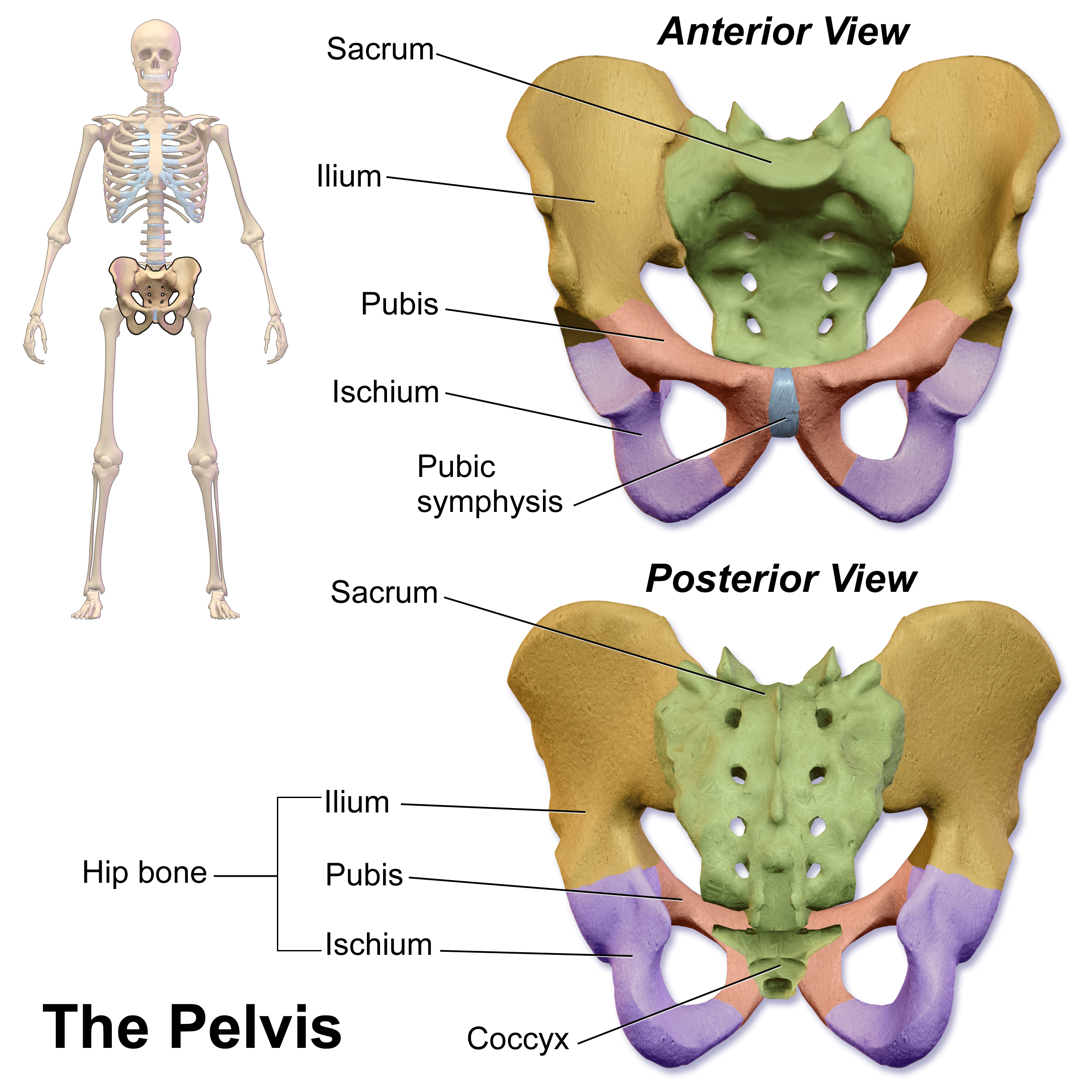
Pubic Symphysis
The pubic symphysis is a secondary cartilaginous joint between the left and right superior rami of the pubis of the hip bones. It is in front of and below the urinary bladder. In males, the suspensory ligament of the penis attaches to the pubic symphysis. In females, the pubic symphysis is close to the clitoris. In most adults it can be moved roughly 2 mm and with 1 degree rotation. This increases for women at the time of childbirth. The name comes from the Greek word ''symphysis'', meaning 'growing together'. Structure The pubic symphysis is a nonsynovial amphiarthrodial joint. The width of the pubic symphysis at the front is 3–5 mm greater than its width at the back. This joint is connected by fibrocartilage and may contain a fluid-filled cavity; the center is avascular, possibly due to the nature of the compressive forces passing through this joint, which may lead to harmful vascular disease. The ends of both pubic bones are covered by a thin layer of hyaline ... [...More Info...] [...Related Items...] OR: [Wikipedia] [Google] [Baidu] |
Xiphoid Process
The xiphoid process , or xiphisternum or metasternum, is a small cartilaginous process (extension) of the inferior (lower) part of the sternum, which is usually ossified in the adult human. It may also be referred to as the ensiform process. Both the Greek-derived ''xiphoid'' and its Latin equivalent ''ensiform'' mean 'swordlike' or 'sword-shaped' Structure The xiphoid process is considered to be at the level of the 9th thoracic vertebra and the T7 dermatome. Development In newborns and young (especially small) infants, the tip of the xiphoid process may be both seen and felt as a lump just below the sternal notch. At 15 to 29 years old, the xiphoid usually fuses to the body of the sternum with a fibrous joint. Unlike the synovial articulation of major joints, this is non-movable. Ossification of the xiphoid process occurs around age 40. Variation The xiphoid process can be naturally bifurcated or sometimes perforated (xiphoidal foramen). These variances in morphology are inher ... [...More Info...] [...Related Items...] OR: [Wikipedia] [Google] [Baidu] |
Aponeurosis
An aponeurosis (; plural: ''aponeuroses'') is a type or a variant of the deep fascia, in the form of a sheet of pearly-white fibrous tissue that attaches sheet-like muscles needing a wide area of attachment. Their primary function is to join muscles and the body parts they act upon, whether bone or other muscles. They have a shiny, whitish-silvery color, are histologically similar to tendons, and are very sparingly supplied with blood vessels and nerves. When dissected, aponeuroses are papery and peel off by sections. The primary regions with thick aponeuroses are in the ventral abdominal region, the dorsal lumbar region, the ventriculus in birds, and the palmar (palms) and plantar (soles) regions. Anatomy Anterior abdominal aponeuroses The anterior abdominal aponeuroses are located just superficial to the rectus abdominis muscle. It has for its borders the external oblique, pectoralis muscles, and the latissimus dorsi. Posterior lumbar aponeuroses The posterior lumbar apo ... [...More Info...] [...Related Items...] OR: [Wikipedia] [Google] [Baidu] |
Spermatic Cord
The spermatic cord is the cord-like structure in males formed by the vas deferens (''ductus deferens'') and surrounding tissue that runs from the deep inguinal ring down to each testicle. Its serosal covering, the tunica vaginalis, is an extension of the peritoneum that passes through the transversalis fascia. Each testicle develops in the lower thoracic and upper lumbar region and migrates into the scrotum. During its descent it carries along with it the vas deferens, its vessels, nerves etc. There is one on each side. Structure The spermatic cord is ensheathed in three layers of tissue: * ''external spermatic fascia'', an extension of the innominate fascia that overlies the aponeurosis of the external oblique muscle. * ''cremasteric muscle and fascia'', formed from a continuation of the internal oblique muscle and its fascia. * ''internal spermatic fascia'', continuous with the transversalis fascia. The normal diameter of the spermatic cord is about 16 mm (range 11 to 22 mm). It ... [...More Info...] [...Related Items...] OR: [Wikipedia] [Google] [Baidu] |
Pectineal Line (pubis)
The pectineal line of the pubis (also pecten pubis) is a ridge on the superior ramus of the pubic bone. It forms part of the pelvic brim. Lying across from the pectineal line are fibers of the pectineal ligament, and the proximal origin of the pectineus muscle. In combination with the arcuate line, it makes the iliopectineal line The iliopectineal line is the border of the iliopubic eminence. It can be defined as a compound structure of the arcuate line (from the ilium) and pectineal line (from the pubis). With the sacral promontory, it makes up the linea terminalis The .... References External links * () {{Authority control Bones of the pelvis Pubis (bone) ... [...More Info...] [...Related Items...] OR: [Wikipedia] [Google] [Baidu] |
Pubic Tubercle
The pubic tubercle is a prominent tubercle on the superior ramus of the pubis bone of the pelvis. Structure The pubic tubercle is a prominent forward-projecting tubercle on the upper border of the medial portion of the superior ramus of the pubis bone. The inguinal ligament attaches to it. Part of the abdominal external oblique muscle inserts onto it. The inferior epigastric artery passes between the pubic tubercle and the anterior superior iliac spine. The pubic spine is a rough ridge that extends from the pubic tubercle to the upper border of the pubic symphysis. Clinical significance The pubic tubercle may be palpated. It serves as a landmark for local anaesthetic of the genital branch of the genitofemoral nerve, which lies slightly lateral to the pubic tubercle. This may also be used for the obturator nerve The obturator nerve in human anatomy arises from the ventral divisions of the second, third, and fourth lumbar nerves in the lumbar plexus; the branch from t ... [...More Info...] [...Related Items...] OR: [Wikipedia] [Google] [Baidu] |


