|
Anterior Scalene
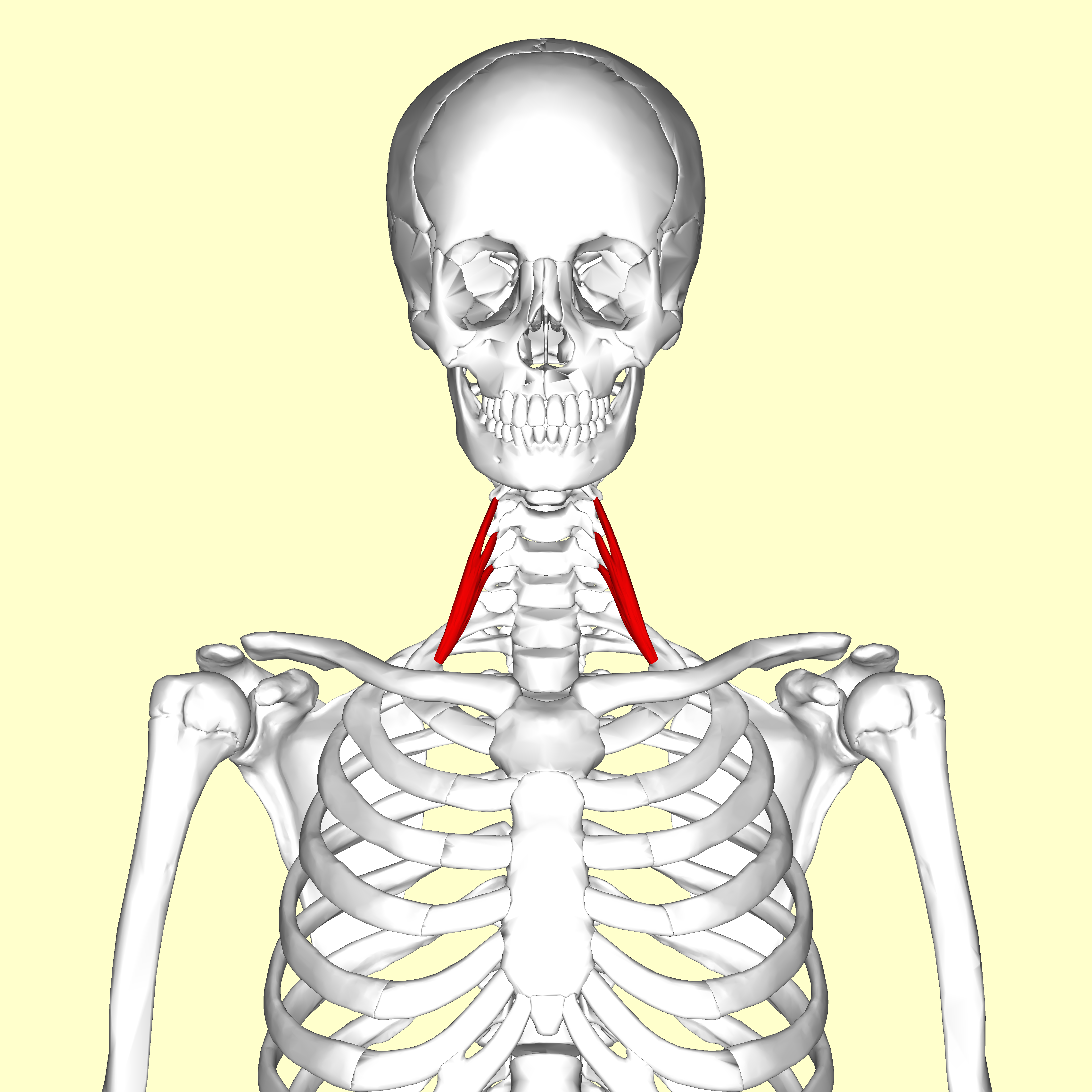
The scalene muscles are a group of three pairs of muscles in the lateral neck, namely the anterior scalene, middle scalene, and posterior scalene. They are innervated by the third to the eight cervical spinal nerves (C3-C8). The anterior and middle scalene muscles lift the first rib and bend the neck to the same side; the posterior scalene lifts the second rib and tilts the neck to the same side. The muscles are named . Structure The scalene muscles originate from the transverse processes from the cervical vertebrae of C2 to C7 and insert onto the first and second ribs. Anterior scalene The anterior scalene muscle ( la, scalenus anterior), lies deeply at the side of the neck, behind the sternocleidomastoid muscle. It arises from the anterior tubercles of the transverse processes of the third, fourth, fifth, and sixth cervical vertebrae, and descending, almost vertically, is inserted by a narrow, flat tendon into the scalene tubercle on the inner border of the first rib, and i ... [...More Info...] [...Related Items...] OR: [Wikipedia] [Google] [Baidu] |
Cervical Vertebrae
In tetrapods, cervical vertebrae (singular: vertebra) are the vertebrae of the neck, immediately below the skull. Truncal vertebrae (divided into thoracic and lumbar vertebrae in mammals) lie caudal (toward the tail) of cervical vertebrae. In sauropsid species, the cervical vertebrae bear cervical ribs. In lizards and saurischian dinosaurs, the cervical ribs are large; in birds, they are small and completely fused to the vertebrae. The vertebral transverse processes of mammals are homologous to the cervical ribs of other amniotes. Most mammals have seven cervical vertebrae, with the only three known exceptions being the manatee with six, the two-toed sloth with five or six, and the three-toed sloth with nine. In humans, cervical vertebrae are the smallest of the true vertebrae and can be readily distinguished from those of the thoracic or lumbar regions by the presence of a foramen (hole) in each transverse process, through which the vertebral artery, vertebral veins, an ... [...More Info...] [...Related Items...] OR: [Wikipedia] [Google] [Baidu] |
Scalenus Anterior01
''Scalenus'' is an Old World genus of round-necked longhorn beetles of the subfamily Cerambycinae. Species '' Scalenus auricomus'' (Ritsema, 1890) '' Scalenus borneensis'' Bentanachs & Drouin, 2014 ''Scalenus cingalensis'' (White, 1855) ''Scalenus fasciatipennis'' (Waterhouse, 1885) ''Scalenus fulvus'' (Bates, 1879) ''Scalenus hefferni'' Bentanachs & Jiroux, 2018 ''Scalenus hemipterus'' (Olivier, 1795) ''Scalenus kalimantanensis'' Bentanachs & Jiroux, 2018 ''Scalenus pejchai'' Bentanachs & Jiroux, 2018 ''Scalenus philippensis'' Bentanachs & Drouin, 2014 ''Scalenus sericeus'' (Saunders, 1853) ''Scalenus skalei'' Bentanachs & Jiroux, 2018 ''Scalenus ysmaeli ''Scalenus'' is an Old World genus of round-necked longhorn beetles of the subfamily Cerambycinae. Species '' Scalenus auricomus'' (Ritsema, 1890) '' Scalenus borneensis'' Bentanachs & Drouin, 2014 ''Scalenus cingalensis'' (White, 1855) ''Sc ...'' Hüdepohl, 1987 References Callichromatini {{Callichromatini ... [...More Info...] [...Related Items...] OR: [Wikipedia] [Google] [Baidu] |
Sternocleidomastoids
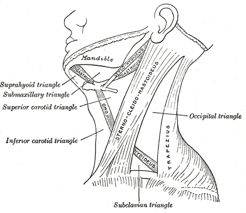
The sternocleidomastoid muscle is one of the largest and most superficial cervical muscles. The primary actions of the muscle are rotation of the head to the opposite side and flexion of the neck. The sternocleidomastoid is innervated by the accessory nerve. Etymology and location It is given the name ''sternocleidomastoid'' because it originates at the manubrium of the sternum (''sterno-'') and the clavicle (''cleido-'') and has an insertion at the mastoid process of the temporal bone of the skull. Structure The sternocleidomastoid muscle originates from two locations: the manubrium of the sternum and the clavicle. It travels obliquely across the side of the neck and inserts at the mastoid process of the temporal bone of the skull by a thin aponeurosis. The sternocleidomastoid is thick and narrow at its centre, and broader and thinner at either end. The sternal head is a round fasciculus, tendinous in front, fleshy behind, arising from the upper part of the front of the manubriu ... [...More Info...] [...Related Items...] OR: [Wikipedia] [Google] [Baidu] |
Accessory Muscles Of Respiration
The muscles of respiration are the muscles that contribute to inhalation and exhalation, by aiding in the expansion and contraction of the thoracic cavity. The diaphragm and, to a lesser extent, the intercostal muscles drive respiration during quiet breathing. The elasticity of these muscles is crucial to the health of the respiratory system and to maximize its functional capabilities. Diaphragm The diaphragm is the major muscle responsible for breathing. It is a thin, dome-shaped muscle that separates the abdominal cavity from the thoracic cavity. During inhalation, the diaphragm contracts, so that its center moves caudally (downward) and its edges move cranially (upward). This compresses the abdominal cavity, raises the ribs upward and outward and thus expands the thoracic cavity. This expansion draws air into the lungs. When the diaphragm relaxes, elastic recoil of the lungs causes the thoracic cavity to contract, forcing air out of the lungs, and returning to its dome-shap ... [...More Info...] [...Related Items...] OR: [Wikipedia] [Google] [Baidu] |
Scalenus Posterior04
''Scalenus'' is an Old World genus of round-necked longhorn beetles of the subfamily Cerambycinae. Species '' Scalenus auricomus'' (Ritsema, 1890) '' Scalenus borneensis'' Bentanachs & Drouin, 2014 ''Scalenus cingalensis'' (White, 1855) ''Scalenus fasciatipennis'' (Waterhouse, 1885) ''Scalenus fulvus'' (Bates, 1879) ''Scalenus hefferni'' Bentanachs & Jiroux, 2018 ''Scalenus hemipterus'' (Olivier, 1795) ''Scalenus kalimantanensis'' Bentanachs & Jiroux, 2018 ''Scalenus pejchai'' Bentanachs & Jiroux, 2018 ''Scalenus philippensis'' Bentanachs & Drouin, 2014 ''Scalenus sericeus'' (Saunders, 1853) ''Scalenus skalei'' Bentanachs & Jiroux, 2018 ''Scalenus ysmaeli ''Scalenus'' is an Old World genus of round-necked longhorn beetles of the subfamily Cerambycinae. Species '' Scalenus auricomus'' (Ritsema, 1890) '' Scalenus borneensis'' Bentanachs & Drouin, 2014 ''Scalenus cingalensis'' (White, 1855) ''Sc ...'' Hüdepohl, 1987 References Callichromatini {{Callichromatini ... [...More Info...] [...Related Items...] OR: [Wikipedia] [Google] [Baidu] |
Subclavian Artery
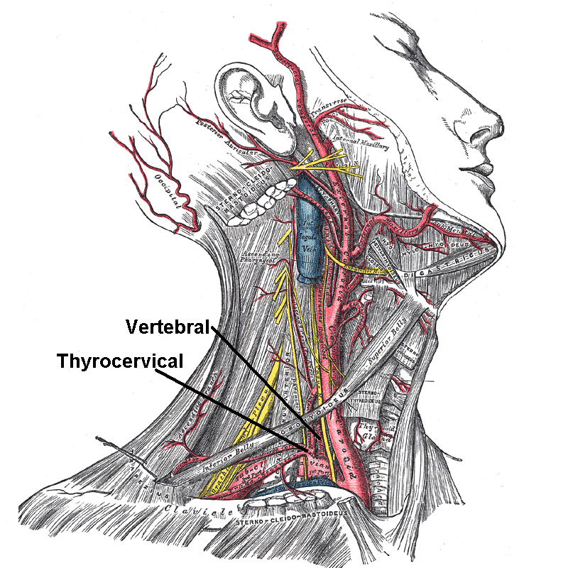
In human anatomy, the subclavian arteries are paired major arteries of the upper thorax, below the clavicle. They receive blood from the aortic arch. The left subclavian artery supplies blood to the left arm and the right subclavian artery supplies blood to the right arm, with some branches supplying the head and thorax. On the left side of the body, the subclavian comes directly off the aortic arch, while on the right side it arises from the relatively short brachiocephalic artery when it bifurcates into the subclavian and the right common carotid artery. The usual branches of the subclavian on both sides of the body are the vertebral artery, the internal thoracic artery, the thyrocervical trunk, the costocervical trunk and the dorsal scapular artery, which may branch off the transverse cervical artery, which is a branch of the thyrocervical trunk. The subclavian becomes the axillary artery at the lateral border of the first rib. Structure From its origin, the subclavian artery t ... [...More Info...] [...Related Items...] OR: [Wikipedia] [Google] [Baidu] |
Brachial Plexus
The brachial plexus is a network () of nerves formed by the anterior rami of the lower four cervical nerves and first thoracic nerve ( C5, C6, C7, C8, and T1). This plexus extends from the spinal cord, through the cervicoaxillary canal in the neck, over the first rib, and into the armpit, it supplies afferent and efferent nerve fibers the to chest, shoulder, arm, forearm, and hand. Structure The brachial plexus is divided into five ''roots'', three ''trunks'', six ''divisions'' (three anterior and three posterior), three ''cords'', and five ''branches''. There are five "terminal" branches and numerous other "pre-terminal" or "collateral" branches, such as the subscapular nerve, the thoracodorsal nerve, and the long thoracic nerve, that leave the plexus at various points along its length. A common structure used to identify part of the brachial plexus in cadaver dissections is the M or W shape made by the musculocutaneous nerve, lateral cord, median nerve, medial cord, and ... [...More Info...] [...Related Items...] OR: [Wikipedia] [Google] [Baidu] |
Vertebral Column
The vertebral column, also known as the backbone or spine, is part of the axial skeleton. The vertebral column is the defining characteristic of a vertebrate in which the notochord (a flexible rod of uniform composition) found in all chordata, chordates has been replaced by a segmented series of bone: vertebrae separated by intervertebral discs. Individual vertebrae are named according to their region and position, and can be used as anatomical landmarks in order to guide procedures such as Lumbar puncture, lumbar punctures. The vertebral column houses the spinal canal, a cavity that encloses and protects the spinal cord. There are about 50,000 species of animals that have a vertebral column. The human vertebral column is one of the most-studied examples. Many different diseases in humans can affect the spine, with spina bifida and scoliosis being recognisable examples. The general structure of human vertebrae is fairly typical of that found in mammals, reptiles, and birds. Th ... [...More Info...] [...Related Items...] OR: [Wikipedia] [Google] [Baidu] |
Cervical Nerve
A spinal nerve is a mixed nerve, which carries motor, sensory, and autonomic signals between the spinal cord and the body. In the human body there are 31 pairs of spinal nerves, one on each side of the vertebral column. These are grouped into the corresponding cervical, thoracic, lumbar, sacral and coccygeal regions of the spine. There are eight pairs of cervical nerves, twelve pairs of thoracic nerves, five pairs of lumbar nerves, five pairs of sacral nerves, and one pair of coccygeal nerves. The spinal nerves are part of the peripheral nervous system. Structure Each spinal nerve is a mixed nerve, formed from the combination of nerve fibers from its dorsal and ventral roots. The dorsal root is the afferent sensory root and carries sensory information to the brain. The ventral root is the efferent motor root and carries motor information from the brain. The spinal nerve emerges from the spinal column through an opening (intervertebral foramen) between adjacent vertebrae. Th ... [...More Info...] [...Related Items...] OR: [Wikipedia] [Google] [Baidu] |
Anterior Ramus
The ventral ramus (pl. ''rami'') (Latin for ''branch'') is the anterior division of a spinal nerve. The ventral rami supply the antero-lateral parts of the trunk and the limbs. They are mainly larger than the dorsal rami. Shortly after a spinal nerve exits the intervertebral foramen, it branches into the dorsal ramus, the ventral ramus, and the ramus communicans. Each of these three structures carries both sensory and motor information. Each spinal nerve carries both sensory and motor information, via efferent and afferent nerve fibers - ultimately via the motor cortex in the frontal lobe and to somatosensory cortex in the parietal lobe - but also through the phenomenon of reflex. Spinal nerves are referred to as "mixed nerves". In the thoracic region they remain distinct from each other and each innervates a narrow strip of muscle and skin along the sides, chest, ribs, and abdominal wall. These rami are called the intercostal nerves. In regions other than the thoracic, ventral ... [...More Info...] [...Related Items...] OR: [Wikipedia] [Google] [Baidu] |
Subclavian Groove (part of the Brachial plexus)
{{Disambig ...
In general, Subclavian means beneath the clavicle, and it may refer to: * Subclavian vein * Subclavian artery * Subclavian nerve The subclavian nerve, also known as the nerve to the subclavius, is small branch of the upper trunk of the brachial plexus. It contains axons from C5 and C6. The subclavian nerve innervates the subclavius muscle. Structure The subclavian ner ... [...More Info...] [...Related Items...] OR: [Wikipedia] [Google] [Baidu] |


