|
Wells' Score For Pulmonary Embolism
The Wells score is a clinical prediction rule used to classify patients suspected of having pulmonary embolism (PE) into risk groups by quantifying the pre-test probability. It is different than Well's score for DVT (deep vein thrombosis). It was originally described by Well's et al. in 1998, using their experience from creating Well's score for DVT in 1995. Today, there are multiple (revised or simplified) versions of the rule, which may lead to ambiguity. The purpose of the rule is to select the best method of investigation (e.g. D-dimer testing, CT angiography) for ruling in or ruling out the diagnosis of PE, and to improve the interpretation and accuracy of subsequent testing, based on a Bayesian framework for the probability of the diagnosis. The rule is more objective than clinician gestalt, but still includes subjective opinion (unlike e.g. Geneva score). Original algorithm Originally it was developed in 1998 to improve the low specificity of V/Q scan results (which ... [...More Info...] [...Related Items...] OR: [Wikipedia] [Google] [Baidu] |
Clinical Prediction Rule
A clinical prediction rule or clinical probability assessment specifies how to use medical signs, symptoms, and other findings to estimate the probability of a specific disease or clinical outcome. Physicians have difficulty in estimated risks of diseases; frequently erring towards overestimation, perhaps due to cognitive biases such as base rate fallacy in which the risk of an adverse outcome is exaggerated. Methods In a prediction rule study, investigators identify a consecutive group of patients who are suspected of having a specific disease or outcome. The investigators then obtain a standard set of clinical observations on each patient and a test or clinical follow-up to define the true state of the patient. They then use statistical methods to identify the best clinical predictors of the patient's true state. The probability of disease will depend on the patient's key clinical predictors. Published methodological standards specify good practices for developing a clinical pred ... [...More Info...] [...Related Items...] OR: [Wikipedia] [Google] [Baidu] |
Pulmonary Embolism
Pulmonary embolism (PE) is a blockage of an pulmonary artery, artery in the lungs by a substance that has moved from elsewhere in the body through the bloodstream (embolism). Symptoms of a PE may include dyspnea, shortness of breath, chest pain particularly upon breathing in, and coughing up blood. Symptoms of a deep vein thrombosis, blood clot in the leg may also be present, such as a erythema, red, warm, swollen, and painful leg. Signs of a PE include low blood oxygen saturation, oxygen levels, tachypnea, rapid breathing, tachycardia, rapid heart rate, and sometimes a mild fever. Severe cases can lead to Syncope (medicine), passing out, shock (circulatory), abnormally low blood pressure, obstructive shock, and cardiac arrest, sudden death. PE usually results from a blood clot in the leg that travels to the lung. The risk of blood clots is increased by advanced age, cancer, prolonged bed rest and immobilization, smoking, stroke, long-haul travel over 4 hours, certain genetics, g ... [...More Info...] [...Related Items...] OR: [Wikipedia] [Google] [Baidu] |
Pre- And Post-test Probability
Pre-test probability and post-test probability (alternatively spelled pretest and posttest probability) are the probabilities of the presence of a condition (such as a disease) before and after a diagnostic test, respectively. ''Post-test probability'', in turn, can be ''positive'' or ''negative'', depending on whether the test falls out as a positive test or a negative test, respectively. In some cases, it is used for the probability of developing the condition of interest in the future. Test, in this sense, can refer to any medical test (but usually in the sense of diagnostic tests), and in a broad sense also including questions and even assumptions (such as assuming that the target individual is a female or male). The ability to make a difference between pre- and post-test probabilities of various conditions is a major factor in the indication of medical tests. Pre-test probability The pre-test probability of an individual can be chosen as one of the following: *The prevalenc ... [...More Info...] [...Related Items...] OR: [Wikipedia] [Google] [Baidu] |
Deep Vein Thrombosis
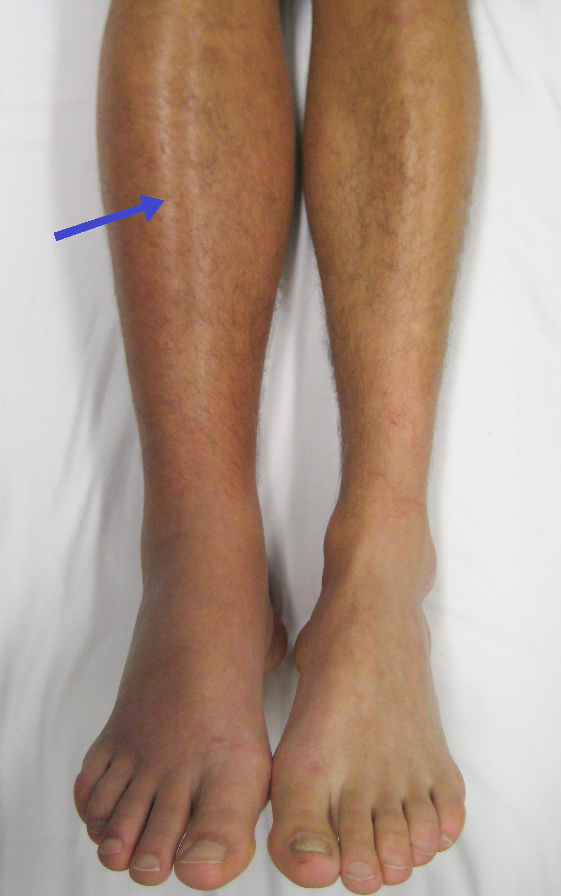
Deep vein thrombosis (DVT) is a type of venous thrombosis involving the formation of a blood clot in a deep vein, most commonly in the legs or pelvis. A minority of DVTs occur in the arms. Symptoms can include pain, swelling, redness, and enlarged veins in the affected area, but some DVTs have no symptoms. The most common life-threatening concern with DVT is the potential for a clot to embolize (detach from the veins), travel as an embolus through the right side of the heart, and become lodged in a pulmonary artery that supplies blood to the lungs. This is called a pulmonary embolism (PE). DVT and PE comprise the cardiovascular disease of venous thromboembolism (VTE). About two-thirds of VTE manifests as DVT only, with one-third manifesting as PE with or without DVT. The most frequent long-term DVT complication is post-thrombotic syndrome, which can cause pain, swelling, a sensation of heaviness, itching, and in severe cases, ulcers. Recurrent VTE occurs in about 30% of those i ... [...More Info...] [...Related Items...] OR: [Wikipedia] [Google] [Baidu] |
Computed Tomography Angiography
Computed tomography angiography (also called CT angiography or CTA) is a computed tomography technique used for angiography—the visualization of arteries and veins—throughout the human body. Using contrast injected into the blood vessels, images are created to look for blockages, aneurysms (dilations of walls), dissections (tearing of walls), and stenosis (narrowing of vessel). CTA can be used to visualize the vessels of the heart, the aorta and other large blood vessels, the lungs, the kidneys, the head and neck, and the arms and legs. CTA can also be used to localise arterial or venous bleed of the gastrointestinal system. Medical uses CTA can be used to examine blood vessels in many key areas of the body including the brain, kidneys, pelvis, and the lungs. Coronary CT angiography Coronary CT angiography (CCTA) is the use of CT angiography to assess the arteries of the heart. The patient receives an intravenous injection of contrast and then the heart is scanned using a ... [...More Info...] [...Related Items...] OR: [Wikipedia] [Google] [Baidu] |
Geneva Score
The Geneva score is a clinical prediction rule used in determining the pre-test probability of pulmonary embolism (PE) based on a patient's risk factors and clinical findings. It has been shown to be as accurate as the Wells Score, and is less reliant on the experience of the doctor applying the rule. The Geneva score has been revised and simplified from its original version. The simplified Geneva score is the newest version for the general population, and predicted to have the same diagnostic utility as the original Geneva score. A version of the revised score was modified to be applicable to pregnant patients. Original Geneva Score The original Geneva score was developed in 2001 in Geneva, Switzerland. It's calculated using 7 risk factors and clinical variables: The score obtained relates to the probability of the patient having had a pulmonary embolism (the lower the score, the lower the probability): * 8 points indicates a high probability of PE. (81%) Revised Geneva Sc ... [...More Info...] [...Related Items...] OR: [Wikipedia] [Google] [Baidu] |
Ventilation/perfusion Scan
A ventilation/perfusion lung scan, also called a V/Q lung scan, or ventilation/perfusion scintigraphy, is a type of medical imaging using scintigraphy and medical isotopes to evaluate the circulation of air and blood within a patient's lungs, in order to determine the ventilation/perfusion ratio. The ventilation part of the test looks at the ability of air to reach all parts of the lungs, while the perfusion part evaluates how well blood circulates within the lungs. As Q in physiology is the letter used to describe bloodflow the term V/Q scan emerged. Uses This test is most commonly done in order to check for the presence of a blood clot or abnormal blood flow inside the lungs (such as a pulmonary embolism (PE) although computed tomography with radiocontrast is now more commonly used for this purpose. The V/Q scan may be used in some circumstances where radiocontrast would be inappropriate, as in Allergy to contrast agent or kidney failure. A V/Q lung scan may be performed in th ... [...More Info...] [...Related Items...] OR: [Wikipedia] [Google] [Baidu] |
Angiography
Angiography or arteriography is a medical imaging technique used to visualize the inside, or lumen, of blood vessels and organs of the body, with particular interest in the arteries, veins, and the heart chambers. Modern angiography is performed by injecting a radio-opaque contrast agent into the blood vessel and imaging using X-ray based techniques such as fluoroscopy. The word itself comes from the Greek words ἀγγεῖον ''angeion'' 'vessel' and γράφειν ''graphein'' 'to write, record'. The film or image of the blood vessels is called an ''angiograph'', or more commonly an ''angiogram''. Though the word can describe both an arteriogram and a venogram, in everyday usage the terms angiogram and arteriogram are often used synonymously, whereas the term venogram is used more precisely. The term angiography has been applied to radionuclide angiography and newer vascular imaging techniques such as CO2 angiography, CT angiography and MR angiography. The term ''isotope a ... [...More Info...] [...Related Items...] OR: [Wikipedia] [Google] [Baidu] |
Compression Ultrasound
Medical ultrasound includes diagnostic techniques (mainly medical imaging, imaging techniques) using ultrasound, as well as therapeutic ultrasound, therapeutic applications of ultrasound. In diagnosis, it is used to create an image of internal body structures such as tendons, muscles, joints, blood vessels, and internal organs, to measure some characteristics (e.g. distances and velocities) or to generate an informative audible sound. Its aim is usually to find a source of disease or to exclude pathology. The usage of ultrasound to produce visual images for medicine is called medical ultrasonography or simply sonography. The practice of examining pregnant women using ultrasound is called obstetric ultrasonography, and was an early development of clinical ultrasonography. Ultrasound is composed of sound waves with frequency, frequencies which are significantly higher than the range of human hearing (>20,000 Hz). Ultrasonic images, also known as sonograms, are created by se ... [...More Info...] [...Related Items...] OR: [Wikipedia] [Google] [Baidu] |
D-dimer
D-dimer (or D dimer) is a fibrin degradation product (or FDP), a small protein fragment present in the blood after a blood clot is degraded by fibrinolysis. It is so named because it contains two D fragments of the fibrin protein joined by a cross-link, hence forming a protein dimer. D-dimer concentration may be determined by a blood test to help diagnose thrombosis. Since its introduction in the 1990s, it has become an important test performed in people with suspected thrombotic disorders, such as venous thromboembolism. While a negative result practically rules out thrombosis, a positive result can indicate thrombosis, but does not exclude other potential causes. Its main use, therefore, is to exclude thromboembolic disease where the probability is low. D-dimer levels are used as a predictive biomarker for the blood disorder, disseminated intravascular coagulation and in the coagulation disorders associated with COVID-19 infection. A four-fold increase in the protein is an ind ... [...More Info...] [...Related Items...] OR: [Wikipedia] [Google] [Baidu] |
Medical Terminology
Medical terminology is a language used to precisely describe the human body including all its components, processes, conditions affecting it, and procedures performed upon it. Medical terminology is used in the field of medicine Medical terminology has quite regular morphology, the same prefixes and suffixes are used to add meanings to different roots. The root of a term often refers to an organ, tissue, or condition. For example, in the disorder known as hypertension, the prefix "hyper-" means "high" or "over", and the root word "tension" refers to pressure, so the word "hypertension" refers to abnormally high blood pressure. The roots, prefixes and suffixes are often derived from Greek or Latin, and often quite dissimilar from their English-language variants. This regular morphology means that once a reasonable number of morphemes are learnt it becomes easy to understand very precise terms assembled from these morphemes. Much medical language is anatomical terminology, concern ... [...More Info...] [...Related Items...] OR: [Wikipedia] [Google] [Baidu] |
Medical Tests
A medical test is a medical procedure performed to detect, diagnose, or monitor diseases, disease processes, susceptibility, or to determine a course of treatment. Medical tests such as, physical and visual exams, diagnostic imaging, genetic testing, chemical and cellular analysis, relating to clinical chemistry and molecular diagnostics, are typically performed in a medical setting. Types of tests By purpose Medical tests can be classified by their purposes, the most common of which are diagnosis, screening and evaluation. Diagnostic A diagnostic test is a procedure performed to confirm or determine the presence of disease in an individual suspected of having a disease, usually following the report of symptoms, or based on other medical test results. This includes posthumous diagnosis. Examples of such tests are: * Using nuclear medicine to examine a patient suspected of having a lymphoma. * Measuring the blood sugar in a person suspected of having diabetes mellitus after ... [...More Info...] [...Related Items...] OR: [Wikipedia] [Google] [Baidu] |


