|
Westphal's Sign
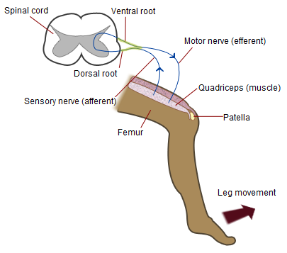
Westphal's sign is the clinical correlate of the absence or decrease of patellar reflex or knee jerk. Patellar reflex or knee jerk is a kind of deep or stretch reflex where an application of a stimulus to the patellar tendon such as strike by a solid object or hammer caused the leg to extend due to such stimulus causes the quadriceps femoris muscle to contract. It is named for Karl Friedrich Otto Westphal (1833-1890). __TOC__ Associated conditions Westphal's sign has a clinical significance used in determining neurodisorders or diseases such as: * receptor damage, peripheral nerve disease, involving the dorsal(sensory) columns of the spinal cord and cerebellar lesions * lesions present within the motor cortex of the brain or the pyramidal tracts which it combined with muscular spasms * complete interruption of sensory and/or motor impulse transmission in the femoral nerve References * Symptoms and signs: Nervous system Examination of the knee {{med-sign-s ... [...More Info...] [...Related Items...] OR: [Wikipedia] [Google] [Baidu] |
Correlate
In statistics, correlation or dependence is any statistical relationship, whether causal or not, between two random variables or bivariate data. Although in the broadest sense, "correlation" may indicate any type of association, in statistics it usually refers to the degree to which a pair of variables are '' linearly'' related. Familiar examples of dependent phenomena include the correlation between the height of parents and their offspring, and the correlation between the price of a good and the quantity the consumers are willing to purchase, as it is depicted in the so-called demand curve. Correlations are useful because they can indicate a predictive relationship that can be exploited in practice. For example, an electrical utility may produce less power on a mild day based on the correlation between electricity demand and weather. In this example, there is a causal relationship, because extreme weather causes people to use more electricity for heating or cooling. H ... [...More Info...] [...Related Items...] OR: [Wikipedia] [Google] [Baidu] |
Patellar Reflex
The patellar reflex, also called the knee reflex or knee-jerk, is a stretch reflex which tests the L2, L3, and L4 segments of the spinal cord. Mechanism Striking of the patellar tendon with a reflex hammer just below the patella stretches the muscle spindle in the quadriceps muscle. This produces a signal which travels back to the spinal cord and synapses (without interneurons) at the level of L3 or L4 in the spinal cord, completely independent of higher centres. From there, an alpha motor neuron conducts an efferent impulse back to the quadriceps femoris muscle, triggering contraction. This contraction, coordinated with the relaxation of the antagonistic flexor hamstring muscle causes the leg to kick. There is a latency of around 18 ms between stretch of the patellar tendon and the beginning of contraction of the quadriceps femoris muscle. This is a reflex of proprioception which helps maintain posture and balance, allowing to keep one's balance with little effort or conscio ... [...More Info...] [...Related Items...] OR: [Wikipedia] [Google] [Baidu] |
Stretch Reflex
The stretch reflex (myotatic reflex), or more accurately "muscle stretch reflex", is a muscle contraction in response to stretching within the muscle. The reflex functions to maintain the muscle at a constant length. The term deep tendon reflex is often used by many health workers and students to refer to this reflex. "Tendons have little to do with the response, other than being responsible for mechanically transmitting the sudden stretch from the reflex hammer to the muscle spindle. In addition, some muscles with stretch reflexes have no tendons (e.g., "jaw jerk" of the masseter muscle)". As an example of a spinal reflex, it results in a fast response that involves an afferent signal into the spinal cord and an efferent signal out to the muscle. The stretch reflex can be a monosynaptic reflex which provides automatic regulation of skeletal muscle length, whereby the signal entering the spinal cord arises from a change in muscle length or velocity. It can also include a polysyna ... [...More Info...] [...Related Items...] OR: [Wikipedia] [Google] [Baidu] |
Patellar Tendon
The patellar tendon is the distal portion of the common tendon of the quadriceps femoris, which is continued from the patella to the tibial tuberosity. It is also sometimes called the patellar ligament as it forms a bone to bone connection when the patella is fully ossified. Structure The patellar tendon is a strong, flat ligament, which originates on the apex of the patella distally and adjoining margins of the patella and the rough depression on its posterior surface; below, it inserts on the tuberosity of the tibia; its superficial fibers are continuous over the front of the patella with those of the tendon of the quadriceps femoris. It is about 4.5 cm long in adults (range from 3 to 6 cm). The medial and lateral portions of the quadriceps tendon pass down on either side of the patella to be inserted into the upper extremity of the tibia on either side of the tuberosity; these portions merge into the capsule, as stated above, forming the medial and lateral patellar retin ... [...More Info...] [...Related Items...] OR: [Wikipedia] [Google] [Baidu] |
Quadriceps Femoris
The quadriceps femoris muscle (, also called the quadriceps extensor, quadriceps or quads) is a large muscle group that includes the four prevailing muscles on the front of the thigh. It is the sole extensor muscle of the knee, forming a large fleshy mass which covers the front and sides of the femur. The name derives . Structure Parts The quadriceps femoris muscle is subdivided into four separate muscles (the 'heads'), with the first superficial to the other three over the femur (from the trochanters to the condyles): *The rectus femoris muscle occupies the middle of the thigh, covering most of the other three quadriceps muscles. It originates on the ilium. It is named for its straight course. *The vastus lateralis muscle is on the ''lateral side'' of the femur (i.e. on the outer side of the thigh). *The vastus medialis muscle is on the ''medial side'' of the femur (i.e. on the inner part thigh). *The vastus intermedius muscle lies between vastus lateralis and vastus med ... [...More Info...] [...Related Items...] OR: [Wikipedia] [Google] [Baidu] |
Karl Friedrich Otto Westphal
Karl Friedrich Otto Westphal (23 March 1833, in Berlin – 27 January 1890, in Kreuzlingen) was a German psychiatrist from Berlin. He was the son of Otto Carl Friedrich Westphal (1800–1879) and Karoline Friederike Heine and the father of Alexander Karl Otto Westphal (1863-1941). He was married to Klara, daughter of the banker Alexander Mendelssohn. Academic career After receiving his doctorate, he worked at the Berlin Charité, and subsequently became an assistant in the department for the mentally ill under Wilhelm Griesinger (1817–1868) and Karl Wilhelm Ideler (1795–1860). In 1869 he became an associate professor of psychiatry, as well as a clinical instructor in the department for mental and nervous diseases, In 1874 he attained the title of full professor of psychiatry. Achievements in medicine Westphal's contributions to medical science are many; in 1871 he coined the term "agoraphobia", when he observed that three male patients of his displayed extreme anxiety an ... [...More Info...] [...Related Items...] OR: [Wikipedia] [Google] [Baidu] |
Dorsal(sensory) Columns
The posterior grey column (posterior cornu, dorsal horn, spinal dorsal horn, posterior horn, sensory horn) of the spinal cord is one of the three grey columns of the spinal cord. It receives several types of sensory information from the body, including fine touch, proprioception, and vibration. This information is sent from receptors of the skin, bones, and joints through sensory neurons whose cell bodies lie in the dorsal root ganglion. Anatomy The posterior grey column is subdivided into six layers termed Rexed laminae I-VI *Marginal nucleus of spinal cord (lamina I) *Substantia gelatinosa of Rolando (lamina II) *Nucleus proprius (laminae III, IV) *Spinal lamina V, the neck of the posterior horn *Spinal lamina VI, the base of the posterior horn. The other four Rexed laminae are located in the other two grey columns in the spinal cord. Additional images File:Gray687.png, Section of the medulla oblongata through the lower part of the decussation of the pyramids See also * Po ... [...More Info...] [...Related Items...] OR: [Wikipedia] [Google] [Baidu] |
Cerebral Cortex
The cerebral cortex, also known as the cerebral mantle, is the outer layer of neural tissue of the cerebrum of the brain in humans and other mammals. The cerebral cortex mostly consists of the six-layered neocortex, with just 10% consisting of allocortex. It is separated into two cortices, by the longitudinal fissure that divides the cerebrum into the left and right cerebral hemispheres. The two hemispheres are joined beneath the cortex by the corpus callosum. The cerebral cortex is the largest site of neural integration in the central nervous system. It plays a key role in attention, perception, awareness, thought, memory, language, and consciousness. The cerebral cortex is part of the brain responsible for cognition. In most mammals, apart from small mammals that have small brains, the cerebral cortex is folded, providing a greater surface area in the confined volume of the cranium. Apart from minimising brain and cranial volume, cortical folding is crucial for ... [...More Info...] [...Related Items...] OR: [Wikipedia] [Google] [Baidu] |
Pyramidal Tracts
The pyramidal tracts include both the corticobulbar tract and the corticospinal tract. These are aggregations of efferent nerve fibers from the upper motor neurons that travel from the cerebral cortex and terminate either in the brainstem (''corticobulbar'') or spinal cord (''corticospinal'') and are involved in the control of motor functions of the body. The corticobulbar tract conducts impulses from the brain to the cranial nerves. These nerves control the muscles of the face and neck and are involved in facial expression, mastication, swallowing, and other motor functions. The corticospinal tract conducts impulses from the brain to the spinal cord. It is made up of a lateral and anterior tract. The corticospinal tract is involved in voluntary movement. The majority of fibres of the corticospinal tract cross over in the medulla oblongata, resulting in muscles being controlled by the opposite side of the brain. The corticospinal tract contains the axons of the pyramidal cells ... [...More Info...] [...Related Items...] OR: [Wikipedia] [Google] [Baidu] |
Femoral Nerve
The femoral nerve is a nerve in the thigh that supplies skin on the upper thigh and inner leg, and the muscles that extend the knee. Structure The femoral nerve is the major nerve supplying the anterior compartment of the thigh. It is the largest branch of the lumbar plexus, and arises from the dorsal divisions of the ventral rami of the second, third, and fourth lumbar nerves (L2, L3, and L4). The nerve enters Scarpa's triangle by passing beneath the inguinal ligament, just lateral to the femoral artery. In the thigh, the nerve lies in a groove between iliacus muscle and psoas major muscle, outside the femoral sheath, and lateral to the femoral artery. After a short course of about 4 cm in the thigh, the nerve is divided into anterior and posterior divisions, separated by lateral femoral circumflex artery. The branches are shown below: Muscular branches * The nerve to the pectineus muscle arises immediately above the inguinal ligament from the medial side of the femora ... [...More Info...] [...Related Items...] OR: [Wikipedia] [Google] [Baidu] |
Symptoms And Signs: Nervous System
Signs and symptoms are the observed or detectable signs, and experienced symptoms of an illness, injury, or condition. A sign for example may be a higher or lower temperature than normal, raised or lowered blood pressure or an abnormality showing on a medical scan. A symptom is something out of the ordinary that is experienced by an individual such as feeling feverish, a headache or other pain or pains in the body. Signs and symptoms Signs A medical sign is an objective observable indication of a disease, injury, or abnormal physiological state that may be detected during a physical examination, examining the patient history, or diagnostic procedure. These signs are visible or otherwise detectable such as a rash or bruise. Medical signs, along with symptoms, assist in formulating diagnostic hypothesis. Examples of signs include elevated blood pressure, nail clubbing of the fingernails or toenails, staggering gait, and arcus senilis and arcus juvenilis of the eyes. Indication ... [...More Info...] [...Related Items...] OR: [Wikipedia] [Google] [Baidu] |


