|
Walthard Cell Rest
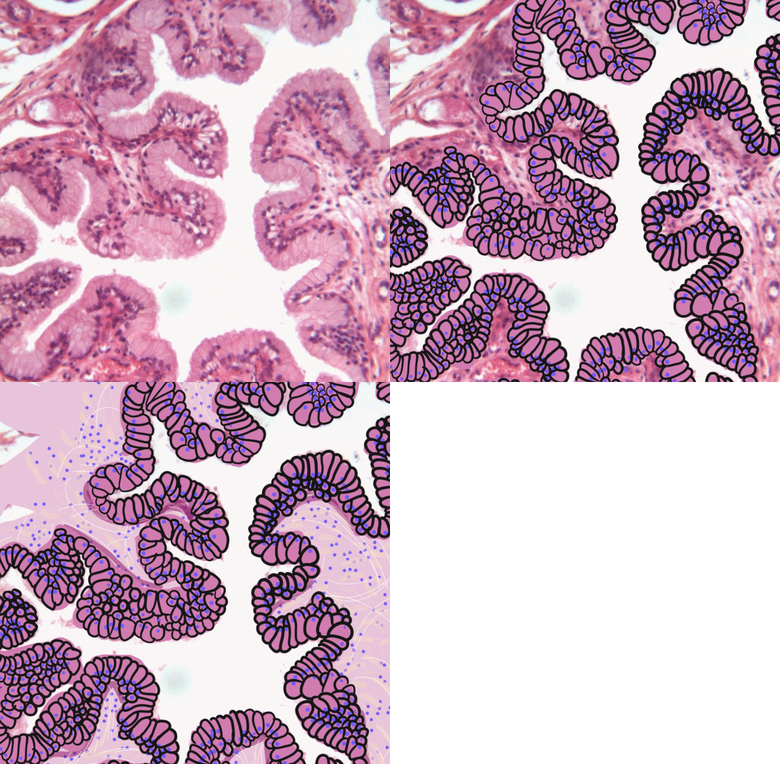
Walthard cell rests, sometimes called Walthard cell nests, are a benign cluster of epithelial cells most commonly found in the connective tissue of the Fallopian tubes, but also seen in the mesovarium, mesosalpinx and ovarian hilus. Appearance They appear as white/yellow cysts or nodules that can reach a size of 2 millimeters. They typically have elliptical nuclei with a long groove (along the major axis) – so-called "coffee bean" nuclei. Pathology It has been suggested that these cell rests are the histogenetic origins of Brenner tumors, due to the histological similarity of the epithelium of Walthard cell rests and Brenner tumors to the urothelium of the lower urinary tract. Also, it has been proposed that Brenner tumors and Walthard cell rests signify urothelial differentiation within the female genital tract. Eponym They are named after Swiss gynecologist Max Walthard (1867–1933), who provided a comprehensive description of them in 1903. Additional images Image:Bre ... [...More Info...] [...Related Items...] OR: [Wikipedia] [Google] [Baidu] |
Walthard Cell Rest - Very Low Mag
Walthard (or Waltaro) (died 12 August 1012) was the Archbishop of Magdeburg very briefly from June to August in 1012. Walthard was the initial archiepiscopal candidate of the cathedral chapter on the death of Archbishop Giseler in 1004, but King Henry II elected to appoint his chaplain Tagino instead. When Tagino died on 9 June 1012, the cathedral elected Walthard, but he died within a few months and they elected one named Otto, who was passed over again by the king in favour of Gero.Reuter, 195. Otto was accepted into the royal chaplaincy as a consolation. Sources *Reuter, Timothy Timothy Alan Reuter (25 January 1947 – 14 October 2002), grandson of the former mayor of Berlin Ernst Reuter, was a German-British historian who specialized in the study of medieval Germany, particularly the social, military and ecclesiastical i .... ''Germany in the Early Middle Ages 800–1056''. New York: Longman, 1991. * Thompson, James Westfall. ''Feudal Germany, Volume II''. New York: ... [...More Info...] [...Related Items...] OR: [Wikipedia] [Google] [Baidu] |
Histology
Histology, also known as microscopic anatomy or microanatomy, is the branch of biology which studies the microscopic anatomy of biological tissues. Histology is the microscopic counterpart to gross anatomy, which looks at larger structures visible without a microscope. Although one may divide microscopic anatomy into ''organology'', the study of organs, ''histology'', the study of tissues, and ''cytology'', the study of cells, modern usage places all of these topics under the field of histology. In medicine, histopathology is the branch of histology that includes the microscopic identification and study of diseased tissue. In the field of paleontology, the term paleohistology refers to the histology of fossil organisms. Biological tissues Animal tissue classification There are four basic types of animal tissues: muscle tissue, nervous tissue, connective tissue, and epithelial tissue. All animal tissues are considered to be subtypes of these four principal tissue types ... [...More Info...] [...Related Items...] OR: [Wikipedia] [Google] [Baidu] |
H&E Stain
Hematoxylin and eosin stain ( or haematoxylin and eosin stain or hematoxylin-eosin stain; often abbreviated as H&E stain or HE stain) is one of the principal tissue stains used in histology. It is the most widely used stain in medical diagnosis and is often the gold standard. For example, when a pathologist looks at a biopsy of a suspected cancer, the histological section is likely to be stained with H&E. H&E is the combination of two histological stains: hematoxylin and eosin. The hematoxylin stains cell nuclei a purplish blue, and eosin stains the extracellular matrix and cytoplasm pink, with other structures taking on different shades, hues, and combinations of these colors. Hence a pathologist can easily differentiate between the nuclear and cytoplasmic parts of a cell, and additionally, the overall patterns of coloration from the stain show the general layout and distribution of cells and provides a general overview of a tissue sample's structure. Thus, pattern recogniti ... [...More Info...] [...Related Items...] OR: [Wikipedia] [Google] [Baidu] |
Coffee Bean
A coffee bean is a seed of the ''Coffea'' plant and the source for coffee. It is the pip inside the red or purple fruit often referred to as a coffee cherry. Just like ordinary cherries, the coffee fruit is also a so-called stone fruit. Even though the coffee beans are not technically beans, they are referred to as such because of their resemblance to true beans. The fruits; cherries or berries, most commonly contain two stones with their flat sides together. A small percentage of cherries contain a single seed, instead of the usual two. This is called a "peaberry". The peaberry occurs only between 10% and 15% of the time, and it is a fairly common (yet scientifically unproven) belief that they have more flavour than normal coffee beans. Like Brazil nuts (a seed) and white rice, coffee beans consist mostly of endosperm. The two most economically important varieties of coffee plant are the Arabica and the Robusta; approximately 60% of the coffee produced worldwide is Arabica and ... [...More Info...] [...Related Items...] OR: [Wikipedia] [Google] [Baidu] |
Micrograph
A micrograph or photomicrograph is a photograph or digital image taken through a microscope or similar device to show a magnified image of an object. This is opposed to a macrograph or photomacrograph, an image which is also taken on a microscope but is only slightly magnified, usually less than 10 times. Micrography is the practice or art of using microscopes to make photographs. A micrograph contains extensive details of microstructure. A wealth of information can be obtained from a simple micrograph like behavior of the material under different conditions, the phases found in the system, failure analysis, grain size estimation, elemental analysis and so on. Micrographs are widely used in all fields of microscopy. Types Photomicrograph A light micrograph or photomicrograph is a micrograph prepared using an optical microscope, a process referred to as ''photomicroscopy''. At a basic level, photomicroscopy may be performed simply by connecting a camera to a microscope, th ... [...More Info...] [...Related Items...] OR: [Wikipedia] [Google] [Baidu] |
Max Walthard
Max or MAX may refer to: Animals * Max (dog) (1983–2013), at one time purported to be the world's oldest living dog * Max (English Springer Spaniel), the first pet dog to win the PDSA Order of Merit (animal equivalent of OBE) * Max (gorilla) (1971–2004), a western lowland gorilla at the Johannesburg Zoo who was shot by a criminal in 1997 Brands and enterprises * Australian Max Beer * Max Hamburgers, a fast-food corporation * MAX Index, a Hungarian domestic government bond index * Max Fashion, an Indian clothing brand Computing * MAX (operating system), a Spanish-language Linux version * Max (software), a music programming language * Commodore MAX Machine * Multimedia Acceleration eXtensions, extensions for HP PA-RISC Films * ''Max'' (1994 film), a Canadian film by Charles Wilkinson * ''Max'' (2002 film), a film about Adolf Hitler * ''Max'' (2015 film), an American war drama film Games * '' Dancing Stage Max'', a 2005 game in the ''Dance Dance Revolution'' series * ''DDRM ... [...More Info...] [...Related Items...] OR: [Wikipedia] [Google] [Baidu] |
Gynecologist
Gynaecology or gynecology (see spelling differences) is the area of medicine that involves the treatment of women's diseases, especially those of the reproductive organs. It is often paired with the field of obstetrics, forming the combined area of obstetrics and gynecology (OB-GYN). The term comes from Greek and means "the science of women". Its counterpart is andrology, which deals with medical issues specific to the male reproductive system. Etymology The word "gynaecology" comes from the oblique stem (γυναικ-) of the Greek word γυνή (''gyne)'' semantically attached to "woman", and ''-logia'', with the semantic attachment "study". The word gynaecology in Kurdish means "jinekolojî", separated word as "jin-ekolojî", so the Kurdish "jin" called like "gyn" and means in Kurdish "woman". History Antiquity The Kahun Gynaecological Papyrus, dated to about 1800 BC, deals with gynaecological diseases, fertility, pregnancy, contraception, etc. The text is divided into thir ... [...More Info...] [...Related Items...] OR: [Wikipedia] [Google] [Baidu] |
Urinary Tract
The urinary system, also known as the urinary tract or renal system, consists of the kidneys, ureters, bladder, and the urethra. The purpose of the urinary system is to eliminate waste from the body, regulate blood volume and blood pressure, control levels of electrolytes and metabolites, and regulate blood pH. The urinary tract is the body's drainage system for the eventual removal of urine. The kidneys have an extensive blood supply via the renal arteries which leave the kidneys via the renal vein. Each kidney consists of functional units called nephrons. Following filtration of blood and further processing, wastes (in the form of urine) exit the kidney via the ureters, tubes made of smooth muscle fibres that propel urine towards the urinary bladder, where it is stored and subsequently expelled from the body by urination (voiding). The female and male urinary system are very similar, differing only in the length of the urethra. Urine is formed in the kidneys through a filtrati ... [...More Info...] [...Related Items...] OR: [Wikipedia] [Google] [Baidu] |
Urothelium
Transitional epithelium also known as urothelium is a type of stratified epithelium. Transitional epithelium is a type of tissue that changes shape in response to stretching (stretchable epithelium). The transitional epithelium usually appears cuboidal when relaxed and squamous when stretched. This tissue consists of multiple layers of epithelial cells which can contract and expand in order to adapt to the degree of distension needed. Transitional epithelium lines the organs of the urinary system and is known here as urothelium. The bladder for example has a need for great distension. Structure The appearance of transitional epithelium differs according to its cell layer. Cells of the basal layer are cuboidal (cube-shaped), or columnar (column-shaped), while the cells of the superficial layer vary in appearance depending on the degree of distension. These cells appear to be cuboidal with a domed apex when the organ or the tube in which they reside is not stretched. When the orga ... [...More Info...] [...Related Items...] OR: [Wikipedia] [Google] [Baidu] |
Epithelium
Epithelium or epithelial tissue is one of the four basic types of animal tissue, along with connective tissue, muscle tissue and nervous tissue. It is a thin, continuous, protective layer of compactly packed cells with a little intercellular matrix. Epithelial tissues line the outer surfaces of organs and blood vessels throughout the body, as well as the inner surfaces of cavities in many internal organs. An example is the epidermis, the outermost layer of the skin. There are three principal shapes of epithelial cell: squamous (scaly), columnar, and cuboidal. These can be arranged in a singular layer of cells as simple epithelium, either squamous, columnar, or cuboidal, or in layers of two or more cells deep as stratified (layered), or ''compound'', either squamous, columnar or cuboidal. In some tissues, a layer of columnar cells may appear to be stratified due to the placement of the nuclei. This sort of tissue is called pseudostratified. All glands are made up of epithe ... [...More Info...] [...Related Items...] OR: [Wikipedia] [Google] [Baidu] |
Brenner Tumor
Brenner tumors are an uncommon subtype of the surface epithelial-stromal tumor group of ovarian neoplasms. The majority are benign, but some can be malignant. They are most frequently found incidentally on pelvic examination or at laparotomy. Brenner tumours very rarely can occur in other locations, including the testes. Presentation On gross pathological examination, they are solid, sharply circumscribed and pale yellow-tan in colour. 90% are unilateral (arising in one ovary, the other is unaffected). The tumours can vary in size from less than to . Borderline and malignant Brenner tumours are possible but each are rare. Diagnosis Histologically, there are nests of transitional epithelial (urothelial) cells with longitudinal nuclear grooves (coffee bean nuclei) lying in abundant fibrous stroma. Also recall that the "coffee bean nuclei" are the nuclear grooves exceptionally pathognomonic to the sex cord stromal tumor, the ovarian granulosa cell tumor, with the fluid-fille ... [...More Info...] [...Related Items...] OR: [Wikipedia] [Google] [Baidu] |
Epithelial
Epithelium or epithelial tissue is one of the four basic types of animal tissue, along with connective tissue, muscle tissue and nervous tissue. It is a thin, continuous, protective layer of compactly packed cells with a little intercellular matrix. Epithelial tissues line the outer surfaces of organs and blood vessels throughout the body, as well as the inner surfaces of cavities in many internal organs. An example is the epidermis, the outermost layer of the skin. There are three principal shapes of epithelial cell: squamous (scaly), columnar, and cuboidal. These can be arranged in a singular layer of cells as simple epithelium, either squamous, columnar, or cuboidal, or in layers of two or more cells deep as stratified (layered), or ''compound'', either squamous, columnar or cuboidal. In some tissues, a layer of columnar cells may appear to be stratified due to the placement of the nuclei. This sort of tissue is called pseudostratified. All glands are made up of epitheli ... [...More Info...] [...Related Items...] OR: [Wikipedia] [Google] [Baidu] |


