|
WHO Classification Of The Tumors Of The Central Nervous System
The following is a simplified (deprecated) version of the 2021 WHO classification of the tumours of the central nervous system. Currently, as of 2021, clinicians are using the WHO grade 5th edition, which incorporates recent advances in molecular pathology. Listed for each tumour are the WHO official name, the ICD-O code (with Arabic numeral, where /0 indicates "benign" tumour, /3 malignant tumour and /1 borderline tumour), and the WHO Grade (a parameter connected with the "aggressiveness" of the tumour), also in Arabic numerals as per the updated 2021 guidelines.See the article Grading of the tumors of the central nervous system. 1. Gliomas, glioneuronal tumors, and neuronal tumours :1.1 Adult-type diffuse gliomas ::1.1.1 Astrocytoma, IDH-mutant ::1.1.2 Oligodendroglioma, IDH-mutant, and 1p/19q-codeleted ::1.1.3 Glioblastoma, IDH-wildtype :1.2 Pediatric-type diffuse low-grade gliomas ::1.2.1 Diffuse astrocytoma, MYB- or MYBL1-altered ::1.2.2 Angiocentric glioma ::1. ... [...More Info...] [...Related Items...] OR: [Wikipedia] [Google] [Baidu] |
Anaplastic Astrocytoma
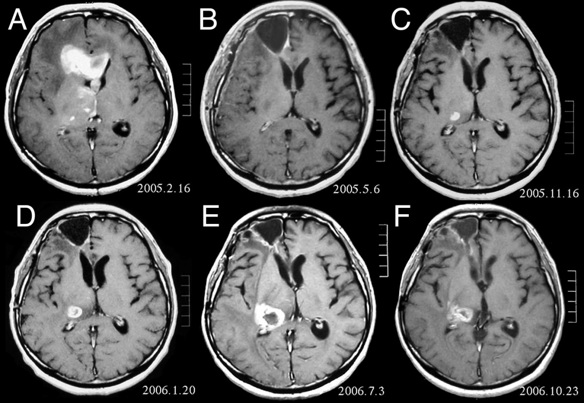
Anaplastic astrocytoma is a rare WHO grade III type of astrocytoma, which is a type of cancer of the brain. In the United States, the annual incidence rate for anaplastic astrocytoma is 0.44 per 100,000 people. Signs and symptoms Initial presenting symptoms most commonly are headache, depressed mental status, focal neurological deficits, and/or seizures. The growth rate and mean interval between onset of symptoms and diagnosis is approximately 1.5–2 years but is highly variable, being intermediate between that of low-grade astrocytomas and glioblastomas. Seizures are less common among patients with anaplastic astrocytomas compared to low-grade lesions. Causes Most high-grade gliomas occur sporadically or without identifiable cause. However, a small proportion (less than 5%) of persons with malignant astrocytoma have a definite or suspected hereditary predisposition. The main hereditary predispositions are mainly neurofibromatosis type I, Li-Fraumeni syndrome, hereditary nonpol ... [...More Info...] [...Related Items...] OR: [Wikipedia] [Google] [Baidu] |
Gangliocytoma
Ganglioglioma is a rare, slow-growing primary central nervous system (CNS) tumor which most frequently occurs in the temporal lobes of children and young adults Classification Gangliogliomas are generally benign WHO grade I tumors; the presence of anaplastic changes in the glial component is considered to represent WHO grade III (anaplastic ganglioglioma). Criteria for WHO grade II have been suggested, but are not established. Malignant transformation of spinal ganglioglioma has been seen in only a select few cases. Poor prognostic factors for adults with gangliogliomas include older age at diagnosis, male sex, and malignant histologic features. Histopathology Histologically, ganglioglioma is composed of both neoplastic glial and ganglion cells which are disorganized, variably cellular, and non-infiltrative. Occasionally, it may be challenging to differentiate ganglion cell tumors from an infiltrating glioma with entrapped neurons. The presence of neoplastic ganglion cells formin ... [...More Info...] [...Related Items...] OR: [Wikipedia] [Google] [Baidu] |
Neurofibroma
A neurofibroma is a benign nerve-sheath tumor in the peripheral nervous system. In 90% of cases, they are found as stand-alone tumors (solitary neurofibroma, solitary nerve sheath tumor or sporadic neurofibroma), while the remainder are found in persons with neurofibromatosis type I (NF1), an autosomal-dominant genetically inherited disease. They can result in a range of symptoms from physical disfiguration and pain to cognitive disability. Neurofibromas arise from nonmyelinating-type Schwann cells that exhibit biallelic inactivation of the ''NF1'' gene that codes for the protein neurofibromin. This protein is responsible for regulating the RAS-mediated cell growth signaling pathway. In contrast to schwannomas, another type of tumor arising from Schwann cells, neurofibromas incorporate many additional types of cells and structural elements in addition to Schwann cells, making it difficult to identify and understand all the mechanisms through which they originate and develop. ... [...More Info...] [...Related Items...] OR: [Wikipedia] [Google] [Baidu] |
Schwannoma
A schwannoma (or neurilemmoma) is a usually benign nerve sheath tumor composed of Schwann cells, which normally produce the insulating myelin sheath covering peripheral nerves. Schwannomas are homogeneous tumors, consisting only of Schwann cells. The tumor cells always stay on the outside of the nerve, but the tumor itself may either push the nerve aside and/or up against a bony structure (thereby possibly causing damage). Schwannomas are relatively slow-growing. For reasons not yet understood, schwannomas are mostly benign and less than 1% become malignant, degenerating into a form of cancer known as neurofibrosarcoma. These masses are generally contained within a capsule, so surgical removal is often successful. Schwannomas can be associated with neurofibromatosis type II, which may be due to a loss-of-function mutation in the protein merlin. They are universally S-100 positive, which is a marker for cells of neural crest cell origin. Schwannomas of the head and neck are a fa ... [...More Info...] [...Related Items...] OR: [Wikipedia] [Google] [Baidu] |
Pinealoblastoma
Pineoblastoma is a malignant tumor of the pineal gland. A pineoblastoma is a supratentorial midline primitive neuroectodermal tumor. Pineoblastoma can present at any age, but is most common in young children. They account for 0.001% of all primary CNS neoplasms. Epidemiology Pineoblastomas typically occur at very young ages. One study found the average age of presentation to be 4.3 years, with peaks at age 3 and 8. Another cites cases to more commonly occur in patients under 2 years of age. Rates of occurrence for males and females are similar, but may be slightly more common in females. One study found incidence of pineoblastoma to be increased in black patients compared to white patients by around 71%. This difference was most apparent in patients aged 5 to 9 years old. Pathophysiology The pineal gland is a small organ in the center of the brain that is responsible for controlling melatonin secretion. Several tumors can occur in the area of the pineal gland, with the most ... [...More Info...] [...Related Items...] OR: [Wikipedia] [Google] [Baidu] |
Pineocytoma
Pineocytoma, is a benign, slowly growing tumor of the pineal gland. Unlike the similar condition pineal gland cyst, it is uncommon. Diagnosis Pineocytomas are diagnosed from tissue, i.e. a brain biopsy.They consist of: * Cytopathology, cytologically benign cells (with nucleus (cell), nuclei of uniform size, regular nuclear membranes, and light chromatin) and, * have the characteristic Pineocytomatous/neurocytic pseudorosettes, pineocytomatous/neurocytic rosettes, which is an irregular circular/flower-like arrangement of cells with a large meshwork of fibers (neuropil) at the centre. Pineocytomatous/neurocytic rosettes are superficially similar to Homer Wright rosettes; however, they differ from Homer Wright rosettes as they have (1) more neuropil at centre of the rosette and, (2) the edge of neuropil meshwork irregular/undulating. Management See also * Pineal gland References External links {{Endocrine gland neoplasia Endocrine neoplasia Brain tumor ... [...More Info...] [...Related Items...] OR: [Wikipedia] [Google] [Baidu] |
Embryonal Tumour With Multilayered Rosettes
Embryonal tumor with multilayered rosettes (ETMR) is an embryonal central nervous system tumor. It is considered an embryonal tumor because it arises from cells partially differentiated or still undifferentiated from birth, usually neuroepithelial cells, stem cells destined to turn into glia or neurons. It can occur anywhere within the brain and can have multiple sites of origins, with a high probability of metastasis through cerebrospinal fluid (CSF). Metastases outside the central nervous system have been reported, but remain rare. Until recently, ETMRs were not recognized as a standalone entity and were grouped together with other CNS primitive neuroectodermal tumors. Histologically, it is similar to other CNS embryonal tumors, such as medulloblastoma, but different regarding genetic factors. It is a rare disease occurring in less than 1 in 700,000 children under the age of 4. Symptoms depend on the location of the tumor and, thus, may vary, but they may include raised in ... [...More Info...] [...Related Items...] OR: [Wikipedia] [Google] [Baidu] |
Cribiform Neuroepithelial Tumour
In mammalian anatomy, the cribriform plate ( Latin for lit. '' sieve-shaped''), horizontal lamina or lamina cribrosa is part of the ethmoid bone. It is received into the ethmoidal notch of the frontal bone and roofs in the nasal cavities. It supports the olfactory bulb, and is perforated by olfactory foramina for the passage of the olfactory nerves to the roof of the nasal cavity to convey smell to the brain. The foramina at the medial part of the groove allow the passage of the nerves to the upper part of the nasal septum while the foramina at the lateral part transmit the nerves to the superior nasal concha. A fractured cribriform plate can result in olfactory dysfunction, septal hematoma, cerebrospinal fluid rhinorrhoea (CSF rhinorrhoea), and possibly infection which can lead to meningitis. CSF rhinorrhoea (clear fluid leaking from the nose) is very serious and considered a medical emergency. Aging can cause the openings in the cribriform plate to close, pinching olf ... [...More Info...] [...Related Items...] OR: [Wikipedia] [Google] [Baidu] |
Atypical Teratoid Rhabdoid Tumor
An atypical teratoid rhabdoid tumor (AT/RT) is a rare tumor usually diagnosed in childhood. Although usually a brain tumor, AT/RT can occur anywhere in the central nervous system (CNS), including the spinal cord. About 60% will be in the posterior cranial fossa (particularly the cerebellum). One review estimated 52% in the posterior fossa, 39% are supratentorial primitive neuroectodermal tumors (sPNET), 5% are in the pineal, 2% are spinal, and 2% are multifocal. In the United States, three children per 1,000,000 or around 30 new AT/RT cases are diagnosed each year. AT/RT represents around 3% of pediatric cancers of the CNS. Around 17% of all pediatric cancers involve the CNS, making these cancers the most common childhood solid tumor. The survival rate for CNS tumors is around 60%. Pediatric brain cancer is the second-leading cause of childhood cancer death, just after leukemia. Recent trends suggest that the rate of overall CNS tumor diagnosis is increasing by about 2. ... [...More Info...] [...Related Items...] OR: [Wikipedia] [Google] [Baidu] |
Medulloblastoma
Medulloblastoma is a common type of primary brain cancer in children. It originates in the part of the brain that is towards the back and the bottom, on the floor of the skull, in the cerebellum, or posterior fossa. The brain is divided into two main parts, the larger cerebrum on top and the smaller cerebellum below towards the back. They are separated by a membrane called the tentorium. Tumors that originate in the cerebellum or the surrounding region below the tentorium are, therefore, called infratentorial. Historically medulloblastomas have been classified as a primitive neuroectodermal tumor (PNET), but it is now known that medulloblastoma is distinct from supratentorial PNETs and they are no longer considered similar entities. Medulloblastomas are invasive, rapidly growing tumors that, unlike most brain tumors, spread through the cerebrospinal fluid and frequently metastasize to different locations along the surface of the brain and spinal cord. Metastasis all the way dow ... [...More Info...] [...Related Items...] OR: [Wikipedia] [Google] [Baidu] |
Choroid Plexus Carcinoma
A choroid plexus carcinoma (WHO Grades of CNS Tumors, WHO grade III) is a type of choroid plexus tumor that affects the choroid plexus of the brain. It is considered the worst of the three grades of chord plexus tumors, having a much poorer prognosis than choroid atypical plexus papilloma (WHO grade II) and choroid plexus papilloma (WHO grade I). The disease creates lesions in the brain and increases cerebrospinal fluid volume, resulting in hydrocephalus. Signs and symptoms The symptoms of choroid plexus carcinoma are similar to those of other brain tumors. They include: * Persistent or new onset headaches * Macrocephaly or bulging fontanels in infants. * Loss of appetite (refusal to take food in infants) * Papilledema * Nausea and emesis * Ataxia * Strabismus * Developmental delays * Altered mental status Cause The cause of choroid plexus carcinomas are relatively unknown, although hereditary factors are suspected. They sometimes occur in conjunction with other hereditary cancers, ... [...More Info...] [...Related Items...] OR: [Wikipedia] [Google] [Baidu] |
Choroid Plexus Papilloma
Choroid plexus papilloma, also known as papilloma of the choroid plexus, is a rare benign neuroepithelial intraventricular WHO grade I lesion found in the choroid plexus. It leads to increased cerebrospinal fluid production, thus causing increased intracranial pressure and hydrocephalus. Choroid plexus papilloma occurs in the lateral ventricles of children and in the fourth ventricle of adults. This is unlike most other pediatric tumors and adult tumors, in which the locations of the tumors is reversed. In children, brain tumors are usually found in the infratentorial region and in adults, brain tumors are usually found in the supratentorial space. The relationship is reversed for choroid plexus papillomas. Epidemiology CPPs are rare tumors of neuroectodermal origin. They make up 0.4 to 0.6 percent of all intracranial neoplasms in children and are the third most prevalent congenital brain tumors after teratomas and gliomas. With a median age upon diagnosis of 3.5 years, th ... [...More Info...] [...Related Items...] OR: [Wikipedia] [Google] [Baidu] |


