|
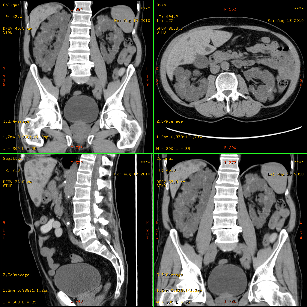
Von Meyenburg Complexes
A bile duct hamartoma or biliary hamartoma, is a benign tumour-like malformation of the liver. They are classically associated with polycystic liver disease, as may be seen in the context of polycystic kidney disease, and represent a malformation of the liver plate. Cause It is a developmental anomaly in which abnormal tissues are present at normal site. Due to failure of regression of embryonic biliary duct. Diagnosis File:Histopathology of a bile duct hamartoma, high magnification.jpg, Histopathology of a bile duct hamartoma, high magnification, H&E stain. It shows typical features of bile duct hamartoma: Topic Completed: 23 November 2020. Minor changes: 23 November 2020 - Small to medium sized, irregularly shaped bile ducts lined by bland cuboidal epithelium (may also be flattened).- Prominent intervening collagenous stroma. - Bile ducts containing eosinophilic debris (may also contain inspissated bile) File:Von Meyenburg complex cropped.tif, von Meyenburg Complex in u ... [...More Info...] [...Related Items...] OR: [Wikipedia] [Google] [Baidu] |
H&E Stain
Hematoxylin and eosin stain ( or haematoxylin and eosin stain or hematoxylin-eosin stain; often abbreviated as H&E stain or HE stain) is one of the principal tissue stains used in histology. It is the most widely used stain in medical diagnosis and is often the gold standard. For example, when a pathologist looks at a biopsy of a suspected cancer, the histological section is likely to be stained with H&E. H&E is the combination of two histological stains: hematoxylin and eosin. The hematoxylin stains cell nuclei a purplish blue, and eosin stains the extracellular matrix and cytoplasm pink, with other structures taking on different shades, hues, and combinations of these colors. Hence a pathologist can easily differentiate between the nuclear and cytoplasmic parts of a cell, and additionally, the overall patterns of coloration from the stain show the general layout and distribution of cells and provides a general overview of a tissue sample's structure. Thus, pattern recogniti ... [...More Info...] [...Related Items...] OR: [Wikipedia] [Google] [Baidu] |
Hamartoma
A hamartoma is a mostly benign, local malformation of cells that resembles a neoplasm of local tissue but is usually due to an overgrowth of multiple aberrant cells, with a basis in a systemic genetic condition, rather than a growth descended from a single mutated cell ( monoclonality), as would typically define a benign neoplasm/tumor. Despite this, many hamartomas are found to have clonal chromosomal aberrations that are acquired through somatic mutations, and on this basis the term ''hamartoma'' is sometimes considered synonymous with neoplasm. Hamartomas are by definition benign, slow-growing or self-limiting, though the underlying condition may still predispose the individual towards malignancies. Hamartomas are usually caused by a genetic syndrome that affects the development cycle of all or at least multiple cells. Many of these conditions are classified as overgrowth syndromes or cancer syndromes. Hamartomas occur in many different parts of the body and are most often asy ... [...More Info...] [...Related Items...] OR: [Wikipedia] [Google] [Baidu] |
Benign
Malignancy () is the tendency of a medical condition to become progressively worse. Malignancy is most familiar as a characterization of cancer. A ''malignant'' tumor contrasts with a non-cancerous benign tumor, ''benign'' tumor in that a malignancy is not self-limited in its growth, is capable of invading into adjacent tissues, and may be capable of spreading to distant tissues. A benign tumor has none of those properties. Malignancy in cancers is characterized by anaplasia, invasiveness, and metastasis. Malignant tumors are also characterized by genome instability, so that cancers, as assessed by whole genome sequencing, frequently have between 10,000 and 100,000 mutations in their entire genomes. Cancers usually show tumour heterogeneity, containing multiple subclones. They also frequently have reduced expression of DNA repair enzymes due to Epigenetics#DNA repair epigenetics in cancer, epigenetic methylation of DNA repair genes or altered MicroRNA#DNA repair and cancer, micr ... [...More Info...] [...Related Items...] OR: [Wikipedia] [Google] [Baidu] |
Tumour
A neoplasm () is a type of abnormal and excessive growth of tissue. The process that occurs to form or produce a neoplasm is called neoplasia. The growth of a neoplasm is uncoordinated with that of the normal surrounding tissue, and persists in growing abnormally, even if the original trigger is removed. This abnormal growth usually forms a mass, when it may be called a tumor. ICD-10 classifies neoplasms into four main groups: benign neoplasms, in situ neoplasms, malignant neoplasms, and neoplasms of uncertain or unknown behavior. Malignant neoplasms are also simply known as cancers and are the focus of oncology. Prior to the abnormal growth of tissue, as neoplasia, cells often undergo an abnormal pattern of growth, such as metaplasia or dysplasia. However, metaplasia or dysplasia does not always progress to neoplasia and can occur in other conditions as well. The word is from Ancient Greek 'new' and 'formation, creation'. Types A neoplasm can be benign, potentially ma ... [...More Info...] [...Related Items...] OR: [Wikipedia] [Google] [Baidu] |
Polycystic Liver Disease
Polycystic liver disease (PLD) usually describes the presence of multiple cysts scattered throughout normal liver tissue. PLD is commonly seen in association with autosomal-dominant polycystic kidney disease, with a prevalence of 1 in 400 to 1000, and accounts for 8–10% of all cases of end-stage renal disease. The much rarer autosomal-dominant polycystic liver disease will progress without any kidney involvement. Presentation Pathophysiology Associations with ''PRKCSH'' and ''SEC63'' have been described. Polycystic liver disease comes in two forms: autosomal dominant polycystic kidney disease (with kidney cysts) and autosomal dominant polycystic liver disease (liver cysts only). Diagnosis Most patients with PLD are asymptomatic with simple cysts found following routine investigations. After confirming the presence of cysts in the liver, laboratory tests may be ordered to check for liver function including bilirubin, alkaline phosphatase, alanine aminotransferase, and prothrombin ... [...More Info...] [...Related Items...] OR: [Wikipedia] [Google] [Baidu] |
Polycystic Kidney Disease
Polycystic kidney disease (PKD or PCKD, also known as polycystic kidney syndrome) is a genetic disorder in which the renal tubules become structurally abnormal, resulting in the development and growth of multiple cysts within the kidney. These cysts may begin to develop in utero, in infancy, in childhood, or in adulthood. Cysts are non-functioning tubules filled with fluid pumped into them, which range in size from microscopic to enormous, crushing adjacent normal tubules and eventually rendering them non-functional as well. PKD is caused by abnormal genes that produce a specific abnormal protein; this protein has an adverse effect on tubule development. PKD is a general term for two types, each having their own pathology and genetic cause: autosomal dominant polycystic kidney disease (ADPKD) and autosomal recessive polycystic kidney disease (ARPKD). The abnormal gene exists in all cells in the body; as a result, cysts may occur in the liver, seminal vesicles, and pancreas. This ... [...More Info...] [...Related Items...] OR: [Wikipedia] [Google] [Baidu] |
CT Scan
A computed tomography scan (CT scan; formerly called computed axial tomography scan or CAT scan) is a medical imaging technique used to obtain detailed internal images of the body. The personnel that perform CT scans are called radiographers or radiology technologists. CT scanners use a rotating X-ray tube and a row of detectors placed in a gantry (medical), gantry to measure X-ray Attenuation#Radiography, attenuations by different tissues inside the body. The multiple X-ray measurements taken from different angles are then processed on a computer using tomographic reconstruction algorithms to produce Tomography, tomographic (cross-sectional) images (virtual "slices") of a body. CT scans can be used in patients with metallic implants or pacemakers, for whom magnetic resonance imaging (MRI) is Contraindication, contraindicated. Since its development in the 1970s, CT scanning has proven to be a versatile imaging technique. While CT is most prominently used in medical diagnosis, ... [...More Info...] [...Related Items...] OR: [Wikipedia] [Google] [Baidu] |
Von Meyenburg Complex
A bile duct hamartoma or biliary hamartoma, is a benign tumour-like malformation of the liver. They are classically associated with polycystic liver disease, as may be seen in the context of polycystic kidney disease, and represent a malformation of the liver plate. Cause It is a developmental anomaly in which abnormal tissues are present at normal site. Due to failure of regression of embryonic biliary duct. Diagnosis File:Histopathology of a bile duct hamartoma, high magnification.jpg, Histopathology of a bile duct hamartoma, high magnification, H&E stain. It shows typical features of bile duct hamartoma: Topic Completed: 23 November 2020. Minor changes: 23 November 2020 - Small to medium sized, irregularly shaped bile ducts lined by bland cuboidal epithelium (may also be flattened).- Prominent intervening collagenous stroma. - Bile ducts containing eosinophilic debris (may also contain inspissated bile) File:Von Meyenburg complex cropped.tif, von Meyenburg Complex in ultras ... [...More Info...] [...Related Items...] OR: [Wikipedia] [Google] [Baidu] |
Hanns Von Meyenburg
Hanns von Meyenburg (actually ''Walter'' but called ''Hanns''; 6 June 1887, in Dresden – 6 November 1971) was a Swiss pathologist. Biography Hanns von Meyenburg was the son of Swiss sculptor Victor von Meyenburg (1834–1893)01384 Viktor von Meyenburg. Matrikeldatenbank. Akademie der Bildenden Künste München. URhttp://matrikel.adbk.de/05ordner/mb_1841-1884/jahr_1856/matrikel-01384 Retrieved 31 October 2009. and his wife Konstanze von May, which belong to the Schaffhausen nobility. He was born in Dresden, where his father had lived since 1869, but returned to his parents' land of origin for his studies and his career. He did his habilitation in 1918 with Otto Busse and became a professor in 1919 at the University of Lausanne. From 1925 to 1953, he was professor at the University of Zürich. From 1932 to 1934, he was Dean of the Faculty of Medicine, and from 1934 to 1936 Rector of the University of Zürich. The von Meyenburg complex, in liver histopathology, in named after ... [...More Info...] [...Related Items...] OR: [Wikipedia] [Google] [Baidu] |
Micrograph
A micrograph or photomicrograph is a photograph or digital image taken through a microscope or similar device to show a magnified image of an object. This is opposed to a macrograph or photomacrograph, an image which is also taken on a microscope but is only slightly magnified, usually less than 10 times. Micrography is the practice or art of using microscopes to make photographs. A micrograph contains extensive details of microstructure. A wealth of information can be obtained from a simple micrograph like behavior of the material under different conditions, the phases found in the system, failure analysis, grain size estimation, elemental analysis and so on. Micrographs are widely used in all fields of microscopy. Types Photomicrograph A light micrograph or photomicrograph is a micrograph prepared using an optical microscope, a process referred to as ''photomicroscopy''. At a basic level, photomicroscopy may be performed simply by connecting a camera to a microscope, th ... [...More Info...] [...Related Items...] OR: [Wikipedia] [Google] [Baidu] |
Trichrome Stain
Trichrome staining is a histological staining method that uses two or more acid dyes in conjunction with a polyacid. Staining differentiates tissues by tinting them in contrasting colours. It increases the contrast of microscopic features in cells and tissues, which makes them easier to see when viewed through a microscope. The word '' trichrome'' means "three colours". The first staining protocol that was described as "trichrome" was Mallory's trichrome stain, which differentially stained erythrocytes to a red colour, muscle tissue to a red colour, and collagen to a blue colour. Some other trichrome staining protocols are the Masson's trichrome stain, Lillie's trichrome, and the Gömöri trichrome stain. Purpose Without trichrome staining, discerning one feature from another can be extremely difficult. Smooth muscle tissue, for example, is hard to differentiate from collagen. A trichrome stain can colour the muscle tissue red, and the collagen fibres green or blue. Liver ... [...More Info...] [...Related Items...] OR: [Wikipedia] [Google] [Baidu] |
Micrograph
A micrograph or photomicrograph is a photograph or digital image taken through a microscope or similar device to show a magnified image of an object. This is opposed to a macrograph or photomacrograph, an image which is also taken on a microscope but is only slightly magnified, usually less than 10 times. Micrography is the practice or art of using microscopes to make photographs. A micrograph contains extensive details of microstructure. A wealth of information can be obtained from a simple micrograph like behavior of the material under different conditions, the phases found in the system, failure analysis, grain size estimation, elemental analysis and so on. Micrographs are widely used in all fields of microscopy. Types Photomicrograph A light micrograph or photomicrograph is a micrograph prepared using an optical microscope, a process referred to as ''photomicroscopy''. At a basic level, photomicroscopy may be performed simply by connecting a camera to a microscope, th ... [...More Info...] [...Related Items...] OR: [Wikipedia] [Google] [Baidu] |


