|
Volar Branch Of Ulnar Nerve
The palmar branch of the ulnar nerve arises about five cm proximal to the wrist from where the ulnar nerve splits into palmar and dorsal branches. It supplies sensory innervation to a small area in the palmar surface of the wrist. The palmar branch represents the continuation of the ulnar nerve as it crosses the flexor retinaculum of the hand on the lateral side of the pisiform bone, medial to and a little behind the ulnar artery. Some sources state that it ends by dividing into a superficial and a deep branch. (Other sources state that the |
Ulnar Nerve
In human anatomy, the ulnar nerve is a nerve that runs near the ulna bone. The ulnar collateral ligament of elbow joint is in relation with the ulnar nerve. The nerve is the largest in the human body unprotected by muscle or bone, so injury is common. This nerve is directly connected to the little finger, and the adjacent half of the ring finger, innervating the palmar aspect of these fingers, including both front and back of the tips, perhaps as far back as the fingernail beds. This nerve can cause an electric shock-like sensation by striking the medial epicondyle of the humerus posteriorly, or inferiorly with the elbow flexed. The ulnar nerve is trapped between the bone and the overlying skin at this point. This is commonly referred to as bumping one's "funny bone". This name is thought to be a pun, based on the sound resemblance between the name of the bone of the upper arm, the humerus, and the word "humorous". Alternatively, according to the Oxford English Dictionary, i ... [...More Info...] [...Related Items...] OR: [Wikipedia] [Google] [Baidu] |
Superficial Branch Of Ulnar Nerve
The superficial branch of the ulnar nerve is a terminal branch of the ulnar nerve. It supplies the palmaris brevis and the skin on the ulnar side of the hand. It also divides into a common palmar digital nerve and a proper palmar digital nerve. The proper digital branches are distributed to the fingers in the same manner as those of the median nerve The median nerve is a nerve in humans and other animals in the upper limb. It is one of the five main nerves originating from the brachial plexus. The median nerve originates from the lateral and medial cords of the brachial plexus, and has contr .... References External links * Nerves of the upper limb {{Neuroanatomy-stub ... [...More Info...] [...Related Items...] OR: [Wikipedia] [Google] [Baidu] |
Deep Branch Of Ulnar Nerve
Deep or The Deep may refer to: Places United States * Deep Creek (Appomattox River tributary), Virginia * Deep Creek (Great Salt Lake), Idaho and Utah * Deep Creek (Mahantango Creek tributary), Pennsylvania * Deep Creek (Mojave River tributary), California * Deep Creek (Pine Creek tributary), Pennsylvania * Deep Creek (Soque River tributary), Georgia * Deep Creek (Texas), a tributary of the Colorado River * Deep Creek (Washington), a tributary of the Spokane River * Deep River (Indiana), a tributary of the Little Calumet River * Deep River (Iowa), a minor tributary of the English River * Deep River (North Carolina) * Deep River (Washington), a minor tributary of the Columbia River * Deep Voll Brook, New Jersey, also known as Deep Brook Elsewhere * Deep Creek (Bahamas) * Deep Creek (Melbourne, Victoria), Australia, a tributary of the Maribyrnong River * Deep River (Western Australia) People * Deep (given name) * Deep (rapper), Punjabi rapper from Houston, Texas * Ravi Deep (b ... [...More Info...] [...Related Items...] OR: [Wikipedia] [Google] [Baidu] |
Wrist
In human anatomy, the wrist is variously defined as (1) the Carpal bones, carpus or carpal bones, the complex of eight bones forming the proximal skeletal segment of the hand; "The wrist contains eight bones, roughly aligned in two rows, known as the carpal bones." (2) the wrist joint or radiocarpal joint, the joint between the radius (bone), radius and the Carpal bones, carpus and; (3) the anatomical region surrounding the carpus including the distal parts of the bones of the forearm and the proximal parts of the metacarpus or five metacarpal bones and the series of joints between these bones, thus referred to as ''wrist joints''. "With the large number of bones composing the wrist (ulna, radius, eight carpas, and five metacarpals), it makes sense that there are many, many joints that make up the structure known as the wrist." This region also includes the carpal tunnel, the anatomical snuff box, bracelet lines, the Flexor retinaculum of the hand, flexor retinaculum, and the ex ... [...More Info...] [...Related Items...] OR: [Wikipedia] [Google] [Baidu] |
Nerve Supply To The Skin
Cutaneous innervation refers to the area of the skin which is supplied by a specific cutaneous nerve. Dermatomes are similar; however, a dermatome only specifies the area served by a spinal nerve. In some cases, the dermatome is less specific (when a spinal nerve is the source for more than one cutaneous nerve), and in other cases it is more specific (when a cutaneous nerve is derived from multiple spinal nerves.) Modern texts are in agreement about which areas of the skin are served by which nerves, but there are minor variations in some of the details. The borders designated by the diagrams in the 1918 edition of Gray's Anatomy are similar, but not identical, to those generally accepted today. Importance of the peripheral nervous system The peripheral nervous system (PNS) is divided into the somatic nervous system, the autonomic nervous system, and the enteric nervous system. However, it is the somatic nervous system, responsible for body movement and the reception of exter ... [...More Info...] [...Related Items...] OR: [Wikipedia] [Google] [Baidu] |
Flexor Retinaculum Of The Hand
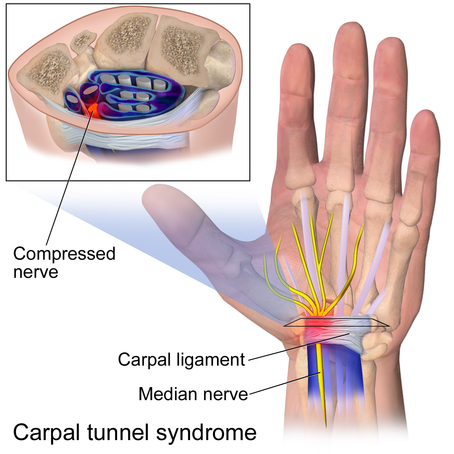
The flexor retinaculum (transverse carpal ligament, or anterior annular ligament) is a fibrous band on the palmar side of the hand near the wrist. It arches over the carpal bones of the hands, covering them and forming the carpal tunnel. Structure The flexor retinaculum is a strong, fibrous band that covers the carpal bones on the palmar side of the hand near the wrist. It attaches to the bones near the radius and ulna. On the ulnar side, the flexor retinaculum attaches to the pisiform bone and the hook of the hamate bone. On the radial side, it attaches to the tubercle of the scaphoid bone, and to the medial part of the palmar surface and the ridge of the trapezium bone. The flexor retinaculum is continuous with the palmar carpal ligament, and deeper with the palmar aponeurosis. The ulnar artery and ulnar nerve, and the cutaneous branches of the median and ulnar nerves, pass on top of the flexor retinaculum. On the radial side of the retinaculum is the tendon of the flexor c ... [...More Info...] [...Related Items...] OR: [Wikipedia] [Google] [Baidu] |
Pisiform
The pisiform bone ( or ), also spelled pisiforme (from the Latin ''pisifomis'', pea-shaped), is a small knobbly, sesamoid bone that is found in the wrist. It forms the ulnar border of the carpal tunnel. Structure The pisiform is a sesamoid bone, with no covering membrane of periosteum. It is the last carpal bone to ossify. The pisiform bone is a small bone found in the proximal row of the wrist (carpus). It is situated where the ulna joins the wrist, within the tendon of the flexor carpi ulnaris muscle. It only has one side that acts as a joint, articulating with the triquetral bone. It is on a plane anterior to the other carpal bones and is spheroidal in form. The pisiform bone has four surfaces: # The ''dorsal surface'' is smooth and oval, and articulates with the triquetral: this facet approaches the superior, but not the inferior border of the bone. # The ''palmar surface'' is rounded and rough, and gives attachment to the transverse carpal ligament, the flexor carpi ulnaris ... [...More Info...] [...Related Items...] OR: [Wikipedia] [Google] [Baidu] |
Ulnar Artery
The ulnar artery is the main blood vessel, with oxygenated blood, of the medial aspects of the forearm. It arises from the brachial artery and terminates in the superficial palmar arch, which joins with the superficial branch of the radial artery. It is palpable on the anterior and medial aspect of the wrist. Along its course, it is accompanied by a similarly named vein or veins, the ulnar vein or ulnar veins. The ulnar artery, the larger of the two terminal branches of the brachial, begins a little below the bend of the elbow in the cubital fossa, and, passing obliquely downward, reaches the ulnar side of the forearm at a point about midway between the elbow and the wrist. It then runs along the ulnar border to the wrist, crosses the transverse carpal ligament on the radial side of the pisiform bone, and immediately beyond this bone divides into two branches, which enter into the formation of the superficial and deep volar arches. Branches Forearm: Anterior ulnar recurrent ... [...More Info...] [...Related Items...] OR: [Wikipedia] [Google] [Baidu] |

