|
Visual Search
Visual search is a type of perception, perceptual task requiring attention that typically involves an active scan of the visual environment for a particular object or feature (the target) among other objects or features (the distractors). Visual search can take place with or without eye movements. The ability to consciously locate an object or target amongst a complex array of stimuli has been extensively studied over the past 40 years. Practical examples of using visual search can be seen in everyday life, such as when one is picking out a product on a supermarket shelf, when animals are searching for food among piles of leaves, when trying to find a friend in a large crowd of people, or simply when playing visual search games such as ''Where's Wally?'' Much previous literature on visual search used reaction time in order to measure the time it takes to detect the target amongst its distractors. An example of this could be a green square (the target) amongst a set of red circles ( ... [...More Info...] [...Related Items...] OR: [Wikipedia] [Google] [Baidu] |
Perception
Perception () is the organization, identification, and interpretation of sensory information in order to represent and understand the presented information or environment. All perception involves signals that go through the nervous system, which in turn result from physical or chemical stimulation of the sensory system.Goldstein (2009) pp. 5–7 Vision involves light striking the retina of the eye; smell is mediated by odor molecules; and hearing involves pressure waves. Perception is not only the passive receipt of these signals, but it is also shaped by the recipient's learning, memory, expectation, and attention. Gregory, Richard. "Perception" in Gregory, Zangwill (1987) pp. 598–601. Sensory input is a process that transforms this low-level information to higher-level information (e.g., extracts shapes for object recognition). The process that follows connects a person's concepts and expectations (or knowledge), restorative and selective mechanisms (such as ... [...More Info...] [...Related Items...] OR: [Wikipedia] [Google] [Baidu] |
Illusory Conjunctions
Illusory conjunctions are psychological effects in which participants combine features of two objects into one object. There are visual illusory conjunctions, auditory illusory conjunctions, and illusory conjunctions produced by combinations of visual and tactile stimuli. Visual illusory conjunctions are thought to occur due to a lack of visual spatial attention, which depends on fixation and (amongst other things) the amount of time allotted to focus on an object. With a short span of time to interpret an object, blending of different aspects within a region of the visual field – like shapes and colors – can occasionally be skewed, which results in visual illusory conjunctions. For example, in a study designed by Anne Treisman and Schmidt, participants were required to view a visual presentation of numbers and shapes in different colors. Some shapes were larger than others but all shapes and numbers were evenly spaced and shown for just 200 ms (followed by a mask). When the par ... [...More Info...] [...Related Items...] OR: [Wikipedia] [Google] [Baidu] |
Superior Colliculus
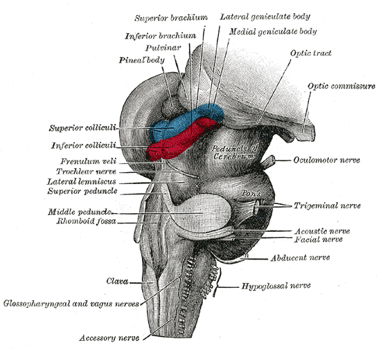
In neuroanatomy, the superior colliculus () is a structure lying on the roof of the mammalian midbrain. In non-mammalian vertebrates, the homologous structure is known as the optic tectum, or optic lobe. The adjective form ''tectal'' is commonly used for both structures. In mammals, the superior colliculus forms a major component of the midbrain. It is a paired structure and together with the paired inferior colliculi forms the corpora quadrigemina. The superior colliculus is a layered structure, with a pattern that is similar to all mammals. The layers can be grouped into the superficial layers ( stratum opticum and above) and the deeper remaining layers. Neurons in the superficial layers receive direct input from the retina and respond almost exclusively to visual stimuli. Many neurons in the deeper layers also respond to other modalities, and some respond to stimuli in multiple modalities. The deeper layers also contain a population of motor-related neurons, capable of activat ... [...More Info...] [...Related Items...] OR: [Wikipedia] [Google] [Baidu] |
V1 Saliency Hypothesis
The V1 Saliency Hypothesis, or V1SH (pronounced‘vish’) is a theory about V1, the primary visual cortex (V1). It proposes that the V1 in primates creates a saliency map of the visual field to guide visual attention or gaze shifts exogenously. Importance V1SH is the only theory so far to not only endow V1 a very important cognitive function, but also to have provided multiple non-trivial theoretical predictions that have been experimentally confirmed subsequently. According to V1SH, V1 creates a saliency map from retinal inputs to guide visual attention or gaze shifts. Anatomically, V1 is the gate for retinal visual inputs to enter neocortex, and is also the largest cortical area devoted to vision. In the 1960s, David Hubel and Torsten Wiesel discovered that V1 neurons are activated by tiny image patches that are large enough to depict a small bar but not a discernible face. This work led to a Nobel prize, and V1 has since been seen as merely serving a back-office function (oi ... [...More Info...] [...Related Items...] OR: [Wikipedia] [Google] [Baidu] |
Superior Colliculus
In neuroanatomy, the superior colliculus () is a structure lying on the roof of the mammalian midbrain. In non-mammalian vertebrates, the homologous structure is known as the optic tectum, or optic lobe. The adjective form ''tectal'' is commonly used for both structures. In mammals, the superior colliculus forms a major component of the midbrain. It is a paired structure and together with the paired inferior colliculi forms the corpora quadrigemina. The superior colliculus is a layered structure, with a pattern that is similar to all mammals. The layers can be grouped into the superficial layers ( stratum opticum and above) and the deeper remaining layers. Neurons in the superficial layers receive direct input from the retina and respond almost exclusively to visual stimuli. Many neurons in the deeper layers also respond to other modalities, and some respond to stimuli in multiple modalities. The deeper layers also contain a population of motor-related neurons, capable of activat ... [...More Info...] [...Related Items...] OR: [Wikipedia] [Google] [Baidu] |
Vision Research
''Vision Research'' is a peer-reviewed scientific journal specializing in the neuroscience and psychology of the visual system of humans and other animals. The journal is abstracted and indexed in PubMed. The journal's impact factor The impact factor (IF) or journal impact factor (JIF) of an academic journal is a scientometric index calculated by Clarivate that reflects the yearly mean number of citations of articles published in the last two years in a given journal, as i ... for 2020 was 1.886 and its 5-year impact factor was 2.823. External links * All articles Vision Neuroscience journals Elsevier academic journals Publications established in 1961 Monthly journals English-language journals Perception journals {{neuroscience-journal-stub ... [...More Info...] [...Related Items...] OR: [Wikipedia] [Google] [Baidu] |
Frontal Eye Field
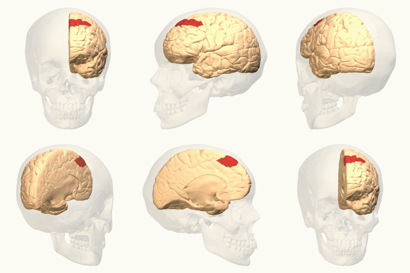
The frontal eye fields (FEF) are a region located in the frontal cortex, more specifically in Brodmann area 8 or BA8, of the primate brain. In humans, it can be more accurately said to lie in a region around the intersection of the middle frontal gyrus with the precentral gyrus, consisting of a frontal and parietal portion. The FEF is responsible for saccadic eye movements for the purpose of visual field perception and awareness, as well as for voluntary eye movement. The FEF communicates with extraocular muscles indirectly via the paramedian pontine reticular formation. Destruction of the FEF causes deviation of the eyes to the ipsilateral side. Function The cortical area called frontal eye field (FEF) plays an important role in the control of visual attention and eye movements. Electrical stimulation in the FEF elicits saccadic eye movements. The FEF have a topographic structure and represents saccade targets in retinotopic coordinates. The frontal eye field is reported to b ... [...More Info...] [...Related Items...] OR: [Wikipedia] [Google] [Baidu] |
Positron Emission Tomography
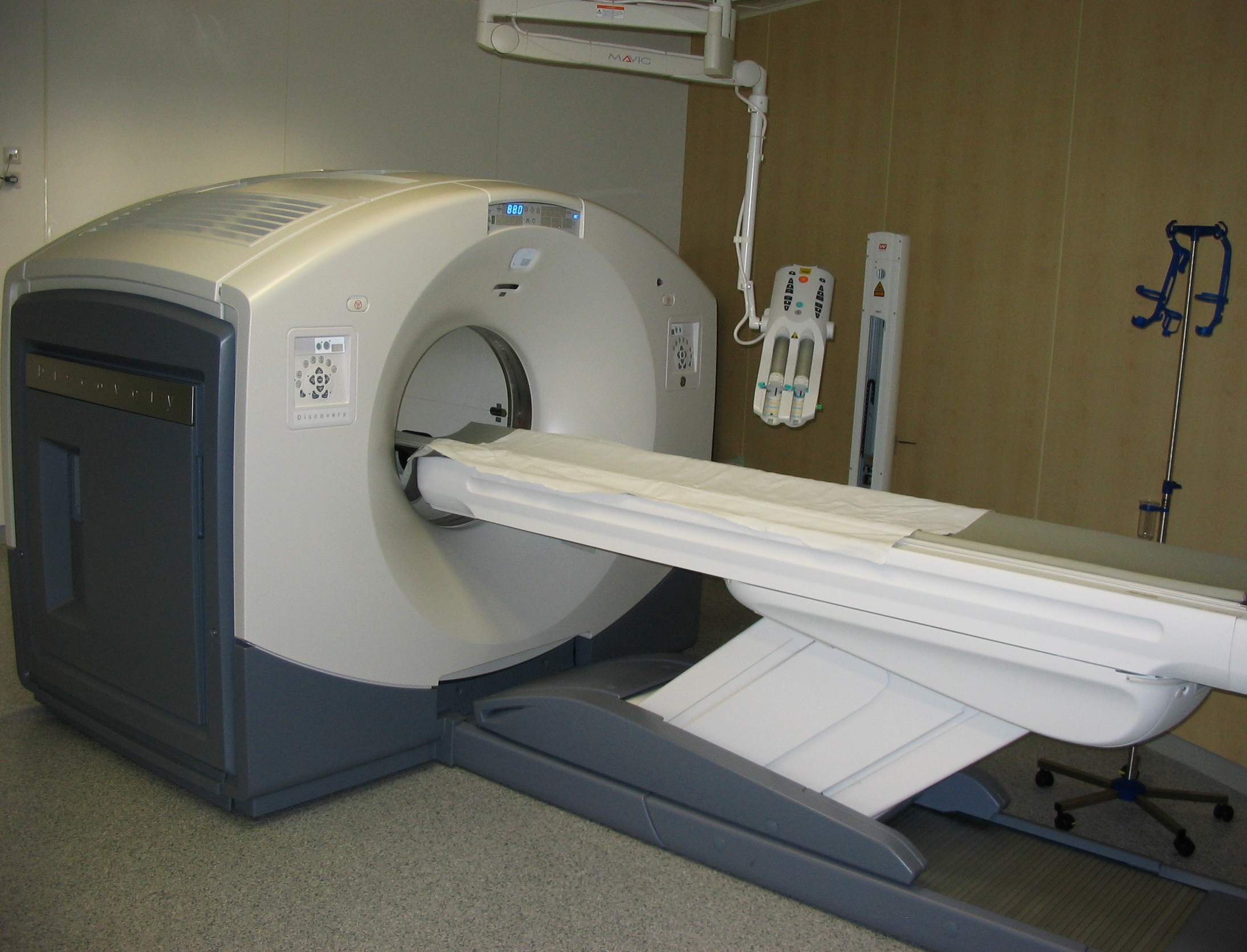
Positron emission tomography (PET) is a functional imaging technique that uses radioactive substances known as radiotracers to visualize and measure changes in Metabolism, metabolic processes, and in other physiological activities including blood flow, regional chemical composition, and absorption. Different tracers are used for various imaging purposes, depending on the target process within the body. For example, 18F-FDG, -FDG is commonly used to detect cancer, Sodium fluoride#Medical imaging, NaF is widely used for detecting bone formation, and Isotopes of oxygen#Oxygen-15, oxygen-15 is sometimes used to measure blood flow. PET is a common medical imaging, imaging technique, a Scintigraphy#Process, medical scintillography technique used in nuclear medicine. A radiopharmaceutical, radiopharmaceutical — a radioisotope attached to a drug — is injected into the body as a radioactive tracer, tracer. When the radiopharmaceutical undergoes beta plus decay, a positron is ... [...More Info...] [...Related Items...] OR: [Wikipedia] [Google] [Baidu] |
Working Memory
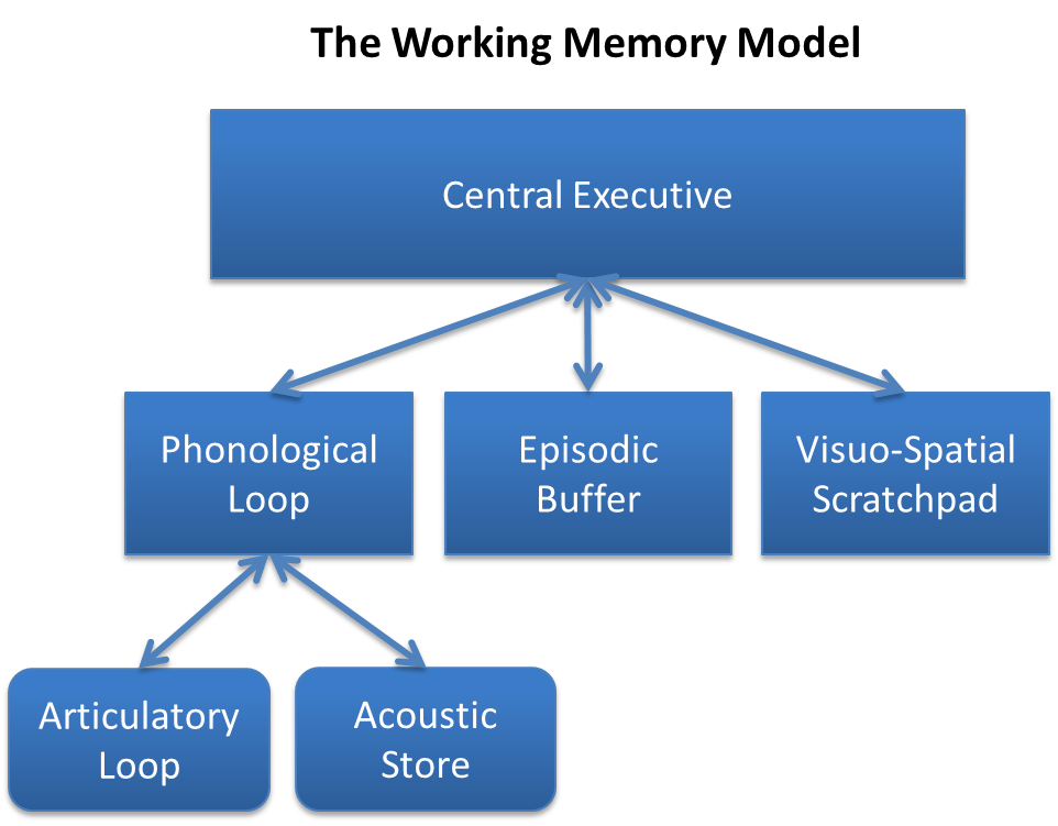
Working memory is a cognitive system with a limited capacity that can hold information temporarily. It is important for reasoning and the guidance of decision-making and behavior. Working memory is often used synonymously with short-term memory, but some theorists consider the two forms of memory distinct, assuming that working memory allows for the manipulation of stored information, whereas short-term memory only refers to the short-term storage of information. Working memory is a theoretical concept central to cognitive psychology, neuropsychology, and neuroscience. History The term "working memory" was coined by Miller, Galanter, and Pribram, and was used in the 1960s in the context of theories that likened the mind to a computer. In 1968, Atkinson and Shiffrin used the term to describe their "short-term store". What we now call working memory was formerly referred to variously as a "short-term store" or short-term memory, primary memory, immediate memory, operant memo ... [...More Info...] [...Related Items...] OR: [Wikipedia] [Google] [Baidu] |
Transcranial Magnetic Stimulation
Transcranial magnetic stimulation (TMS) is a noninvasive form of brain stimulation in which a changing magnetic field is used to induce an electric current at a specific area of the brain through electromagnetic induction. An electric pulse generator, or stimulator, is connected to a magnetic coil connected to the scalp. The stimulator generates a changing electric current within the coil which creates a varying magnetic field, inducing a current within a region in the brain itself.NICE. January 201Transcranial magnetic stimulation for treating and preventing migraine/ref>Michael Craig Miller for Harvard Health Publications. July 26, 201Magnetic stimulation: a new approach to treating depression?/ref> TMS has shown diagnostic and therapeutic potential in the central nervous system with a wide variety of disease states in neurology and mental health, with research still evolving. Adverse effects of TMS appear rare and include fainting and seizure. Other potential issues include ... [...More Info...] [...Related Items...] OR: [Wikipedia] [Google] [Baidu] |
Electroencephalography
Electroencephalography (EEG) is a method to record an electrogram of the spontaneous electrical activity of the brain. The biosignals detected by EEG have been shown to represent the postsynaptic potentials of pyramidal neurons in the neocortex and allocortex. It is typically non-invasive, with the EEG electrodes placed along the scalp (commonly called "scalp EEG") using the International 10-20 system, or variations of it. Electrocorticography, involving surgical placement of electrodes, is sometimes called " intracranial EEG". Clinical interpretation of EEG recordings is most often performed by visual inspection of the tracing or quantitative EEG analysis. Voltage fluctuations measured by the EEG bioamplifier and electrodes allow the evaluation of normal brain activity. As the electrical activity monitored by EEG originates in neurons in the underlying brain tissue, the recordings made by the electrodes on the surface of the scalp vary in accordance with their orientation and ... [...More Info...] [...Related Items...] OR: [Wikipedia] [Google] [Baidu] |


