|
Vincula Tendina
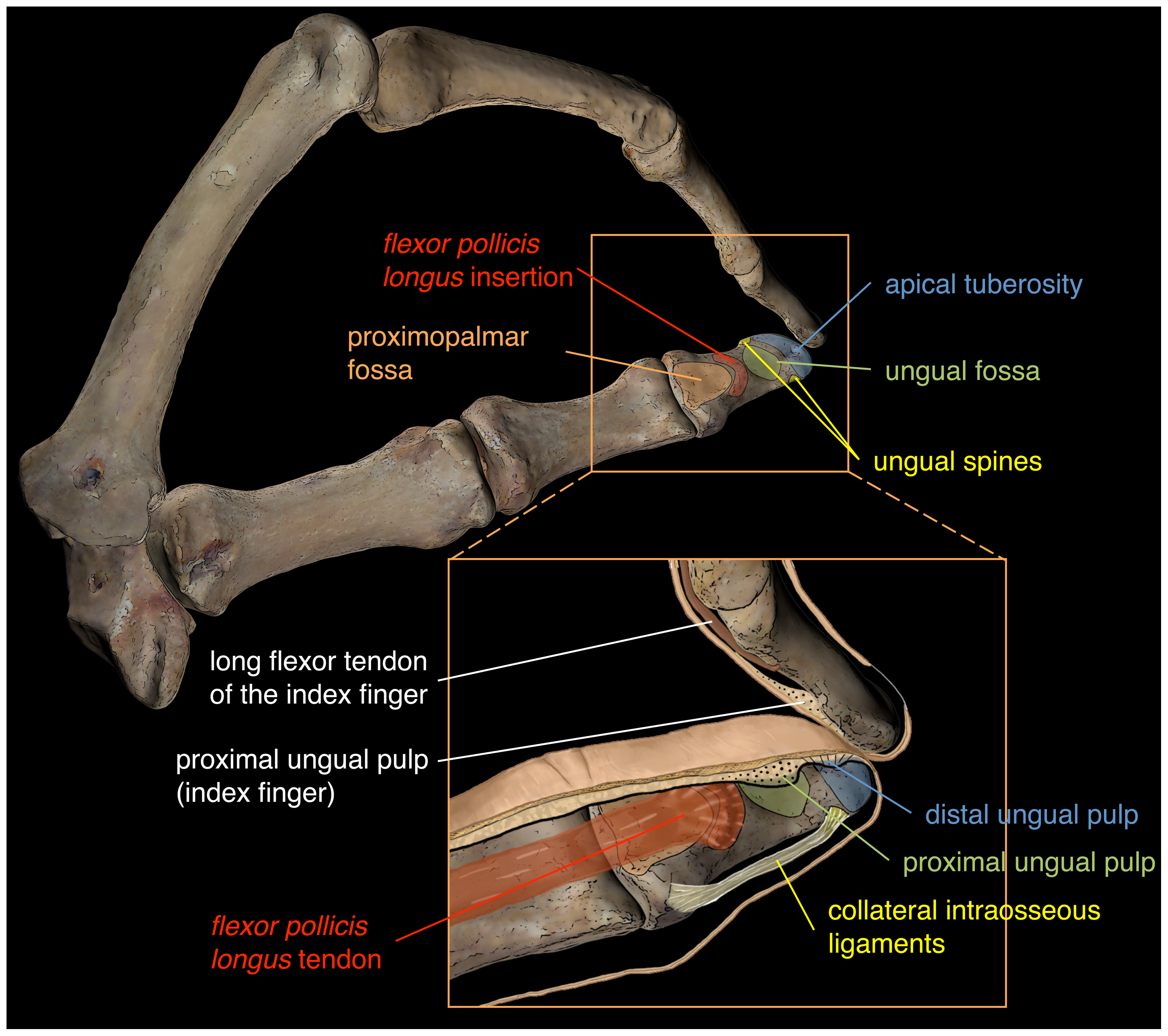
Within each osseo-aponeurotic canal, the tendons of the flexor digitorum superficialis and flexor digitorum profundus are connected to each other, and to the phalanges, by slender, tendinous bands, called vincula tendina. Structure There are around three to seven vincula for each flexor tendon. Vincula tendina can be classified into two types according to their morphology. The ''vincula brevia'' (short), which are two in number in each finger, and consist of triangular bands of fibers, one connecting the tendon of the flexor digitorum superficialis to the front of the first interphalangeal joint and head of the first phalanx, and the other the tendon of the flexor digitorum profundus The flexor digitorum profundus is a muscle in the forearm of humans that flexes the fingers (also known as digits). It is considered an extrinsic hand muscle because it acts on the hand while its muscle belly is located in the forearm. Together t ... to the front of the second interphalangeal j ... [...More Info...] [...Related Items...] OR: [Wikipedia] [Google] [Baidu] |
Forefinger
The index finger (also referred to as forefinger, first finger, second finger, pointer finger, trigger finger, digitus secundus, digitus II, and many other terms) is the second digit of a human hand. It is located between the thumb and the middle finger. It is usually the most dextrous and sensitive digit of the hand, though not the longest. It is shorter than the middle finger, and may be shorter or longer than the ring finger (see digit ratio). Anatomy "Index finger" literally means "pointing finger", from the same Latin source as '' indicate;'' its anatomical names are "index finger" and "second digit". The index finger has three phalanges. It does not contain any muscles, but is controlled by muscles in the hand by attachments of tendons to the bones. Uses A lone index finger held vertically is often used to represent the number 1 (but finger counting differs across cultures), or when held up or moved side to side (finger-wagging), it can be an admonitory gesture. Wit ... [...More Info...] [...Related Items...] OR: [Wikipedia] [Google] [Baidu] |
Flexor Digitorum Superficialis
Flexor digitorum superficialis (''flexor digitorum sublimis'') is an extrinsic flexor muscle of the fingers at the proximal interphalangeal joints. It is in the anterior compartment of the forearm. It is sometimes considered to be the deepest part of the superficial layer of this compartment, and sometimes considered to be a distinct, "intermediate layer" of this compartment. It is relatively common for the Flexor digitorum superficialis to be missing from the little finger, bilaterally and unilaterally, which can cause problems when diagnosing a little finger injury. Structure The muscle has two classically described heads – the humeroulnar and radial – and it is between these heads that the median nerve and ulnar artery pass. The ulnar collateral ligament of elbow joint gives its origin to part of this muscle. Four long tendons come off this muscle near the wrist and travel through the carpal tunnel formed by the flexor retinaculum. These tendons, along with those of fle ... [...More Info...] [...Related Items...] OR: [Wikipedia] [Google] [Baidu] |
Flexor Digitorum Profundus
The flexor digitorum profundus is a muscle in the forearm of humans that flexes the fingers (also known as digits). It is considered an extrinsic hand muscle because it acts on the hand while its muscle belly is located in the forearm. Together the flexor pollicis longus, pronator quadratus, and flexor digitorum profundus form the deep layer of ventral forearm muscles.Platzer 2004, p 162 The muscle is named . Structure Flexor digitorum profundus originates in the upper 3/4 of the anterior and medial surfaces of the ulna, interosseous membrane and deep fascia of the forearm. The muscle fans out into four tendons (one to each of the second to fifth fingers) to the palmar base of the distal phalanx. Along with the flexor digitorum superficialis, it has long tendons that run down the arm and through the carpal tunnel and attach to the palmar side of the phalanges of the fingers. Flexor digitorum profundus lies deep to the superficialis, but it attaches more distally. Therefore, ... [...More Info...] [...Related Items...] OR: [Wikipedia] [Google] [Baidu] |
Phalanges
The phalanges (singular: ''phalanx'' ) are digital bones in the hands and feet of most vertebrates. In primates, the thumbs and big toes have two phalanges while the other digits have three phalanges. The phalanges are classed as long bones. Structure The phalanges are the bones that make up the fingers of the hand and the toes of the foot. There are 56 phalanges in the human body, with fourteen on each hand and foot. Three phalanges are present on each finger and toe, with the exception of the thumb and large toe, which possess only two. The middle and far phalanges of the fifth toes are often fused together (symphalangism). The phalanges of the hand are commonly known as the finger bones. The phalanges of the foot differ from the hand in that they are often shorter and more compressed, especially in the proximal phalanges, those closest to the torso. A phalanx is named according to whether it is proximal, middle, or distal and its associated finger or toe. The proximal ... [...More Info...] [...Related Items...] OR: [Wikipedia] [Google] [Baidu] |
Interphalangeal Articulations Of Hand
The interphalangeal joints of the hand are the hinge joints between the phalanges of the fingers that provide flexion towards the palm of the hand. There are two sets in each finger (except in the thumb, which has only one joint): * "proximal interphalangeal joints" (PIJ or PIP), those between the first (also called proximal) and second (intermediate) phalanges * "distal interphalangeal joints" (DIJ or DIP), those between the second (intermediate) and third (distal) phalanges Anatomically, the proximal and distal interphalangeal joints are very similar. There are some minor differences in how the palmar plates are attached proximally and in the segmentation of the flexor tendon sheath, but the major differences are the smaller dimension and reduced mobility of the distal joint. Joint structure The PIP joint exhibits great lateral stability. Its transverse diameter is greater than its antero-posterior diameter and its thick collateral ligaments are tight in all positions duri ... [...More Info...] [...Related Items...] OR: [Wikipedia] [Google] [Baidu] |
Phalanx Bones
The phalanges (singular: ''phalanx'' ) are digital bones in the hands and feet of most vertebrates. In primates, the thumbs and big toes have two phalanges while the other digits have three phalanges. The phalanges are classed as long bones. Structure The phalanges are the bones that make up the fingers of the hand and the toes of the foot. There are 56 phalanges in the human body, with fourteen on each hand and foot. Three phalanges are present on each finger and toe, with the exception of the thumb and large toe, which possess only two. The middle and far phalanges of the fifth toes are often fused together (symphalangism). The phalanges of the hand are commonly known as the finger bones. The phalanges of the foot differ from the hand in that they are often shorter and more compressed, especially in the proximal phalanges, those closest to the torso. A phalanx is named according to whether it is proximal, middle, or distal and its associated finger or toe. The proximal ... [...More Info...] [...Related Items...] OR: [Wikipedia] [Google] [Baidu] |


