|
Vertebrobasilar System
The basilar artery () is one of the arteries that supplies the brain with oxygen-rich blood. The two vertebral arteries and the basilar artery are known as the vertebral basilar system, which supplies blood to the posterior part of the circle of Willis and joins with blood supplied to the anterior part of the circle of Willis from the internal carotid arteries. Structure The basilar artery arises from the union of the two vertebral arteries at the junction between the medulla oblongata and the pons between the abducens nerves (CN VI). The diameter of the basilar artery range from 1.5 to 6.6 mm. It ascends superiorly in the basilar sulcus of the ventral pons and divides at the junction of the midbrain and pons into the posterior cerebral arteries. Its branches from caudal to rostral include: *anterior inferior cerebellar artery *labyrinthine artery (<15% of people, usually branches from the anterior inferior cerebellar artery) * |
Brainstem
The brainstem (or brain stem) is the posterior stalk-like part of the brain that connects the cerebrum with the spinal cord. In the human brain the brainstem is composed of the midbrain, the pons, and the medulla oblongata. The midbrain is continuous with the thalamus of the diencephalon through the tentorial notch, and sometimes the diencephalon is included in the brainstem. The brainstem is very small, making up around only 2.6 percent of the brain's total weight. It has the critical roles of regulating cardiac, and respiratory function, helping to control heart rate and breathing rate. It also provides the main motor and sensory nerve supply to the face and neck via the cranial nerves. Ten pairs of cranial nerves come from the brainstem. Other roles include the regulation of the central nervous system and the body's sleep cycle. It is also of prime importance in the conveyance of motor and sensory pathways from the rest of the brain to the body, and from the body back to t ... [...More Info...] [...Related Items...] OR: [Wikipedia] [Google] [Baidu] |
Circle Of Willis
The circle of Willis (also called Willis' circle, loop of Willis, cerebral arterial circle, and Willis polygon) is a circulatory anastomosis that supplies blood to the brain and surrounding structures in reptiles, birds and mammals, including humans. It is named after Thomas Willis (1621–1675), an English physician. Structure The circle of Willis is a part of the cerebral circulation and is composed of the following arteries: * Anterior cerebral artery (left and right) * Anterior communicating artery * Internal carotid artery (left and right) * Posterior cerebral artery (left and right) * Posterior communicating artery (left and right) The middle cerebral arteries, supplying the brain, are not considered part of the circle of Willis. Origin of arteries The left and right internal carotid arteries arise from the left and right common carotid arteries. The posterior communicating artery is given off as a branch of the internal carotid artery just before it divides into its termi ... [...More Info...] [...Related Items...] OR: [Wikipedia] [Google] [Baidu] |
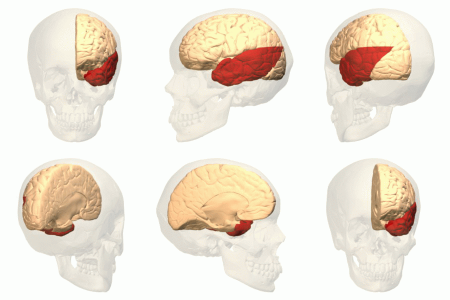
Temporal Lobe
The temporal lobe is one of the four Lobes of the brain, major lobes of the cerebral cortex in the brain of mammals. The temporal lobe is located beneath the lateral fissure on both cerebral hemispheres of the mammalian brain. The temporal lobe is involved in processing sensory input into derived meanings for the appropriate retention of visual memory, language comprehension, and emotion association. ''Temporal'' refers to the head's Temple (anatomy), temples. Structure The Temple (anatomy)#Etymology, temporal Lobe (anatomy), lobe consists of structures that are vital for declarative or long-term memory. Declarative memory, Declarative (denotative) or Explicit memory, explicit memory is conscious memory divided into semantic memory (facts) and episodic memory (events). Medial temporal lobe structures that are critical for long-term memory include the hippocampus, along with the surrounding Hippocampal formation, hippocampal region consisting of the Perirhinal cortex, perirhinal, ... [...More Info...] [...Related Items...] OR: [Wikipedia] [Google] [Baidu] |
Locked-in Syndrome
Locked-in syndrome (LIS), also known as pseudocoma, is a condition in which a patient is aware but cannot move or communicate verbally due to complete paralysis of nearly all voluntary muscles in the body except for vertical eye movements and blinking. The individual is conscious and sufficiently intact cognitively to be able to communicate with eye movements. Electroencephalography results are normal in locked-in syndrome. Total locked-in syndrome, or completely locked-in state (CLIS), is a version of locked-in syndrome wherein the eyes are paralyzed as well. Fred Plum and Jerome B. Posner coined the term for this disorder in 1966. Signs and symptoms Locked-in syndrome is usually characterized by quadriplegia (loss of limb function) and the inability to speak in otherwise cognitively intact individuals. Those with locked-in syndrome may be able to communicate with others through coded messages by blinking or moving their eyes, which are often not affected by the paralysis. The ... [...More Info...] [...Related Items...] OR: [Wikipedia] [Google] [Baidu] |
Pontine Arteries
The pontine arteries are a number of small arteries which come off at right angles from either side of the basilar artery and supply the pons and adjacent parts of the brain. The pontine arteries include the paramedian arteries, the short circumferential, and the long circumferential arteries. See also * Superior cerebellar artery The superior cerebellar artery (SCA) is an artery of the head. It arises near the end of the basilar artery. It is a branch of the basilar artery. It supplies parts of the cerebellum, the midbrain, and other nearby structures. It is the cause of ... Notes References Additional images Image:Gray516.png, The arteries of the base of the brain. Image:Human brainstem blood supply description.JPG, Human brainstem blood supply {{Authority control Arteries of the head and neck ... [...More Info...] [...Related Items...] OR: [Wikipedia] [Google] [Baidu] |
Labyrinthine Artery
The labyrinthine artery (auditory artery, internal auditory artery) is a branch of either the anterior inferior cerebellar artery or the basilar artery. It accompanies the vestibulocochlear nerve (CN VIII) through the internal acoustic meatus. It supplies blood to the internal ear. Structure The labyrinthine artery is a branch of either the anterior inferior cerebellar artery (AICA) or the basilar artery. It accompanies the vestibulocochlear nerve (CN VIII) through the internal acoustic meatus. It divides into a cochlear branch and a labyrinthine (or anterior vestibular) branch. Function The labyrinthine artery supplies blood to the inner ear. It also supplies the vestibulocochlear nerve (CN VIII) along its length. Clinical significance The labyrinthine artery may become occluded. This can cause loss of hearing and balance on the affected side. History The labyrinthine artery may also be known as the internal auditory artery or the auditory artery. See also * Intern ... [...More Info...] [...Related Items...] OR: [Wikipedia] [Google] [Baidu] |
Basilar Sulcus
The basilar sulcus (groove for basilar artery) is a groove in the pons, part of the brainstem. The basilar sulcus is vertical directed and lies in the midline of the pons on its anterior (front) surface. The basilar artery The basilar artery () is one of the arteries that supplies the brain with oxygen-rich blood. The two vertebral arteries and the basilar artery are known as the vertebral basilar system, which supplies blood to the posterior part of the circle of W ... runs within the basilar sulcus. The basilar sulcus is bounded on either side by an eminence caused by the descent of the cerebrospinal fibers through the substance of the pons. Additional images File:Lower pons horizontal KB.svg, A cross section of the lower pons showing the basilar sulcus at the bottom. References External links * https://web.archive.org/web/20100426123803/http://anatomy.med.umich.edu/atlas/n2a4p1.html Pons {{neuroanatomy-stub ... [...More Info...] [...Related Items...] OR: [Wikipedia] [Google] [Baidu] |
Abducens Nerve
The abducens nerve or abducent nerve, also known as the sixth cranial nerve, cranial nerve VI, or simply CN VI, is a cranial nerve in humans and various other animals that controls the movement of the lateral rectus muscle, one of the extraocular muscles responsible for outward gaze. It is a somatic efferent nerve. Structure Nucleus The abducens nucleus is located in the pons, on the floor of the fourth ventricle, at the level of the facial colliculus. Axons from the facial nerve loop around the abducens nucleus, creating a slight bulge (the facial colliculus) that is visible on the dorsal surface of the floor of the fourth ventricle. The abducens nucleus is close to the midline, like the other motor nuclei that control eye movements (the oculomotor and trochlear nuclei). Motor axons leaving the abducens nucleus run ventrally and caudally through the pons. They pass lateral to the corticospinal tract (which runs longitudinally through the pons at this level) before exiting t ... [...More Info...] [...Related Items...] OR: [Wikipedia] [Google] [Baidu] |
Pons
The pons (from Latin , "bridge") is part of the brainstem that in humans and other bipeds lies inferior to the midbrain, superior to the medulla oblongata and anterior to the cerebellum. The pons is also called the pons Varolii ("bridge of Varolius"), after the Italian anatomist and surgeon Costanzo Varolio (1543–75). This region of the brainstem includes neural pathways and tracts that conduct signals from the brain down to the cerebellum and medulla, and tracts that carry the sensory signals up into the thalamus.Saladin Kenneth S.(2007) Anatomy & physiology the unity of form and function. Dubuque, IA: McGraw-Hill Structure The pons is in the brainstem situated between the midbrain and the medulla oblongata, and in front of the cerebellum. A separating groove between the pons and the medulla is the inferior pontine sulcus. The superior pontine sulcus separates the pons from the midbrain. The pons can be broadly divided into two parts: the basilar part of the pons (ventral ... [...More Info...] [...Related Items...] OR: [Wikipedia] [Google] [Baidu] |
Medulla Oblongata
The medulla oblongata or simply medulla is a long stem-like structure which makes up the lower part of the brainstem. It is anterior and partially inferior to the cerebellum. It is a cone-shaped neuronal mass responsible for autonomic (involuntary) functions, ranging from vomiting to sneezing. The medulla contains the cardiac, respiratory, vomiting and vasomotor centers, and therefore deals with the autonomic functions of breathing, heart rate and blood pressure as well as the sleep–wake cycle. During embryonic development, the medulla oblongata develops from the myelencephalon. The myelencephalon is a secondary vesicle which forms during the maturation of the rhombencephalon, also referred to as the hindbrain. The bulb is an archaic term for the medulla oblongata. In modern clinical usage, the word bulbar (as in bulbar palsy) is retained for terms that relate to the medulla oblongata, particularly in reference to medical conditions. The word bulbar can refer to the nerves ... [...More Info...] [...Related Items...] OR: [Wikipedia] [Google] [Baidu] |
Internal Carotid Artery
The internal carotid artery (Latin: arteria carotis interna) is an artery in the neck which supplies the anterior circulation of the brain. In human anatomy, the internal and external carotids arise from the common carotid arteries, where these bifurcate at cervical vertebrae C3 or C4. The internal carotid artery supplies the brain, including the eyes, while the external carotid nourishes other portions of the head, such as the face, scalp, skull, and meninges. Classification Terminologia Anatomica in 1998 subdivided the artery into four parts: "cervical", "petrous", "cavernous", and "cerebral". However, in clinical settings, the classification system of the internal carotid artery usually follows the 1996 recommendations by Bouthillier, describing seven anatomical segments of the internal carotid artery, each with a corresponding alphanumeric identifier—C1 cervical, C2 petrous, C3 lacerum, C4 cavernous, C5 clinoid, C6 ophthalmic, and C7 communicating. The Bouthillier nomenclat ... [...More Info...] [...Related Items...] OR: [Wikipedia] [Google] [Baidu] |
Vertebral Artery
The vertebral arteries are major arteries An artery (plural arteries) () is a blood vessel in humans and most animals that takes blood away from the heart to one or more parts of the body (tissues, lungs, brain etc.). Most arteries carry oxygenated blood; the two exceptions are the pu ... of the neck. Typically, the vertebral arteries originate from the subclavian arteries. Each vessel courses superiorly along each side of the neck, merging within the skull to form the single, midline basilar artery. As the supplying component of the ''vertebrobasilar vascular system'', the vertebral arteries supply blood to the upper spinal cord, brainstem, cerebellum, and Cerebral circulation#Posterior cerebral circulation, posterior part of brain. Structure The vertebral arteries usually arise from the posterosuperior aspect of the central subclavian arteries on each side of the body, then enter deep to the transverse process at the level of the 6th cervical vertebrae (C6), or occasio ... [...More Info...] [...Related Items...] OR: [Wikipedia] [Google] [Baidu] |


