|
Ventrolateral Prefrontal Cortex
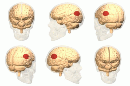
The ventrolateral prefrontal cortex (VLPFC) is a section of the prefrontal cortex located on the inferior frontal gyrus, bounded superiorly by the inferior frontal sulcus and inferiorly by the lateral sulcus. It is attributed to the anatomical structures of Brodmann's area (BA) 47, 45 and 44 (considered the subregions of the VLPFC – the anterior, mid and posterior subregions). Specific functional distinctions have been presented between the three Brodmann subregions of the VLPFC. There are also specific functional differences in activity in the right and left VLPFC. Neuroimaging studies employing various cognitive tasks have shown that the right VLPFC region is a critical substrate of control. At present, two prominent theories feature the right VLPFC as a key functional region. From one perspective, the right VLPFC is thought to play a critical role in motor inhibition, where control is engaged to stop or override motor responses. Alternatively, Corbetta and Shulm ... [...More Info...] [...Related Items...] OR: [Wikipedia] [Google] [Baidu] |
VLPFC BA Pial 20131213 Alxhng
The ventrolateral prefrontal cortex (VLPFC) is a section of the prefrontal cortex located on the inferior frontal gyrus, bounded superiorly by the inferior frontal sulcus and inferiorly by the lateral sulcus. It is attributed to the anatomical structures of Brodmann's area (BA) Brodmann area 47, 47, Brodmann area 45, 45 and Brodmann area 44, 44 (considered the subregions of the VLPFC – the anterior, mid and posterior subregions). Specific functional distinctions have been presented between the three Brodmann subregions of the VLPFC. There are also specific functional differences in activity in the right and left VLPFC. Neuroimaging studies employing various cognitive tasks have shown that the right VLPFC region is a critical substrate of control. At present, two prominent theories feature the right VLPFC as a key functional region. From one perspective, the right VLPFC is thought to play a critical role in motor inhibition, where control is engaged to stop or override ... [...More Info...] [...Related Items...] OR: [Wikipedia] [Google] [Baidu] |
Inferior Parietal Lobule
The inferior parietal lobule (subparietal district) lies below the horizontal portion of the intraparietal sulcus, and behind the lower part of the postcentral sulcus. Also known as Geschwind's territory after Norman Geschwind, an American neurologist, who in the early 1960s recognised its importance. It is a part of the parietal lobe. Structure It is divided from rostral to caudal into two gyri: * One, the supramarginal gyrus, arches over the upturned end of the lateral fissure; it is continuous in front with the postcentral gyrus, and behind with the superior temporal gyrus. * The second, the angular gyrus, arches over the posterior end of the superior temporal sulcus, behind which it is continuous with the middle temporal gyrus. In macaque neuroanatomy, this region is often divided into caudal and rostral portions, cIPL and rIPL, respectively. The cIPL is further divided into areas Opt and PG whereas rIPL is divided into PFG and PF areas. Function Inferior parietal lobule has ... [...More Info...] [...Related Items...] OR: [Wikipedia] [Google] [Baidu] |
Wisconsin Card Sorting Test
The Wisconsin Card Sorting Test (WCST) is a neuropsychological test of set-shifting, which is the capability to show flexibility when exposed to changes in reinforcement.E. A. Berg. (1948). A simple objective technique for measuring flexibility in thinking J. Gen. Psychol. 39: 15-22. The WCST was written by David A. Grant and Esta A. Berg. ''The Professional Manual for the WCST'' was written by Robert K. Heaton, Gordon J. Chelune, Jack L. Talley, Gary G. Kay, and Glenn Curtiss. Method Stimulus cards are shown to the participant and the participant is then instructed to match the cards. They are not given instructions on how to match the cards but are given feedback when the matches they make are right or wrong. When the test was first released the method of showing the cards was done with an evaluator using paper cards with the evaluator on one side of the desk facing the participant on the other. The test takes approximately 12–20 minutes to carry out using manual scoring which ... [...More Info...] [...Related Items...] OR: [Wikipedia] [Google] [Baidu] |
Mesocortical Pathway
The mesocortical pathway is a dopaminergic pathway that connects the ventral tegmentum to the prefrontal cortex. It is one of the four major dopamine pathways in the brain. It is essential to the normal cognitive function of the dorsolateral prefrontal cortex (part of the frontal lobe), and is thought to be involved in cognitive control, motivation, and emotional response. This pathway may be the brain system that is abnormal or functioning abnormally in psychoses, such as schizophrenia.Diaz, Jaime. How Drugs Influence Behavior. Englewood Cliffs: Prentice Hall, 1996. It is thought to be associated with the negative symptoms of schizophrenia, which include avolition, alogia and flat affect. This pathway is closely associated with the mesolimbic pathway, which is also known as the mesolimbic reward pathway. Other dopamine pathways Other major dopamine pathways include: * mesolimbic pathway * nigrostriatal pathway * tuberoinfundibular pathway See also * Dopamine * Schizophrenia ... [...More Info...] [...Related Items...] OR: [Wikipedia] [Google] [Baidu] |
Dorsolateral Prefrontal Cortex
The dorsolateral prefrontal cortex (DLPFC or DL-PFC) is an area in the prefrontal cortex of the primate brain. It is one of the most recently derived parts of the human brain. It undergoes a prolonged period of maturation which lasts until adulthood. The DLPFC is not an anatomical structure, but rather a functional one. It lies in the middle frontal gyrus of humans (i.e., lateral part of Brodmann's area (BA) 9 and 46). In macaque monkeys, it is around the principal sulcus (i.e., in Brodmann's area 46). Other sources consider that DLPFC is attributed anatomically to BA 9 and 46 and BA 8, 9 and 10. The DLPFC has connections with the orbitofrontal cortex, as well as the thalamus, parts of the basal ganglia (specifically, the dorsal caudate nucleus), the hippocampus, and primary and secondary association areas of neocortex (including posterior temporal, parietal, and occipital areas). The DLPFC is also the end point for the dorsal pathway (stream), which is concerned with how to ... [...More Info...] [...Related Items...] OR: [Wikipedia] [Google] [Baidu] |
Cognitive Control
In cognitive science and neuropsychology, executive functions (collectively referred to as executive function and cognitive control) are a set of cognitive processes that are necessary for the cognitive control of behavior: selecting and successfully monitoring behaviors that facilitate the attainment of chosen goals. Executive functions include basic cognitive processes such as attentional control, cognitive inhibition, inhibitory control, working memory, and cognitive flexibility. Higher-order executive functions require the simultaneous use of multiple basic executive functions and include planning and fluid intelligence (e.g., reasoning and problem-solving). Executive functions gradually develop and change across the lifespan of an individual and can be improved at any time over the course of a person's life. Similarly, these cognitive processes can be adversely affected by a variety of events which affect an individual. Both neuropsychological tests (e.g., the Stroop test ... [...More Info...] [...Related Items...] OR: [Wikipedia] [Google] [Baidu] |
Attentional Shift
Attentional shift (or shift of attention) occurs when directing attention to a point increases the efficiency of processing of that point and includes inhibition to decrease attentional resources to unwanted or irrelevant inputs. Shifting of attention is needed to allocate attentional resources to more efficiently process information from a stimulus. Research has shown that when an object or area is attended, processing operates more efficiently. Task switching costs occur when performance on a task suffers due to the increased effort added in shifting attention. There are competing theories that attempt to explain why and how attention is shifted as well as how attention is moved through space. Unitary resource and multiple resource models According to the unitary resource model of attention, there is a single resource of attention divided among different tasks in different amounts, and attention is voluntarily shifted when demands on attention needed exceeds the limited supply of ... [...More Info...] [...Related Items...] OR: [Wikipedia] [Google] [Baidu] |
Attention Versus Memory In Prefrontal Cortex
In mammalian brain anatomy, the prefrontal cortex (PFC) covers the front part of the frontal lobe of the cerebral cortex. The PFC contains the Brodmann areas BA8, BA9, BA10, BA11, BA12, BA13, BA14, BA24, BA25, BA32, BA44, BA45, BA46, and BA47. The basic activity of this brain region is considered to be orchestration of thoughts and actions in accordance with internal goals. Many authors have indicated an integral link between a person's will to live, personality, and the functions of the prefrontal cortex. This brain region has been implicated in executive functions, such as planning, decision making, short-term memory, personality expression, moderating social behavior and controlling certain aspects of speech and language. Executive function relates to abilities to differentiate among conflicting thoughts, determine good and bad, better and best, same and different, future consequences of current activities, working toward a defined goal, prediction of outcomes, e ... [...More Info...] [...Related Items...] OR: [Wikipedia] [Google] [Baidu] |
Ventral Stream
The two-streams hypothesis is a model of the neural processing of vision as well as hearing. The hypothesis, given its initial characterisation in a paper by David Milner and Melvyn A. Goodale in 1992, argues that humans possess two distinct visual systems. Recently there seems to be evidence of two distinct auditory systems as well. As visual information exits the occipital lobe, and as sound leaves the phonological network, it follows two main pathways, or "streams". The ventral stream (also known as the "what pathway") leads to the temporal lobe, which is involved with object and visual identification and recognition. The dorsal stream (or, "where pathway") leads to the parietal lobe, which is involved with processing the object's spatial location relative to the viewer and with speech repetition. History Several researchers had proposed similar ideas previously. The authors themselves credit the inspiration of work on blindsight by Weiskrantz, and previous neuroscientific vis ... [...More Info...] [...Related Items...] OR: [Wikipedia] [Google] [Baidu] |
Temporoparietal Junction
The temporoparietal junction (TPJ) is an area of the brain where the temporal and parietal lobes meet, at the posterior end of the lateral sulcus (Sylvian fissure). The TPJ incorporates information from the thalamus and the limbic system as well as from the visual, auditory, and somatosensory systems. The TPJ also integrates information from both the external environment as well as from within the body. The TPJ is responsible for collecting all of this information and then processing it. This area is also known to play a crucial role in self–other distinctions processes and theory of mind (ToM). Furthermore, damage to the TPJ has been implicated in having adverse effects on an individual's ability to make moral decisions and has been known to produce out-of-body experiences (OBEs). Electromagnetic stimulation of the TPJ can also cause these effects. Apart from these diverse roles that the TPJ plays, it is also known for its involvement in a variety of widespread disorders incl ... [...More Info...] [...Related Items...] OR: [Wikipedia] [Google] [Baidu] |
VLPFC BA Regions
The ventrolateral prefrontal cortex (VLPFC) is a section of the prefrontal cortex located on the inferior frontal gyrus, bounded superiorly by the inferior frontal sulcus and inferiorly by the lateral sulcus. It is attributed to the anatomical structures of Brodmann's area (BA) 47, 45 and 44 (considered the subregions of the VLPFC – the anterior, mid and posterior subregions). Specific functional distinctions have been presented between the three Brodmann subregions of the VLPFC. There are also specific functional differences in activity in the right and left VLPFC. Neuroimaging studies employing various cognitive tasks have shown that the right VLPFC region is a critical substrate of control. At present, two prominent theories feature the right VLPFC as a key functional region. From one perspective, the right VLPFC is thought to play a critical role in motor inhibition, where control is engaged to stop or override motor responses. Alternatively, Corbetta and Shulm ... [...More Info...] [...Related Items...] OR: [Wikipedia] [Google] [Baidu] |
Brodmann Area 44
Brodmann area 44, or BA44, is part of the frontal cortex in the human brain. Situated just anterior to premotor cortex ( BA6) and on the lateral surface, inferior to BA9. This area is also known as pars opercularis (of the inferior frontal gyrus), and it refers to a subdivision of the cytoarchitecturally defined frontal region of cerebral cortex. In the human it corresponds approximately to the opercular part of the inferior frontal gyrus. Thus, it is bounded caudally by the inferior precentral sulcus (H) and rostrally by the anterior ascending limb of lateral sulcus (H). It surrounds the diagonal sulcus (H). In the depth of the lateral sulcus it borders on the insula. Cytoarchitectonically it is bounded caudally and dorsally by the agranular frontal area 6, dorsally by the granular frontal area 9 and rostrally by the triangular part of inferior frontal gyrus (Brodmann area 45 BA 45). Functions Together with left-hemisphere BA45, the left hemisphere BA44 comprises Broca ... [...More Info...] [...Related Items...] OR: [Wikipedia] [Google] [Baidu] |


