|
Ventricle (heart)
A ventricle is one of two large chambers toward the bottom of the heart that collect and expel blood towards the peripheral beds within the body and lungs. The blood pumped by a ventricle is supplied by an atrium (heart), atrium, an adjacent chamber in the upper heart that is smaller than a ventricle. Interventricular means between the ventricles (for example the interventricular septum), while intraventricular means within one ventricle (for example an intraventricular block). In a four-chambered heart, such as that in humans, there are two ventricles that operate in a double circulatory system: the right ventricle pumps blood into the pulmonary circulation to the lungs, and the left ventricle pumps blood into the systemic circulation through the aorta. Structure Ventricles have thicker walls than atria and generate higher blood pressures. The physiological load on the ventricles requiring pumping of blood throughout the body and lungs is much greater than the pressure generated ... [...More Info...] [...Related Items...] OR: [Wikipedia] [Google] [Baidu] |
Heart
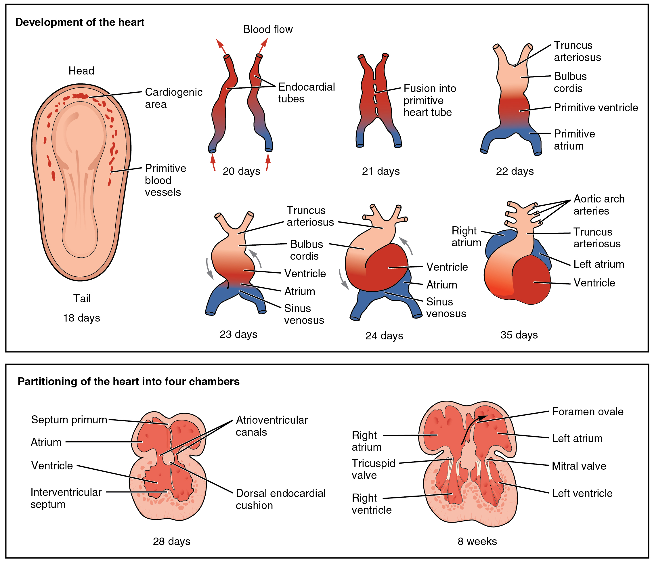
The heart is a muscular organ found in most animals. This organ pumps blood through the blood vessels of the circulatory system. The pumped blood carries oxygen and nutrients to the body, while carrying metabolic waste such as carbon dioxide to the lungs. In humans, the heart is approximately the size of a closed fist and is located between the lungs, in the middle compartment of the chest, called the mediastinum. In humans, other mammals, and birds, the heart is divided into four chambers: upper left and right atria and lower left and right ventricles. Commonly, the right atrium and ventricle are referred together as the right heart and their left counterparts as the left heart. Fish, in contrast, have two chambers, an atrium and a ventricle, while most reptiles have three chambers. In a healthy heart, blood flows one way through the heart due to heart valves, which prevent backflow. The heart is enclosed in a protective sac, the pericardium, which also contains ... [...More Info...] [...Related Items...] OR: [Wikipedia] [Google] [Baidu] |
Chordae Tendinae
The chordae tendineae (tendinous cords), colloquially known as the heart strings, are inelastic cords of fibrous connective tissue that connect the papillary muscles to the tricuspid valve and the mitral valve in the heart. Structure The chordae tendineae connect the atrioventricular valves ( tricuspid and mitral), to the papillary muscles within the ventricles. Multiple chordae tendineae attach to each leaflet or cusp of the valves. Chordae tendineae contain elastin in a delicate structure notably at their periphery. Tendon of Todaro The ''tendon of Todaro'' is a continuation of the Eustachian valve of the inferior vena cava and the valve of the coronary sinus. Along with the opening of the coronary sinus and the septal cusp of the tricuspid valve, it makes up Koch's triangle. The apex of Koch's triangle is the location of the atrioventricular node. Function During atrial systole, blood flows from the atria to the ventricles down the pressure gradient. Chordae tendinea ... [...More Info...] [...Related Items...] OR: [Wikipedia] [Google] [Baidu] |
Cardiology
Cardiology () is a branch of medicine that deals with disorders of the heart and the cardiovascular system. The field includes medical diagnosis and treatment of congenital heart defects, coronary artery disease, heart failure, valvular heart disease and electrophysiology. Physicians who specialize in this field of medicine are called cardiologists, a specialty of internal medicine. Pediatric cardiologists are pediatricians who specialize in cardiology. Physicians who specialize in cardiac surgery are called cardiothoracic surgeons or cardiac surgeons, a specialty of general surgery. Specializations All cardiologists study the disorders of the heart, but the study of adult and child heart disorders each require different training pathways. Therefore, an adult cardiologist (often simply called "cardiologist") is inadequately trained to take care of children, and pediatric cardiologists are not trained to treat adult heart disease. Surgical aspects are not included in ca ... [...More Info...] [...Related Items...] OR: [Wikipedia] [Google] [Baidu] |
Pulmonary Valve
The pulmonary valve (sometimes referred to as the pulmonic valve) is a valve of the heart that lies between the right ventricle and the pulmonary artery and has three cusps. It is one of the four valves of the heart and one of the two semilunar valves, the other being the aortic valve. Similar to the aortic valve, the pulmonary valve opens in ventricular systole, when the pressure in the right ventricle rises above the pressure in the pulmonary artery. At the end of ventricular systole, when the pressure in the right ventricle falls rapidly, the pressure in the pulmonary artery will close the pulmonary valve. The closure of the pulmonary valve contributes the P2 component of the second heart sound (S2). The right heart is a low-pressure system, so the P2 component of the second heart sound is usually softer than the A2 component of the second heart sound. However, it is physiologically normal in some young people to hear both components separated during inhalation. Descript ... [...More Info...] [...Related Items...] OR: [Wikipedia] [Google] [Baidu] |
Pulmonary Vein
The pulmonary veins are the veins that transfer oxygenated blood from the lungs to the heart. The largest pulmonary veins are the four ''main pulmonary veins'', two from each lung that drain into the left atrium of the heart. The pulmonary veins are part of the pulmonary circulation. Structure There are four main pulmonary veins, two from each lung – an inferior and a superior main vein, emerging from each hilum. The main pulmonary veins receive blood from three or four feeding veins in each lung, and drain into the left atrium. The peripheral feeding veins do not follow the bronchial tree. They run between the pulmonary segments from which they drain the blood. At the root of the lung, the right superior pulmonary vein lies in front of and a little below the pulmonary artery; the inferior is situated at the lowest part of the lung hilum. Behind the pulmonary artery is the bronchus. The right main pulmonary veins (contains oxygenated blood) pass behind the right atriu ... [...More Info...] [...Related Items...] OR: [Wikipedia] [Google] [Baidu] |
Aortic Valve
The aortic valve is a valve in the heart of humans and most other animals, located between the left ventricle and the aorta. It is one of the four valves of the heart and one of the two semilunar valves, the other being the pulmonary valve. The aortic valve normally has three cusps or leaflets, although in 1–2% of the population it is found to congenitally have two leaflets. The aortic valve is the last structure in the heart the blood travels through before stopping the flow through the systemic circulation. Structure The aortic valve normally has three cusps however there is some discrepancy in their naming. They may be called the left coronary, right coronary and non-coronary cusp. Some sources also advocate they be named as a left, right and posterior cusp. Anatomists have traditionally named them the left posterior (origin of left coronary), anterior (origin of the right coronary) and right posterior. The three cusps, when the valve is closed, contain a sinus called an ... [...More Info...] [...Related Items...] OR: [Wikipedia] [Google] [Baidu] |
Left Atrium
The atrium ( la, ātrium, , entry hall) is one of two upper chambers in the heart that receives blood from the circulatory system. The blood in the atria is pumped into the heart ventricles through the atrioventricular valves. There are two atria in the human heart – the left atrium receives blood from the pulmonary circulation, and the right atrium receives blood from the venae cavae of the systemic circulation. During the cardiac cycle the atria receive blood while relaxed in diastole, then contract in systole to move blood to the ventricles. Each atrium is roughly cube-shaped except for an ear-shaped projection called an atrial appendage, sometimes known as an auricle. All animals with a closed circulatory system have at least one atrium. The atrium was formerly called the 'auricle'. That term is still used to describe this chamber in some other animals, such as the ''Mollusca''. They have thicker muscular walls than the atria do. Structure Humans have a four-ch ... [...More Info...] [...Related Items...] OR: [Wikipedia] [Google] [Baidu] |
Diastole
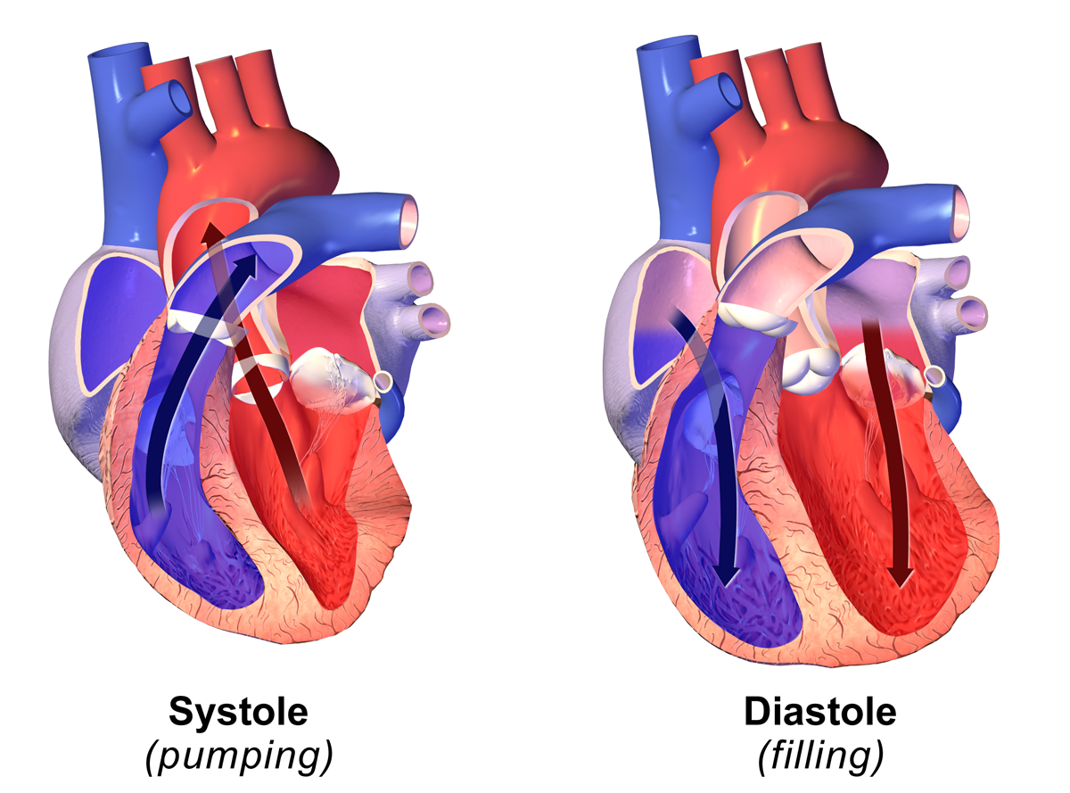
Diastole ( ) is the relaxed phase of the cardiac cycle when the chambers of the heart are re-filling with blood. The contrasting phase is systole when the heart chambers are contracting. Atrial diastole is the relaxing of the atria, and ventricular diastole the relaxing of the ventricles. The term originates from the Greek word (''diastolē''), meaning "dilation", from (''diá'', "apart") + (''stéllein'', "to send"). Role in cardiac cycle A typical heart rate is 75 beats per minute (bpm), which means that the cardiac cycle that produces one heartbeat, lasts for less than one second. The cycle requires 0.3 sec in ventricular systole (contraction)—pumping blood to all body systems from the two ventricles; and 0.5 sec in diastole (dilation), re-filling the four chambers of the heart, for a total of 0.8 sec to complete the cycle. Early ventricular diastole During early ventricular diastole, pressure in the two ventricles begins to drop from the peak reached during syst ... [...More Info...] [...Related Items...] OR: [Wikipedia] [Google] [Baidu] |
Systole (medicine)
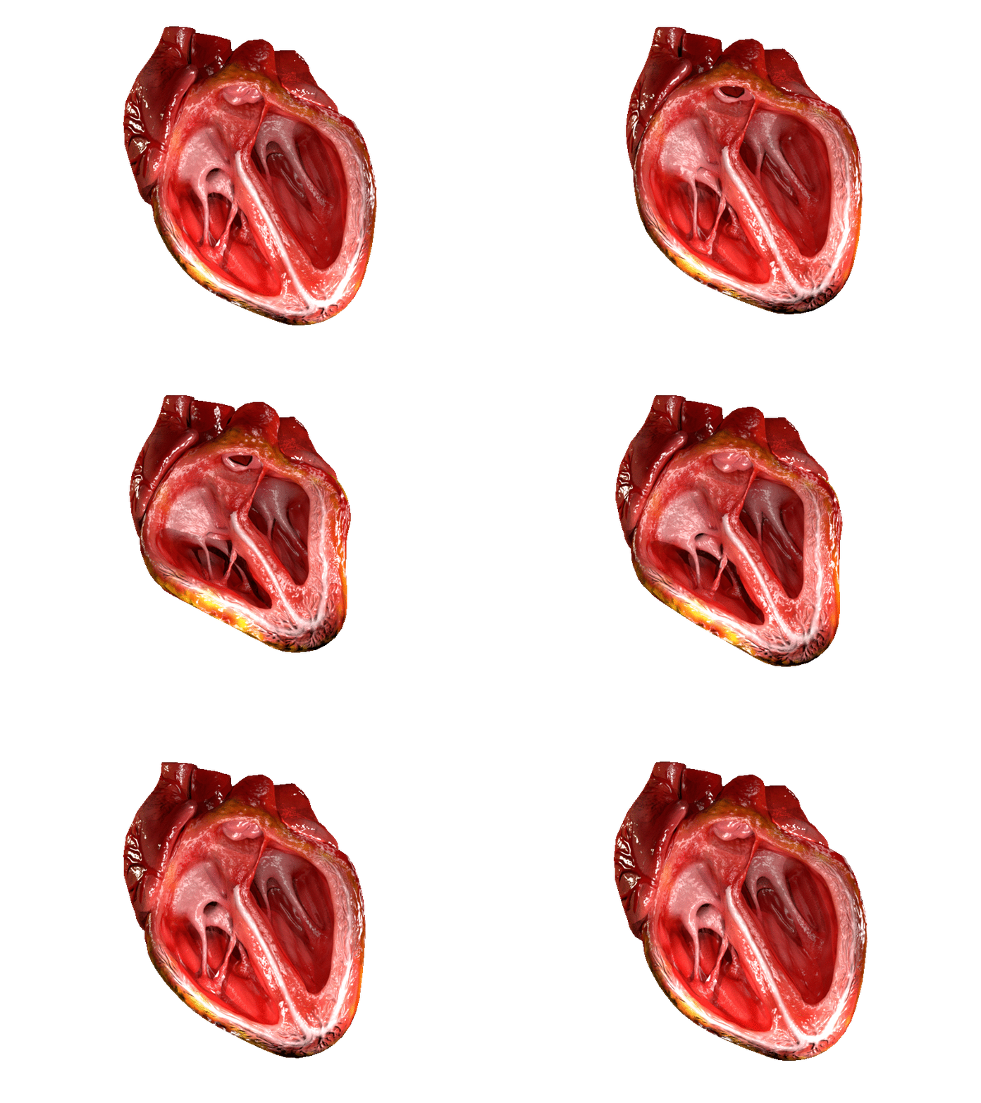
Systole ( ) is the part of the cardiac cycle during which some chambers of the heart contract after refilling with blood. The term originates, via New Latin, from Ancient Greek (''sustolē''), from (''sustéllein'' 'to contract'; from ''sun'' 'together' + ''stéllein'' 'to send'), and is similar to the use of the English term ''to squeeze''. The mammalian heart has four chambers: the left atrium above the left ventricle (lighter pink, see graphic), which two are connected through the mitral (or bicuspid) valve; and the right atrium above the right ventricle (lighter blue), connected through the tricuspid valve. The atria are the receiving blood chambers for the circulation of blood and the ventricles are the discharging chambers. In late ventricular diastole, the atrial chambers contract and send blood to the larger, lower ventricle chambers. This flow fills the ventricles with blood, and the resulting pressure closes the valves to the atria. The ventricles now perfo ... [...More Info...] [...Related Items...] OR: [Wikipedia] [Google] [Baidu] |
Apex Of The Heart
The heart is a muscular organ found in most animals. This organ pumps blood through the blood vessels of the circulatory system. The pumped blood carries oxygen and nutrients to the body, while carrying metabolic waste such as carbon dioxide to the lungs. In humans, the heart is approximately the size of a closed fist and is located between the lungs, in the middle compartment of the chest, called the mediastinum. In humans, other mammals, and birds, the heart is divided into four chambers: upper left and right atria and lower left and right ventricles. Commonly, the right atrium and ventricle are referred together as the right heart and their left counterparts as the left heart. Fish, in contrast, have two chambers, an atrium and a ventricle, while most reptiles have three chambers. In a healthy heart, blood flows one way through the heart due to heart valves, which prevent backflow. The heart is enclosed in a protective sac, the pericardium, which also contains a sma ... [...More Info...] [...Related Items...] OR: [Wikipedia] [Google] [Baidu] |
Ventricular Septum
The interventricular septum (IVS, or ventricular septum, or during development septum inferius) is the stout wall separating the ventricles, the lower chambers of the heart, from one another. The ventricular septum is directed obliquely backward to the right and curved with the convexity toward the right ventricle; its margins correspond with the anterior and posterior interventricular sulci. The lower part of the septum, which is the major part, is thick and muscular, and its much smaller upper part is thin and membraneous. During each cardiac cycle the interventricular septum contracts by shortening longitudinally and becoming thicker. Structure The interventricular septum is the stout wall separating the ventricles, the lower chambers of the heart, from one another. The ventricular septum is directed obliquely backward to the right and curved with the convexity toward the right ventricle; its margins correspond with the anterior and posterior longitudinal sulci. The gr ... [...More Info...] [...Related Items...] OR: [Wikipedia] [Google] [Baidu] |
Sternocostal
The sternocostal joints, also known as sternochondral joints or costosternal articulations, are synovial plane joints of the costal cartilages of the true ribs with the sternum. The only exception is the first rib, which has a synchondrosis A synchondrosis (or primary cartilaginous joint) is a type of cartilaginous joint where hyaline cartilage completely joins together two bones. Synchondroses are different than symphyses (secondary cartilaginous joints) which are formed of fibrocar ... joint since the cartilage is directly united with the sternum. The sternocostal joints are important for thoracic wall mobility. The ligaments connecting them are: * Articular capsules * Interarticular sternocostal ligament * Radiate sternocostal ligaments * Costoxiphoid ligaments Clinical significance Ankylosis, joint stiffness caused by ossification, may occur at the sternocostal joints. See also * Costochondritis References External links Joints Thorax (human anato ... [...More Info...] [...Related Items...] OR: [Wikipedia] [Google] [Baidu] |


