|
Venous
Veins are blood vessels in humans and most other animals that carry blood towards the heart. Most veins carry deoxygenated blood from the tissues back to the heart; exceptions are the pulmonary and umbilical veins, both of which carry oxygenated blood to the heart. In contrast to veins, arteries carry blood away from the heart. Veins are less muscular than arteries and are often closer to the skin. There are valves (called ''pocket valves'') in most veins to prevent backflow. Structure Veins are present throughout the body as tubes that carry blood back to the heart. Veins are classified in a number of ways, including superficial vs. deep, pulmonary vs. systemic, and large vs. small. * Superficial veins are those closer to the surface of the body, and have no corresponding arteries. * Deep veins are deeper in the body and have corresponding arteries. * Perforator veins drain from the superficial to the deep veins. These are usually referred to in the lower limbs and feet. * ... [...More Info...] [...Related Items...] OR: [Wikipedia] [Google] [Baidu] |
Venous Blood
Venous blood is deoxygenated blood which travels from the peripheral blood vessels, through the venous system into the right atrium of the heart. Deoxygenated blood is then pumped by the right ventricle to the lungs via the pulmonary artery which is divided in two branches, left and right to the left and right lungs respectively. Blood is oxygenated in the lungs and returns to the left atrium through the pulmonary veins. Venous blood is typically colder than arterial blood, and has a lower oxygen content and pH. It also has lower concentrations of glucose and other nutrients, and has higher concentrations of urea and other waste products. The difference in the oxygen content of arterial blood and venous blood is known as the arteriovenous oxygen difference. Most medical laboratory tests are conducted on venous blood, with the exception of arterial blood gas tests. Venous blood is obtained for lab work by venipuncture In medicine, venipuncture or venepuncture is the pr ... [...More Info...] [...Related Items...] OR: [Wikipedia] [Google] [Baidu] |
Deoxygenated Blood
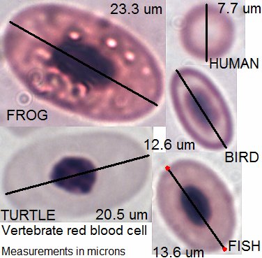
Blood is a body fluid in the circulatory system of humans and other vertebrates that delivers necessary substances such as nutrients and oxygen to the cells, and transports metabolic waste products away from those same cells. Blood in the circulatory system is also known as ''peripheral blood'', and the blood cells it carries, ''peripheral blood cells''. Blood is composed of blood cells suspended in blood plasma. Plasma, which constitutes 55% of blood fluid, is mostly water (92% by volume), and contains proteins, glucose, mineral ions, hormones, carbon dioxide (plasma being the main medium for excretory product transportation), and blood cells themselves. Albumin is the main protein in plasma, and it functions to regulate the colloidal osmotic pressure of blood. The blood cells are mainly red blood cells (also called RBCs or erythrocytes), white blood cells (also called WBCs or leukocytes) and platelets (also called thrombocytes). The most abundant cells in vertebrate blood ar ... [...More Info...] [...Related Items...] OR: [Wikipedia] [Google] [Baidu] |
Blood
Blood is a body fluid in the circulatory system of humans and other vertebrates that delivers necessary substances such as nutrients and oxygen to the cells, and transports metabolic waste products away from those same cells. Blood in the circulatory system is also known as ''peripheral blood'', and the blood cells it carries, ''peripheral blood cells''. Blood is composed of blood cells suspended in blood plasma. Plasma, which constitutes 55% of blood fluid, is mostly water (92% by volume), and contains proteins, glucose, mineral ions, hormones, carbon dioxide (plasma being the main medium for excretory product transportation), and blood cells themselves. Albumin is the main protein in plasma, and it functions to regulate the colloidal osmotic pressure of blood. The blood cells are mainly red blood cells (also called RBCs or erythrocytes), white blood cells (also called WBCs or leukocytes) and platelets (also called thrombocytes). The most abundant cells in verte ... [...More Info...] [...Related Items...] OR: [Wikipedia] [Google] [Baidu] |
Perforator Vein
Perforator veins are so called because they perforate the deep fascia of muscles, to connect the superficial veins to the deep veins where they drain. Perforator veins play an essential role in maintaining normal blood draining. They have valves which prevent blood flowing back ( regurgitation) from deep to superficial veins in muscular systole Systole ( ) is the part of the cardiac cycle during which some chambers of the heart contract after refilling with blood. The term originates, via New Latin, from Ancient Greek (''sustolē''), from (''sustéllein'' 'to contract'; from ' ... or contraction. They exist along the length of the lower limb, in greater number in the leg (anatomical ref to below knee) than in the thigh. Some veins are named after the physician who first described them: *Dodd's perforator at the inferior 1/3 of the thigh *Boyd's perforator at the knee level *Cockett's perforators at the inferior 2/3 of the leg (usually there are three: superior mediu ... [...More Info...] [...Related Items...] OR: [Wikipedia] [Google] [Baidu] |
Blood Vessel
Blood vessels are the structures of the circulatory system that transport blood throughout the human body. These vessels transport blood cells, nutrients, and oxygen to the tissues of the body. They also take waste and carbon dioxide away from the tissues. Blood vessels are needed to sustain life, because all of the body's tissues rely on their functionality. There are five types of blood vessels: the arteries, which carry the blood away from the heart; the arterioles; the capillaries, where the exchange of water and chemicals between the blood and the tissues occurs; the venules; and the veins, which carry blood from the capillaries back towards the heart. The word ''vascular'', meaning relating to the blood vessels, is derived from the Latin ''vas'', meaning vessel. Some structures – such as cartilage, the epithelium, and the lens and cornea of the eye – do not contain blood vessels and are labeled ''avascular''. Etymology * artery: late Middle English; from ... [...More Info...] [...Related Items...] OR: [Wikipedia] [Google] [Baidu] |
Atrium (heart)
The atrium ( la, ātrium, , entry hall) is one of two upper chambers in the heart that receives blood from the circulatory system. The blood in the atria is pumped into the heart ventricles through the atrioventricular valves. There are two atria in the human heart – the left atrium receives blood from the pulmonary circulation, and the right atrium receives blood from the venae cavae of the systemic circulation. During the cardiac cycle the atria receive blood while relaxed in diastole, then contract in systole to move blood to the ventricles. Each atrium is roughly cube-shaped except for an ear-shaped projection called an atrial appendage, sometimes known as an auricle. All animals with a closed circulatory system have at least one atrium. The atrium was formerly called the 'auricle'. That term is still used to describe this chamber in some other animals, such as the ''Mollusca''. They have thicker muscular walls than the atria do. Structure Humans have a four-c ... [...More Info...] [...Related Items...] OR: [Wikipedia] [Google] [Baidu] |
Superior Vena Cava
The superior vena cava (SVC) is the anatomical terms of location#Superior and inferior, superior of the two venae cavae, the great vein, venous trunks that return deoxygenated blood from the circulatory system, systemic circulation to the atrium (heart), right atrium of the heart. It is a large-diameter (24 mm) short length vein that receives venous return from the upper half of the body, above the thoracic diaphragm, diaphragm. Venous return from the lower half, below the diaphragm, flows through the inferior vena cava. The SVC is located in the anterior right superior mediastinum. It is the typical site of central venous access via a central venous catheter or a peripherally inserted central catheter. Mentions of "the cava" without further specification usually refer to the SVC. Structure The superior vena cava is formed by the left and right brachiocephalic veins, which receive blood from the upper limbs, head and neck, behind the lower border of the first right costal ca ... [...More Info...] [...Related Items...] OR: [Wikipedia] [Google] [Baidu] |
Communicating Veins
Communicating veins are veins that communicate two different points of the venous system. They can communicate the great saphenous vein with the small saphenous vein, (for example the Giacomini vein) or the superficial venous system with the deep one. In this case they are called perforator vein Perforator veins are so called because they perforate the deep fascia of muscles, to connect the superficial veins to the deep veins where they drain. Perforator veins play an essential role in maintaining normal blood draining. They have valves ...s and have a very important role in the venous system hemodynamics. References Veins {{circulatory-stub ... [...More Info...] [...Related Items...] OR: [Wikipedia] [Google] [Baidu] |
Inferior Vena Cava
The inferior vena cava is a large vein that carries the deoxygenated blood from the lower and middle body into the right atrium of the heart. It is formed by the joining of the right and the left common iliac veins, usually at the level of the fifth lumbar vertebra. The inferior vena cava is the lower ("inferior") of the two venae cavae, the two large veins that carry deoxygenated blood from the body to the right atrium of the heart: the inferior vena cava carries blood from the lower half of the body whilst the superior vena cava carries blood from the upper half of the body. Together, the venae cavae (in addition to the coronary sinus, which carries blood from the muscle of the heart itself) form the venous counterparts of the aorta. It is a large retroperitoneal vein that lies posterior to the abdominal cavity and runs along the right side of the vertebral column. It enters the right auricle at the lower right, back side of the heart. The name derives from la, ven ... [...More Info...] [...Related Items...] OR: [Wikipedia] [Google] [Baidu] |
Major Veins
Major (commandant in certain jurisdictions) is a military rank of commissioned officer status, with corresponding ranks existing in many military forces throughout the world. When used unhyphenated and in conjunction with no other indicators, major is one rank above captain, and one rank below lieutenant colonel. It is considered the most junior of the field officer ranks. Background Majors are typically assigned as specialised executive or operations officers for battalion-sized units of 300 to 1,200 soldiers while in some nations, like Germany, majors are often in command of a company. When used in hyphenated or combined fashion, the term can also imply seniority at other levels of rank, including ''general-major'' or ''major general'', denoting a low-level general officer, and ''sergeant major'', denoting the most senior non-commissioned officer (NCO) of a military unit. The term ''major'' can also be used with a hyphen to denote the leader of a military band such as ... [...More Info...] [...Related Items...] OR: [Wikipedia] [Google] [Baidu] |
Venae Cavae
In anatomy, the venae cavae (; singular: vena cava ; ) are two large veins (great vessels) that return deoxygenated blood from the body into the heart. In humans they are the superior vena cava and the inferior vena cava, and both empty into the right atrium. They are located slightly off-center, toward the right side of the body. The right atrium receives deoxygenated blood through coronary sinus and two large veins called venae cavae. The inferior vena cava (or caudal vena cava in some animals) travels up alongside the abdominal aorta The aorta ( ) is the main and largest artery in the human body, originating from the left ventricle of the heart and extending down to the abdomen, where it splits into two smaller arteries (the common iliac arteries). The aorta distributes ... with blood from the lower part of the body. It is the largest vein in the human body. [...More Info...] [...Related Items...] OR: [Wikipedia] [Google] [Baidu] |
Duckbill Valve
A duckbill valve is a check valve, usually manufactured from rubber or synthetic elastomer, and has 2 (or more) flaps, usually shaped like the beak of a duck. It is commonly used in medical applications to prevent contamination due to backflow. One end of the valve is stretched over the outlet of a supply line, conforming itself to the shape of the line, usually round. The other end, the duckbill, retains its natural flattened shape. When a fluid is pumped through the supply line and therefore the duckbill, the flattened end opens to permit the pressurized fluid to pass. When pressure is removed, however, the duckbill end returns to its flattened shape, preventing backflow. The duckbill valve is similar in function to the mitral valve in the heart. See also Heimlich valve. A trifold form of this valve, known as a joker valve, is used in one popular marine toilet The head (pl. heads) is a ship's toilet. The name derives from sailing ships in which the toilet area for the ... [...More Info...] [...Related Items...] OR: [Wikipedia] [Google] [Baidu] |


