|
Urinate
Urination, also known as micturition, is the release of urine from the urinary bladder through the urethra to the outside of the body. It is the urinary system's form of excretion. It is also known medically as micturition, voiding, uresis, or, rarely, emiction, and known colloquially by various names including peeing, weeing, and pissing. In healthy humans (and many other animals), the process of urination is under voluntary control. In infants, some elderly individuals, and those with neurological injury, urination may occur as a reflex. It is normal for adult humans to urinate up to seven times during the day. In some animals, in addition to expelling waste material, urination can mark territory or express submissiveness. Physiologically, urination involves coordination between the central, autonomic, and somatic nervous systems. Brain centres that regulate urination include the pontine micturition center, periaqueductal gray, and the cerebral cortex. In placental ma ... [...More Info...] [...Related Items...] OR: [Wikipedia] [Google] [Baidu] |
Urine
Urine is a liquid by-product of metabolism in humans and in many other animals. Urine flows from the kidneys through the ureters to the urinary bladder. Urination results in urine being excretion, excreted from the body through the urethra. Cell (biology), Cellular metabolism generates many by-products that are rich in nitrogen and must be clearance (medicine), cleared from the Circulatory system, bloodstream, such as urea, uric acid, and creatinine. These by-products are expelled from the body during urination, which is the primary method for excreting water-soluble chemicals from the body. A urinalysis can detect nitrogenous wastes of the mammalian body. Urine plays an important role in the earth's nitrogen cycle. In balanced ecosystems, urine fertilizes the soil and thus helps plants to grow. Therefore, Reuse of excreta, urine can be used as a fertilizer. Some animals use it to territory (animal)#Scent marking, mark their territories. Historically, aged or fermented urine (kn ... [...More Info...] [...Related Items...] OR: [Wikipedia] [Google] [Baidu] |
Urinary Bladder
The urinary bladder, or simply bladder, is a hollow organ in humans and other vertebrates that stores urine from the kidneys before disposal by urination. In humans the bladder is a distensible organ that sits on the pelvic floor. Urine enters the bladder via the ureters and exits via the urethra. The typical adult human bladder will hold between 300 and (10.14 and ) before the urge to empty occurs, but can hold considerably more. The Latin phrase for "urinary bladder" is ''vesica urinaria'', and the term ''vesical'' or prefix ''vesico -'' appear in connection with associated structures such as vesical veins. The modern Latin word for "bladder" – ''cystis'' – appears in associated terms such as cystitis (inflammation of the bladder). Structure In humans, the bladder is a hollow muscular organ situated at the base of the pelvis. In gross anatomy, the bladder can be divided into a broad , a body, an apex, and a neck. The apex (also called the vertex) is directed forward ... [...More Info...] [...Related Items...] OR: [Wikipedia] [Google] [Baidu] |
Penis
A penis (plural ''penises'' or ''penes'' () is the primary sexual organ that male animals use to inseminate females (or hermaphrodites) during copulation. Such organs occur in many animals, both vertebrate and invertebrate, but males do not bear a penis in every animal species, and in those species in which the male does bear a so-called penis, the penises in the various species are not necessarily homologous. The term ''penis'' applies to many intromittent organs, but not to all. As an example, the intromittent organ of most cephalopoda is the hectocotylus, a specialized arm, and male spiders use their pedipalps. Even within the Vertebrata there are morphological variants with specific terminology, such as Hemipenis, hemipenes. In most species of animals in which there is an organ that might reasonably be described as a penis, it has no major function other than intromission, or at least conveying the sperm to the female, but in the Eutheria, placental mammals the peni ... [...More Info...] [...Related Items...] OR: [Wikipedia] [Google] [Baidu] |
Detrusor
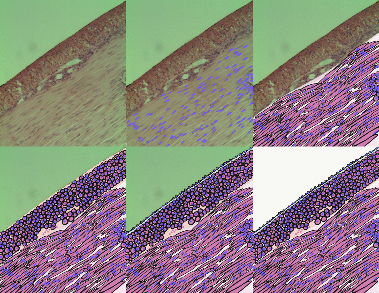
The detrusor muscle, also detrusor urinae muscle, muscularis propria of the urinary bladder and (less precise) muscularis propria, is smooth muscle found in the wall of the bladder. The detrusor muscle remains relaxed to allow the bladder to store urine, and contracts during urination to release urine. Related are the urethral sphincter muscles which envelop the urethra to control the flow of urine when they contract. Structure The fibers of the detrusor muscle arise from the posterior surface of the body of the pubis in both sexes (musculi pubovesicales), and in the male from the adjacent part of the prostate. These fibers pass, in a more or less longitudinal manner, up the inferior surface of the bladder, over its apex, and then descend along its fundus to become attached to the prostate in the male, and to the front of the vagina in the female. At the sides of the bladder the fibers are arranged obliquely and intersect one another. The 3 layers of muscles are arranged lon ... [...More Info...] [...Related Items...] OR: [Wikipedia] [Google] [Baidu] |
Pontine Micturition Center
The Pontine micturition center (PMC, also known as Barrington's nucleus) is a collection of neuronal cell bodies located in the rostral pons in the brainstem involved in the supraspinal regulation of micturition. When activated, the PMC relaxes the urethral sphincter allowing for micturition to occur. The PMC coordinates with other brain centers, including the medial frontal cortex, insular cortex, hypothalamus and periaqueductal gray (PAG). The PAG acts as a relay station for ascending bladder information from the spinal cord and incoming signals from higher brain areas. Regulation In humans and other mammals, neurons in the PMC send descending excitatory projections to spinally located parasympathetic neurons controlling the detrusor muscle of the bladder and inhibitory interneurons regulating Onuf's nucleus. Additionally, the PMC receives ascending input from the level of the lumbosacral spinal cord. During bladder filling, neurons within the PMC are turned off. However, at a ... [...More Info...] [...Related Items...] OR: [Wikipedia] [Google] [Baidu] |
Lumbar
In tetrapod anatomy, lumbar is an adjective that means ''of or pertaining to the abdominal segment of the torso, between the diaphragm and the sacrum.'' The lumbar region is sometimes referred to as the lower spine, or as an area of the back in its proximity. In human anatomy the five lumbar vertebrae (vertebrae in the lumbar region of the back) are the largest and strongest in the movable part of the spinal column, and can be distinguished by the absence of a foramen in the transverse process, and by the absence of facets on the sides of the body. In most mammals, the lumbar region of the spine curves outward. The actual spinal cord terminates between vertebrae one and two of this series, called L1 and L2. The nervous tissue that extends below this point are individual strands that collectively form the cauda equina. In between each lumbar vertebra a nerve root exits, and these nerve roots come together again to form the largest single nerve in the human body, the sciatic n ... [...More Info...] [...Related Items...] OR: [Wikipedia] [Google] [Baidu] |
Smooth Muscle
Smooth muscle is an involuntary non-striated muscle, so-called because it has no sarcomeres and therefore no striations (''bands'' or ''stripes''). It is divided into two subgroups, single-unit and multiunit smooth muscle. Within single-unit muscle, the whole bundle or sheet of smooth muscle cells contracts as a syncytium. Smooth muscle is found in the walls of hollow organs, including the stomach, intestines, bladder and uterus; in the walls of passageways, such as blood, and lymph vessels, and in the tracts of the respiratory, urinary, and reproductive systems. In the eyes, the ciliary muscles, a type of smooth muscle, dilate and contract the iris and alter the shape of the lens. In the skin, smooth muscle cells such as those of the arrector pili cause hair to stand erect in response to cold temperature or fear. Structure Gross anatomy Smooth muscle is grouped into two types: single-unit smooth muscle, also known as visceral smooth muscle, and multiunit smooth muscle. ... [...More Info...] [...Related Items...] OR: [Wikipedia] [Google] [Baidu] |
Sympathetic Nervous System
The sympathetic nervous system (SNS) is one of the three divisions of the autonomic nervous system, the others being the parasympathetic nervous system and the enteric nervous system. The enteric nervous system is sometimes considered part of the autonomic nervous system, and sometimes considered an independent system. The autonomic nervous system functions to regulate the body's unconscious actions. The sympathetic nervous system's primary process is to stimulate the body's fight or flight response. It is, however, constantly active at a basic level to maintain homeostasis. The sympathetic nervous system is described as being antagonistic to the parasympathetic nervous system which stimulates the body to "feed and breed" and to (then) "rest-and-digest". Structure There are two kinds of neurons involved in the transmission of any signal through the sympathetic system: pre-ganglionic and post-ganglionic. The shorter preganglionic neurons originate in the thoracolumbar division o ... [...More Info...] [...Related Items...] OR: [Wikipedia] [Google] [Baidu] |
Spinal Cord
The spinal cord is a long, thin, tubular structure made up of nervous tissue, which extends from the medulla oblongata in the brainstem to the lumbar region of the vertebral column (backbone). The backbone encloses the central canal of the spinal cord, which contains cerebrospinal fluid. The brain and spinal cord together make up the central nervous system (CNS). In humans, the spinal cord begins at the occipital bone, passing through the foramen magnum and then enters the spinal canal at the beginning of the cervical vertebrae. The spinal cord extends down to between the first and second lumbar vertebrae, where it ends. The enclosing bony vertebral column protects the relatively shorter spinal cord. It is around long in adult men and around long in adult women. The diameter of the spinal cord ranges from in the cervical and lumbar regions to in the thoracic area. The spinal cord functions primarily in the transmission of nerve signals from the motor cortex to the body, ... [...More Info...] [...Related Items...] OR: [Wikipedia] [Google] [Baidu] |
Sacrum
The sacrum (plural: ''sacra'' or ''sacrums''), in human anatomy, is a large, triangular bone at the base of the spine that forms by the fusing of the sacral vertebrae (S1S5) between ages 18 and 30. The sacrum situates at the upper, back part of the pelvic cavity, between the two wings of the pelvis. It forms joints with four other bones. The two projections at the sides of the sacrum are called the alae (wings), and articulate with the ilium at the L-shaped sacroiliac joints. The upper part of the sacrum connects with the last lumbar vertebra (L5), and its lower part with the coccyx (tailbone) via the sacral and coccygeal cornua. The sacrum has three different surfaces which are shaped to accommodate surrounding pelvic structures. Overall it is concave (curved upon itself). The base of the sacrum, the broadest and uppermost part, is tilted forward as the sacral promontory internally. The central part is curved outward toward the posterior, allowing greater room for the pel ... [...More Info...] [...Related Items...] OR: [Wikipedia] [Google] [Baidu] |

.jpg)


