|
Ureteric Balloon Catheter
A ureteric balloon catheter is a balloon catheter intended for treating strictures of the ureter. In fact it is a double J stent on which a balloon is mounted. It is connected to a delivery device (pusher) to introduce it from the bladder into the ureter. The system comprises a non-return valve device, and a pusher with a stylet and two ports. The side port is for injecting contrast agent to inflate the balloon, while the straight port is for the guidewire. The catheter has a relatively large-diameter central lumen and a shaft of 2 mm (6 Fr.). The balloon is in two sections: a long narrow section or shaft and a larger cranial bulb. The larger cranial bulb prevents distal migration, while the longer narrow section maintains the increased diameter of a predilated stricture in the ureter section. Stent placement and removal A guide wire has to be placed in the ureter. After dilatation of the ureteric stricture with a high pressure dilatation balloon the guidewire remains in ... [...More Info...] [...Related Items...] OR: [Wikipedia] [Google] [Baidu] |
Balloon Catheter
A balloon catheter is a type of "soft" catheter with an inflatable "balloon" at its tip which is used during a catheterization procedure to enlarge a narrow opening or passage within the body. The deflated balloon catheter is positioned, then inflated to perform the necessary procedure, and deflated again in order to be removed. Some common uses include: *angioplasty or balloon septostomy, via cardiac catheterization (heart cath) *tuboplasty via uterine catheterization *pyeloplasty using a detachable inflatable balloon stent positioned via a cystocopic transvesicular approach. Angioplasty balloon catheters Balloon catheters used in angioplasty are either of Over-the-Wire (OTW) or Rapid Exchange (Rx) design. Rx catheters nowadays are about 90% of the Coronary Intervention market. While OTW Catheters may still be useful in highly tortuous vascular pathways, they sacrifice deflation time and pushability. When a balloon catheter is used to compress plaque within a clogged coronary ... [...More Info...] [...Related Items...] OR: [Wikipedia] [Google] [Baidu] |
Ureteric Stricture
Ureteric stricture (ureteral stricture) is the pathological narrowing of the ureter which may lead to serious complications such as kidney failure. Pathophysiology Several conditions have been identified to cause strictures such as impacted ureteric stones, iatrogenic injuries, tumours and radiotherapy. Iatrogenic injuries Endoscopic urological procedures are common with the advancement of medical technology which allows endoscopic procedures to be among the main operative modalities in urology. However, complications can occur, and iatrogenic injury is disruption of healthy tissue which might lead to local inflammation in the process of healing and scarring which can result in ureteric stricture. Non-endoscopic open surgical procedures such as colon surgery where the operation field is very close to the adjacent retroperitoneal ureter is a well-known procedure where ureters can be injured, especially when surgical plans are distorted in conditions such as tethered colon ... [...More Info...] [...Related Items...] OR: [Wikipedia] [Google] [Baidu] |
Double J Stent
A ureteral stent (pronounced you-REE-ter-ul), or ureteric stent, is a thin tube inserted into the ureter to prevent or treat obstruction of the urine flow from the kidney. The length of the stents used in adult patients varies between 24 and 30 cm. Additionally, stents come in differing diameters or gauges, to fit different size ureters. The stent is usually inserted with the aid of a cystoscope. One or both ends of the stent may be coiled to prevent it from moving out of place; this is called a JJ stent, double J stent or pig-tail stent. Stent placement Ureteral stents are used to ensure the openness of a ureter, which may be compromised, for example, by a kidney stone or a procedure. This method is sometimes used as a temporary measure, to prevent damage to a blocked kidney, until a procedure to remove the stone can be performed. Indwelling times of 12 months or longer are indicated to hold ureters open, which are compressed by tumors in the neighbourhood of the ureter or b ... [...More Info...] [...Related Items...] OR: [Wikipedia] [Google] [Baidu] |
Bladder
The urinary bladder, or simply bladder, is a hollow organ in humans and other vertebrates that stores urine from the kidneys before disposal by urination. In humans the bladder is a distensible organ that sits on the pelvic floor. Urine enters the bladder via the ureters and exits via the urethra. The typical adult human bladder will hold between 300 and (10.14 and ) before the urge to empty occurs, but can hold considerably more. The Latin phrase for "urinary bladder" is ''vesica urinaria'', and the term ''vesical'' or prefix ''vesico -'' appear in connection with associated structures such as vesical veins. The modern Latin word for "bladder" – ''cystis'' – appears in associated terms such as cystitis (inflammation of the bladder). Structure In humans, the bladder is a hollow muscular organ situated at the base of the pelvis. In gross anatomy, the bladder can be divided into a broad , a body, an apex, and a neck. The apex (also called the vertex) is directed forw ... [...More Info...] [...Related Items...] OR: [Wikipedia] [Google] [Baidu] |
Ureter
The ureters are tubes made of smooth muscle that propel urine from the kidneys to the urinary bladder. In a human adult, the ureters are usually long and around in diameter. The ureter is lined by urothelial cells, a type of transitional epithelium, and has an additional smooth muscle layer that assists with peristalsis in its lowest third. The ureters can be affected by a number of diseases, including urinary tract infections and kidney stone. is when a ureter is narrowed, due to for example chronic inflammation. Congenital abnormalities that affect the ureters can include the development of two ureters on the same side or abnormally placed ureters. Additionally, reflux of urine from the bladder back up the ureters is a condition commonly seen in children. The ureters have been identified for at least two thousand years, with the word "ureter" stemming from the stem relating to urinating and seen in written records since at least the time of Hippocrates. It is, ho ... [...More Info...] [...Related Items...] OR: [Wikipedia] [Google] [Baidu] |
Urothelium
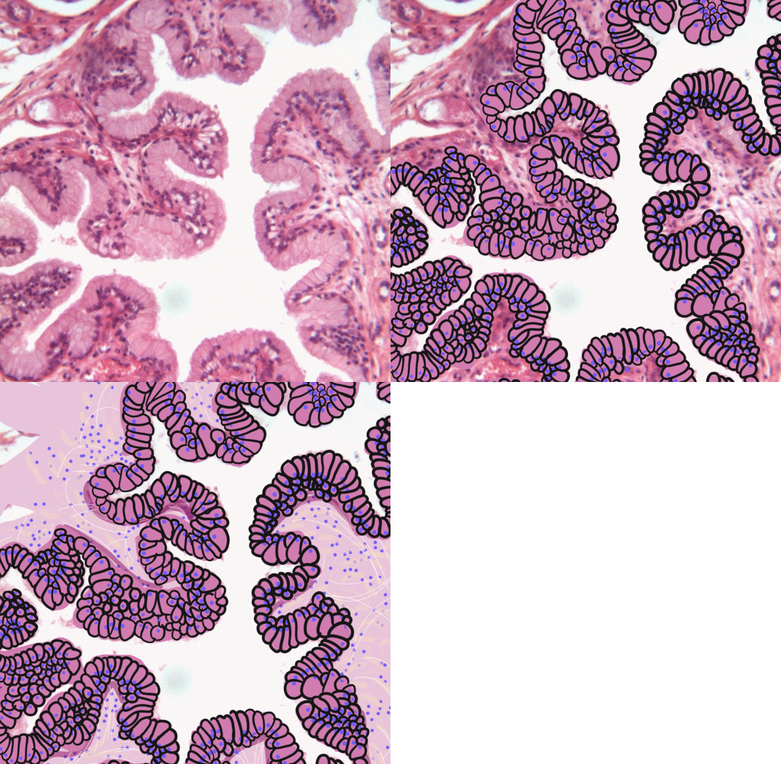
Transitional epithelium also known as urothelium is a type of stratified epithelium. Transitional epithelium is a type of tissue that changes shape in response to stretching (stretchable epithelium). The transitional epithelium usually appears cuboidal when relaxed and squamous when stretched. This tissue consists of multiple layers of epithelial cells which can contract and expand in order to adapt to the degree of distension needed. Transitional epithelium lines the organs of the urinary system and is known here as urothelium. The bladder for example has a need for great distension. Structure The appearance of transitional epithelium differs according to its cell layer. Cells of the basal layer are cuboidal (cube-shaped), or columnar (column-shaped), while the cells of the superficial layer vary in appearance depending on the degree of distension. These cells appear to be cuboidal with a domed apex when the organ or the tube in which they reside is not stretched. When the org ... [...More Info...] [...Related Items...] OR: [Wikipedia] [Google] [Baidu] |
Fibrosis
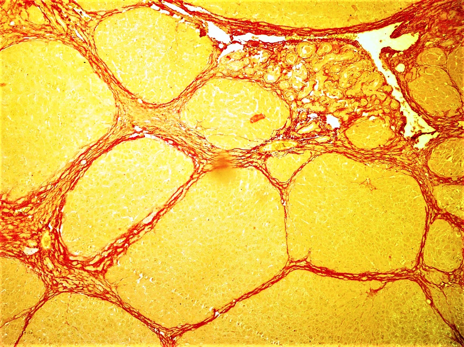
Fibrosis, also known as fibrotic scarring, is a pathological wound healing in which connective tissue replaces normal parenchymal tissue to the extent that it goes unchecked, leading to considerable tissue remodelling and the formation of permanent scar tissue. Repeated injuries, chronic inflammation and repair are susceptible to fibrosis where an accidental excessive accumulation of extracellular matrix components, such as the collagen is produced by fibroblasts, leading to the formation of a permanent fibrotic scar. In response to injury, this is called scarring, and if fibrosis arises from a single cell line, this is called a fibroma. Physiologically, fibrosis acts to deposit connective tissue, which can interfere with or totally inhibit the normal architecture and function of the underlying organ or tissue. Fibrosis can be used to describe the pathological state of excess deposition of fibrous tissue, as well as the process of connective tissue deposition in healing. Define ... [...More Info...] [...Related Items...] OR: [Wikipedia] [Google] [Baidu] |
Hypertrophic
Hypertrophy is the increase in the volume of an organ or tissue due to the enlargement of its component cells. It is distinguished from hyperplasia, in which the cells remain approximately the same size but increase in number.Updated by Linda J. Vorvick. 8/14/1Hyperplasia/ref> Although hypertrophy and hyperplasia are two distinct processes, they frequently occur together, such as in the case of the hormonally-induced proliferation and enlargement of the cells of the uterus during pregnancy. Eccentric hypertrophy is a type of hypertrophy where the walls and chamber of a hollow organ undergo growth in which the overall size and volume are enlarged. It is applied especially to the left ventricle of heart. Sarcomeres are added in series, as for example in dilated cardiomyopathy (in contrast to hypertrophic cardiomyopathy, a type of concentric hypertrophy, where sarcomeres are added in parallel). Gallery File:*+ * Photographic documentation on sexual education - Hypertrophy of br ... [...More Info...] [...Related Items...] OR: [Wikipedia] [Google] [Baidu] |
Ileal Conduit
An ileal conduit urinary diversion is one of various surgical techniques for urinary diversion. It has sometimes been referred to as the Bricker ileal conduit after its inventor, Eugene M. Bricker. It is a form of incontinent urostomy, and was developed during the 1940s and is still one of the most used techniques for the diversion of urine after a patient has had their bladder removed, due to its low complication rate and high patient satisfaction level. It is usually used in conjunction with radical cystectomy in order to control invasive bladder cancer. To create an ileal conduit, the ureters are surgically resected from the bladder and a ureteroenteric anastomosis is made in order to drain the urine into a detached section of ileum at the distal small intestine, though the distal most 25 cm of terminal ileum are avoided as this is where bile salts are reabsorbed. The end of the ileum is then brought out through an opening (a stoma) in the abdominal wall. The residual small bowe ... [...More Info...] [...Related Items...] OR: [Wikipedia] [Google] [Baidu] |
Urinary Diversion
Urinary diversion is any one of several surgical procedures to reroute urine flow from its normal pathway. It may be necessary for diseased or defective ureters, bladder or urethra, either temporarily or permanently. Some diversions result in a stoma. Types * Nephrostomy from the renal pelvis * Urostomy from more distal origins along the urinary tract, with subtypes including: ** Ileal conduit urinary diversion (Bricker conduit) ** Indiana pouch * Neobladder to urethra diversion Ureteroenteric anastomosis A common feature of the three first, and most common, types of urinary diversion is the ureteroenteric anastomosis. This is the joining site of the ureters and the section of intestine used for the diversion. The ureteroenteric anastomosis can be created in a number of different ways. There is the option of a refluxing or a non-refluxing type, and the two ureters can be joined into the intestinal segment either together or separately. The non-refluxing type has been as ... [...More Info...] [...Related Items...] OR: [Wikipedia] [Google] [Baidu] |
Anastomotic
An anastomosis (, plural anastomoses) is a connection or opening between two things (especially cavities or passages) that are normally diverging or branching, such as between blood vessels, leaf veins, or streams. Such a connection may be normal (such as the foramen ovale in a fetus's heart) or abnormal (such as the patent foramen ovale in an adult's heart); it may be acquired (such as an arteriovenous fistula) or innate (such as the arteriovenous shunt of a metarteriole); and it may be natural (such as the aforementioned examples) or artificial (such as a surgical anastomosis). The reestablishment of an anastomosis that had become blocked is called a reanastomosis. Anastomoses that are abnormal, whether congenital or acquired, are often called fistulas. The term is used in medicine, biology, mycology, geology, and geography. Etymology Anastomosis: medical or Modern Latin, from Greek ἀναστόμωσις, anastomosis, "outlet, opening", Gr ana- "up, on, upon", stoma "mout ... [...More Info...] [...Related Items...] OR: [Wikipedia] [Google] [Baidu] |
Stenosis
A stenosis (from Ancient Greek στενός, "narrow") is an abnormal narrowing in a blood vessel or other tubular organ or structure such as foramina and canals. It is also sometimes called a stricture (as in urethral stricture). ''Stricture'' as a term is usually used when narrowing is caused by contraction of smooth muscle (e.g. achalasia, prinzmetal angina); ''stenosis'' is usually used when narrowing is caused by lesion that reduces the space of lumen (e.g. atherosclerosis). The term coarctation is another synonym, but is commonly used only in the context of aortic coarctation. Restenosis is the recurrence of stenosis after a procedure. Types The resulting syndrome depends on the structure affected. Examples of vascular stenotic lesions include: * Intermittent claudication (peripheral artery stenosis) * Angina (coronary artery stenosis) * Carotid artery stenosis which predispose to ( strokes and transient ischaemic episodes) * Renal artery stenosis The types ... [...More Info...] [...Related Items...] OR: [Wikipedia] [Google] [Baidu] |


