|
U Wave
The 'U' wave is a wave on an electrocardiogram (ECG). It comes after the T wave of ventricular repolarization and may not always be observed as a result of its small size. 'U' waves are thought to represent repolarization of the Purkinje fibers. However, the exact source of the U wave remains unclear. The most common theories for the origin are: * Delayed repolarization of Purkinje fibers * Prolonged re-polarisation of mid-myocardial M-cells * After-potentials resulting from mechanical forces in the ventricular wall * The repolarization of the papillary muscle. Description According to V. Gorshkov-Cantacuzene: "The U wave is the momentum carried by the blood in the coronary arteries and blood vessels". The resistivity of stationary blood is expressed as \left(\text\right) = , \text \cdot (1 + \alpha \text), where \alpha is a coefficient, and \text is the hematocrit; at that time, as during acceleration of the blood flow occurs a sharp decrease in the longitudinal resistance w ... [...More Info...] [...Related Items...] OR: [Wikipedia] [Google] [Baidu] |
QRS Complex
The QRS complex is the combination of three of the graphical deflections seen on a typical electrocardiogram (ECG or EKG). It is usually the central and most visually obvious part of the tracing. It corresponds to the depolarization of the right and left ventricles of the heart and contraction of the large ventricular muscles. In adults, the QRS complex normally lasts ; in children it may be shorter. The Q, R, and S waves occur in rapid succession, do not all appear in all leads, and reflect a single event and thus are usually considered together. A Q wave is any downward deflection immediately following the P wave. An R wave follows as an upward deflection, and the S wave is any downward deflection after the R wave. The T wave follows the S wave, and in some cases, an additional U wave follows the T wave. To measure the QRS interval start at the end of the PR interval (or beginning of the Q wave) to the end of the S wave. Normally this interval is 0.08 to 0.10 seconds. Wh ... [...More Info...] [...Related Items...] OR: [Wikipedia] [Google] [Baidu] |
Left Anterior Descending Coronary Artery
The left anterior descending artery (also LAD, anterior interventricular branch of left coronary artery, or anterior descending branch) is a branch of the left coronary artery. Blockage of this artery is often called the ''widow-maker infarction'' due to a high death risk. Structure It passes at first behind the pulmonary artery and then comes forward between that vessel and the left atrium to reach the anterior interventricular sulcus, along which it descends to the notch of cardiac apex. Although rare, multiple anomalous courses of the LAD have been described. These include the origin of the artery from the right aortic sinus. In 78% of cases, it reaches the apex of the heart. Branches The LAD gives off two types of branches: ''septals'' and ''diagonals''. * Septals originate from the LAD at 90 degrees to the surface of the heart, perforating and supplying the anterior 2/3 of the interventricular septum. * Diagonals run along the surface of the heart and supply the lateral ... [...More Info...] [...Related Items...] OR: [Wikipedia] [Google] [Baidu] |
Myocardial Ischemia
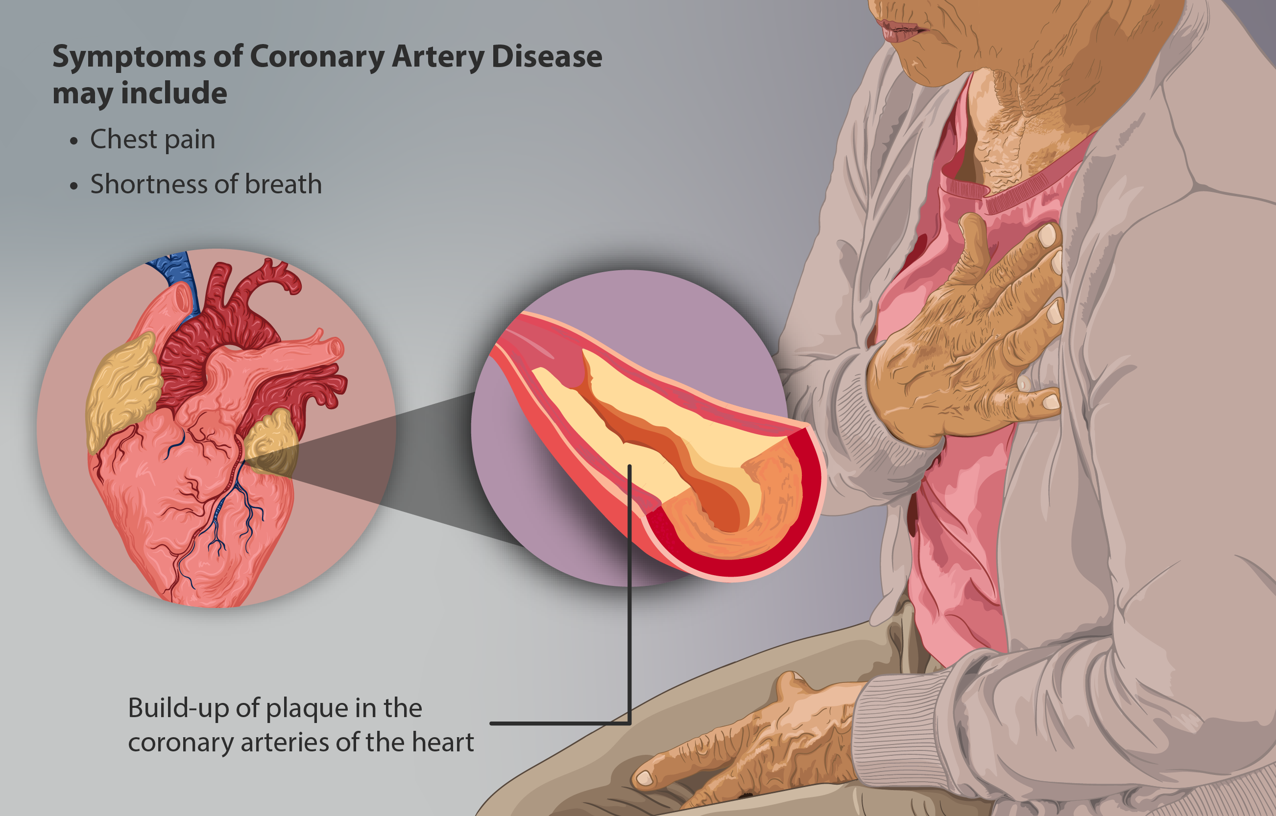
Coronary artery disease (CAD), also called coronary heart disease (CHD), ischemic heart disease (IHD), myocardial ischemia, or simply heart disease, involves the reduction of blood flow to the heart muscle due to build-up of atherosclerotic plaque in the arteries of the heart. It is the most common of the cardiovascular diseases. Types include stable angina, unstable angina, myocardial infarction, and sudden cardiac death. A common symptom is chest pain or discomfort which may travel into the shoulder, arm, back, neck, or jaw. Occasionally it may feel like heartburn. Usually symptoms occur with exercise or emotional stress, last less than a few minutes, and improve with rest. Shortness of breath may also occur and sometimes no symptoms are present. In many cases, the first sign is a heart attack. Other complications include heart failure or an abnormal heartbeat. Risk factors include high blood pressure, smoking, diabetes, lack of exercise, obesity, high blood cholesterol, p ... [...More Info...] [...Related Items...] OR: [Wikipedia] [Google] [Baidu] |
Intracranial Hemorrhage
Intracranial hemorrhage (ICH), also known as intracranial bleed, is hemorrhage, bleeding internal bleeding, within the Human skull, skull. Subtypes are intracerebral bleeds (intraventricular bleeds and intraparenchymal bleeds), subarachnoid bleeds, epidural bleeds, and subdural bleeds. More often than not it ends in a death, lethal outcome. Intracerebral bleeding affects 2.5 per 10,000 people each year. Signs and symptoms Intracranial hemorrhage is a serious medical emergency because the buildup of blood within the skull can lead to increases in intracranial pressure, which can crush delicate brain tissue or limit its blood supply. Severe increases in intracranial pressure (ICP) can cause brain herniation, in which parts of the brain are squeezed past structures in the skull. Causes Trauma is the most common cause of intracranial hemorrhage. It can cause epidural hemorrhage, subdural hemorrhage, and subarachnoid hemorrhage. Other condition such as hemorrhagic parenchymal con ... [...More Info...] [...Related Items...] OR: [Wikipedia] [Google] [Baidu] |
Long QT Syndrome
Long QT syndrome (LQTS) is a condition affecting repolarization (relaxing) of the heart after a heartbeat, giving rise to an abnormally lengthy QT interval. It results in an increased risk of an irregular heartbeat which can result in fainting, drowning, seizures, or sudden death. These episodes can be triggered by exercise or stress. Some rare forms of LQTS are associated with other symptoms and signs including deafness and periods of muscle weakness. Long QT syndrome may be present at birth or develop later in life. The inherited form may occur by itself or as part of larger genetic disorder. Onset later in life may result from certain medications, low blood potassium, low blood calcium, or heart failure. Medications that are implicated include certain antiarrhythmics, antibiotics, and antipsychotics. LQTS can be diagnosed using an electrocardiogram (EKG) if a corrected QT interval of greater than 480–500 milliseconds is found, but clinical findings, other EKG featur ... [...More Info...] [...Related Items...] OR: [Wikipedia] [Google] [Baidu] |
Antiarrhythmics
Antiarrhythmic agents, also known as cardiac dysrhythmia medications, are a group of pharmaceuticals that are used to suppress abnormally fast rhythms ( tachycardias), such as atrial fibrillation, supraventricular tachycardia and ventricular tachycardia. Many attempts have been made to classify antiarrhythmic agents. Many of the antiarrhythmic agents have multiple modes of action, which makes any classification imprecise. Vaughan Williams classification The Vaughan Williams classification was introduced in 1970 by Miles Vaughan Williams.Vaughan Williams, EM (1970) "Classification of antiarrhythmic drugs". In ''Symposium on Cardiac Arrhythmias'' (Eds. Sandoe E; Flensted-Jensen E; Olsen KH). Astra, Elsinore. Denmark (1970) Vaughan Williams was a pharmacology tutor at Hertford College, Oxford. One of his students, Bramah N. Singh, contributed to the development of the classification system. The system is therefore sometimes known as the Singh-Vaughan Williams classification. Th ... [...More Info...] [...Related Items...] OR: [Wikipedia] [Google] [Baidu] |
Epinephrine
Adrenaline, also known as epinephrine, is a hormone and medication which is involved in regulating visceral functions (e.g., respiration). It appears as a white microcrystalline granule. Adrenaline is normally produced by the adrenal glands and by a small number of neurons in the medulla oblongata. It plays an essential role in the fight-or-flight response by increasing blood flow to muscles, heart output by acting on the SA node, pupil dilation response, and blood sugar level. It does this by binding to alpha and beta receptors. It is found in many animals, including humans, and some single-celled organisms. It has also been isolated from the plant ''Scoparia dulcis'' found in Northern Vietnam. Medical uses As a medication, it is used to treat several conditions, including allergic reaction anaphylaxis, cardiac arrest, and superficial bleeding. Inhaled adrenaline may be used to improve the symptoms of croup. It may also be used for asthma when other treatments are not ef ... [...More Info...] [...Related Items...] OR: [Wikipedia] [Google] [Baidu] |
Digitalis
''Digitalis'' ( or ) is a genus of about 20 species of herbaceous perennial plants, shrubs, and biennials, commonly called foxgloves. ''Digitalis'' is native to Europe, western Asia, and northwestern Africa. The flowers are tubular in shape, produced on a tall spike, and vary in colour with species, from purple to pink, white, and yellow. The scientific name means "finger". The genus was traditionally placed in the figwort family, Scrophulariaceae, but phylogenetic research led taxonomists to move it to the Veronicaceae in 2001. More recent phylogenetic work has placed it in the much enlarged family Plantaginaceae. The best-known species is the common foxglove, '' Digitalis purpurea''. This biennial is often grown as an ornamental plant due to its vivid flowers which range in colour from various purple tints through pink and purely white. The flowers can also possess various marks and spottings. Other garden-worthy species include ''D. ferruginea'', ''D. grandiflora'', '' ... [...More Info...] [...Related Items...] OR: [Wikipedia] [Google] [Baidu] |
Thyrotoxicosis
Hyperthyroidism is the condition that occurs due to excessive production of thyroid hormones by the thyroid gland. Thyrotoxicosis is the condition that occurs due to excessive thyroid hormone of any cause and therefore includes hyperthyroidism. Some, however, use the terms interchangeably. Signs and symptoms vary between people and may include irritability, muscle weakness, sleeping problems, a fast heartbeat, heat intolerance, diarrhea, enlargement of the thyroid, hand tremor, and weight loss. Symptoms are typically less severe in the elderly and during pregnancy. An uncommon complication is thyroid storm in which an event such as an infection results in worsening symptoms such as confusion and a high temperature and often results in death. The opposite is hypothyroidism, when the thyroid gland does not make enough thyroid hormone. Graves' disease is the cause of about 50% to 80% of the cases of hyperthyroidism in the United States. Other causes include multinodular goite ... [...More Info...] [...Related Items...] OR: [Wikipedia] [Google] [Baidu] |
Hypercalcemia
Hypercalcemia, also spelled hypercalcaemia, is a high calcium (Ca2+) level in the blood serum. The normal range is 2.1–2.6 mmol/L (8.8–10.7 mg/dL, 4.3–5.2 mEq/L), with levels greater than 2.6 mmol/L defined as hypercalcemia. Those with a mild increase that has developed slowly typically have no symptoms. In those with greater levels or rapid onset, symptoms may include abdominal pain, bone pain, confusion, depression, weakness, kidney stones or an abnormal heart rhythm including cardiac arrest. Most outpatient cases are due to primary hyperparathyroidism and inpatient cases due to cancer. Other causes of hypercalcemia include sarcoidosis, tuberculosis, Paget disease, multiple endocrine neoplasia (MEN), vitamin D toxicity, familial hypocalciuric hypercalcaemia and certain medications such as lithium and hydrochlorothiazide. Diagnosis should generally include either a corrected calcium or ionized calcium level and be confirmed after a week. Specif ... [...More Info...] [...Related Items...] OR: [Wikipedia] [Google] [Baidu] |
Hypokalemia
Hypokalemia is a low level of potassium (K+) in the blood serum. Mild low potassium does not typically cause symptoms. Symptoms may include feeling tired, leg cramps, weakness, and constipation. Low potassium also increases the risk of an abnormal heart rhythm, which is often too slow and can cause cardiac arrest. Causes of hypokalemia include vomiting, diarrhea, medications like furosemide and steroids, dialysis, diabetes insipidus, hyperaldosteronism, hypomagnesemia, and not enough intake in the diet. Normal potassium levels are between 3.5 and 5.0 mmol/L (3.5 and 5.0 mEq/L) with levels below 3.5 mmol/L defined as hypokalemia. It is classified as severe when levels are less than 2.5 mmol/L. Low levels may also be suspected based on an electrocardiogram (ECG). Hyperkalemia is a high level of potassium in the blood serum. The speed at which potassium should be replaced depends on whether or not there are symptoms or abnormalities on an electrocardiogram. Pot ... [...More Info...] [...Related Items...] OR: [Wikipedia] [Google] [Baidu] |


.jpg)


